EFFECT OF CURCUMIN ON AN ANIMAL MODEL WITH HEPATIC FIBROSIS VIA TGF
1,2
Rongrong Wu,
2Yongxiang Li,
2Mingyang Zhang,
1
Zhejiang Provincial Key Laboratory for Technology & Application of Model Organisms, School of Life
Sciences, Wenzhou Medical University, Wenzhou, Zhejiang, 325035, China
2
Department of Clinical Laboratory, Jiaxing Maternity and Child Health Care Hospital,
University Affiliated Women and Children Hospital,
3
Obstetrics Department, Jiaxing Maternity and Child Health Care Hospital,
Women and Children Hospital
ARTICLEINFO ABSTRACT
To study the protective effects and associated mechanisms of curcumin on hepatic fibrosis disease in rats. Thirty
curcumin group.
combination with a high fat diet to induce a hepatic fibrosis model, and the curcumin group followed the meth
weeks, the effects of curcumin on lipid accumulation, hepatic fibrosis gene expression and Smad activities were evaluated by biochemical analysis, HE
western blot.Curcumin treatment significantly alleviated steatosis, fibrosis and cell necrosis in CCl
the mRNA levels of
proteins in the hepatic fibrosis livers. Curcumin was effective in preventing CCl hepatic fibrosis. The effect was related to
inhibiting the TGF
Copyright©2019, Rongrong Wu et al. This is an open access distribution, and reproduction in any medium, provided
INTRODUCTION
The development of liver fibrosis is regulated by a variety of cytokines and their molecular signaling pathways
Therefore, it is practical to block the progression from liver fibrosis to cirrhosis by interfering with signaling pathways for the study and prevention of liver fibrosis. At present, the molecular signaling pathway has become an important target for drug treatment. Furthermore, the study of how the active components of traditional Chinese medicine interfere with the molecular signaling pathway of liver fibrosis has become of interest recently. Curcumin is a kind of plant polyphenol compound extracted from turmeric rhizomes. The main chain contains unsaturated aliphatic and aromatic groups. To date, several studies have found that curcumin can alleviate liver damage caused by a high-fat diet, ischemia
through antioxidative stress as well as
anti-lipid-lowering effects (Badria et al., 2015; Afrin
ISSN: 0975-833X
Article History:
Received 19th February, 2019 Received in revised form 26th March, 2019 Accepted 28th April, 2019 Published online 30th May, 2019
Citation: Rongrong Wu, Yongxiang Li, Mingyang Zhang, LiliXue, Zhen Zhou, Li Shen and Jimin Gao
hepatic fibrosis via tgf-β/smad signaling pathway”, International
Key Words:
Curcumin, Liver Fibrosis, TGF-β, Smad3, α-SMA.
*Corresponding author: Jimin Gao
RESEARCH ARTICLE
EFFECT OF CURCUMIN ON AN ANIMAL MODEL WITH HEPATIC FIBROSIS VIA TGF
SIGNALING PATHWAY
Mingyang Zhang,
3LiliXue,
2Zhen Zhou,
2Li Shen and
Zhejiang Provincial Key Laboratory for Technology & Application of Model Organisms, School of Life
Sciences, Wenzhou Medical University, Wenzhou, Zhejiang, 325035, China
Department of Clinical Laboratory, Jiaxing Maternity and Child Health Care Hospital,
University Affiliated Women and Children Hospital, Jiaxing 314001, China
Jiaxing Maternity and Child Health Care Hospital, Jiaxing Universi
Women and Children Hospital, Jiaxing 314001, China
ABSTRACT
To study the protective effects and associated mechanisms of curcumin on hepatic fibrosis disease in Thirty SD rats were randomly divided into three groups: a control group,
curcumin group. The control group was fed a normal diet, the model group
combination with a high fat diet to induce a hepatic fibrosis model, and the curcumin group followed the methods of the model group but was orally administered curcumin starting at 10
weeks, the effects of curcumin on lipid accumulation, hepatic fibrosis gene expression and Smad activities were evaluated by biochemical analysis, HE-staining, qua
western blot.Curcumin treatment significantly alleviated the serum liver fibrosis index, hepatic steatosis, fibrosis and cell necrosis in CCl4-induced hepatic fibrosis rats. Moreover, curcumin reduced
the mRNA levels of collagen I, α-SMA, and Smad3 as well as the levels of proteins in the hepatic fibrosis livers. Curcumin was effective in preventing CCl
hepatic fibrosis. The effect was related to decreased lipid deposition, and it relieved liver fibrosis via inhibiting the TGF-β/Smad signaling pathway in the hepatic fibrosis livers.
access article distributed under the Creative Commons Attribution the original work is properly cited.
fibrosis is regulated by a variety of cytokines and their molecular signaling pathways (Lee, 2015). Therefore, it is practical to block the progression from liver fibrosis to cirrhosis by interfering with signaling pathways for liver fibrosis. At present, the molecular signaling pathway has become an important target for drug treatment. Furthermore, the study of how the active components of traditional Chinese medicine interfere with the sis has become of Curcumin is a kind of plant polyphenol compound extracted from turmeric rhizomes. The main chain contains unsaturated aliphatic and aromatic groups. To date, several studies have found that curcumin can alleviate liver fat diet, ischemia-reperfusion, etc. -inflammatory and
Afrin et al., 2015).
In a model of acute liver injury induced by CCl
curcumin can reduce inflammatory factors and inhibit the expression of NF-κB by increasing the level of glutathione peroxidase (GSH-Px) in the liver, which can protect the liver through anti-inflammatory effects and antioxidative stress. The mechanism of action of curcumin in rat antifibrosis has not been fully elucidated. This experiment intends to induce a rat model of liver fibrosis through a high
tetrachloride. Drug intervention with curcumin will be observed by the liver fibrosis index a
as well as α-SMA and Smad3 protein expression. The molecular mechanisms of the protective effects of curcumin on liver fibrosis in rats was also studied.
MATERIALS AND METHODS
Reagents: Curcumin was purchased from Sigma. Laminin
(LN), type III procollagen (PIIINP), hyaluronic acid (HA) and type IV collagen (IV-C) assay kits were purchased from
International Journal of Current Research Vol. 11, Issue, 05, pp.3972-3976, May, 2019
DOI: https://doi.org/10.24941/ijcr.35324.05.2019
Rongrong Wu, Yongxiang Li, Mingyang Zhang, LiliXue, Zhen Zhou, Li Shen and Jimin Gao. 2019. “Effect of International Journal of Current Research, 11, (04),3972-3976.
EFFECT OF CURCUMIN ON AN ANIMAL MODEL WITH HEPATIC FIBROSIS VIA TGF-Β/SMAD
Li Shen and
1,*Jimin Gao
Zhejiang Provincial Key Laboratory for Technology & Application of Model Organisms, School of Life
Sciences, Wenzhou Medical University, Wenzhou, Zhejiang, 325035, China
Department of Clinical Laboratory, Jiaxing Maternity and Child Health Care Hospital, Jiaxing
Jiaxing 314001, China
Jiaxing University Affiliated
, Jiaxing 314001, China
To study the protective effects and associated mechanisms of curcumin on hepatic fibrosis disease in control group, a model group and a model group was administered CCl4 in
combination with a high fat diet to induce a hepatic fibrosis model, and the curcumin group followed ods of the model group but was orally administered curcumin starting at 10 weeks. After 14 weeks, the effects of curcumin on lipid accumulation, hepatic fibrosis gene expression and Smad-3 staining, quantitative real-time PCR and serum liver fibrosis index, hepatic induced hepatic fibrosis rats. Moreover, curcumin reduced SMA, and Smad3 as well as the levels of α-SMA and Smad3 proteins in the hepatic fibrosis livers. Curcumin was effective in preventing CCl4-induced increases in
lipid deposition, and it relieved liver fibrosis via β/Smad signaling pathway in the hepatic fibrosis livers.
License, which permits unrestricted use,
In a model of acute liver injury induced by CCl4 in rats, min can reduce inflammatory factors and inhibit the κB by increasing the level of glutathione Px) in the liver, which can protect the liver matory effects and antioxidative stress. The of curcumin in rat antifibrosis has not This experiment intends to induce a rat model of liver fibrosis through a high-fat diet and carbon tetrachloride. Drug intervention with curcumin will be observed by the liver fibrosis index and liver histomorphology SMA and Smad3 protein expression. The molecular mechanisms of the protective effects of curcumin on liver fibrosis in rats was also studied.
MATERIALS AND METHODS
Curcumin was purchased from Sigma. Laminin (LN), type III procollagen (PIIINP), hyaluronic acid (HA) and C) assay kits were purchased from
INTERNATIONAL JOURNAL OFCURRENTRESEARCH
Suzhou Changguang HYBIOME company. Aspartate aminotransferase (AST) and alanine aminotransferase (ALT) were purchased from Abbott Laboratories, USA. Hematoxylin and eosin were purchased from Beijing Suobao Biotech company. TRIZOL was purchased from Invitrogen, USA. The reverse transcription kit and SYBR Green PCR kit were purchased from TOYOTA Corporation, Japan. The primer sequence was synthesized by the Shanghai Synthetic Department of Invitrogen (Table 1). α-SMA protein antibody was purchased from Abcam, USA. p-Smad3 antibody was purchased from Cell Signaling Technology, USA. The secondary antibody was purchased from Pepro Tech, USA.
Instruments: Fully automatic chemiluminescence
immunoassay analyzer AE-180 (Suzhou Changguang
HYBIOME company); automatic biochemical analyzer C16000 (Abbott Company, USA); optical microscope (Olympus, Japan); NanoDrop instrument (Thermo Scientific, USA); 7500 PCR instrument (ABI, USA); Gel Doc XR gel imaging system (Bio-Rad, USA).
Animal experiments: Male 10-week-old Sprague-Dawley
rats, weighing 220-250 g, were provided by the Experimental Animal Center of Zhejiang Medical College, animal use license number: SYXY (Zhejiang) 2013-019. The animals were raised in a standard environment (temperature (24 ± 2) °C, 12hlight:dark cycle, free diet).After 1 week of adaptive feeding, the rats were randomly divided into three groups: a control group of 10 rats fed a normal diet for 14 weeks; a model group of 10 rats fed a high-fat diet for 14 weeks and injected with CCl4 into the abdomen; and a curcumin group of
10 rats with a high-fat diet and CCl4 intraperitoneal injection.
Starting from the 10th week, 100 mg·kg-1 curcumin was
administered intragastrically daily for 4 weeks in the curcumin group. After the rats were sacrificed, the livers were collected. Half of the fresh liver tissue was frozen in liquid nitrogen and half was placed in formalin. The experimental procedure was in accordance with the Regulations on the Administration of Laboratory Animals outlined by the Chinese Science and Technology Commission in 1988 and approved by the Experimental Animal Ethics Committee of the Jiaxing Maternal and Child Health Hospital.
Serum test: The serum test was conducted according
to the kit instructions. ALT and AST were detected by an automatic biochemical analyzer C16000, and LN, PIIINP, HA and IV-C were detected by an automatic chemiluminescence immunoassay analyzer.
HE staining: Fresh liver tissue was fixed with 10%
formalin solution for 24 h and embedded in paraffin with a 4μm thickness. Then, the samples were dehydrated with alcohol. Routine HE was used to stain the tissues, and pathological changes in liver tissue were observed under a light microscope.
Real-time PCR detection: Total RNA was extracted
from liver tissue by TRIzolreagent, and RNA concentrations were detected with a Nano Drop spectrophotometer. The RNA was reverse transcribed into cDNA using a reverse transcription kit, and
real-time PCR was performed following the
manufacturer’s protocol (50 °C for 2 min; 95 °C for 10 min; followed by 40 cycles of 95 °Cfor 15 s, 60 °C for 1 min, 72 °C for 30 s; and finally 72 °C for a full extension of 10 min). β-actin was used as an internal
Western blot detection: The liver tissue was
removed from the -80 °C refrigerator. Total protein was extracted using RIPA lysate, and the protein was quantified by the BCA method. The protein was separated using SDS-PAGE (12% separating gel), and the target proteins were transferred onto PVDF. The membranes were blocked with 5% bovine serum albumin and then primary antibody (α-SMA diluted 1:500, p-Smad3 diluted 1:1000) was added at 4 °C and incubated overnight. The secondary antibody (diluted 1:2000) was incubated for 1 h, developed, exposed, and photographed with Image Lab. The ratio of the gray values of the target protein and GAPDH were used as the relative expression amount of the target protein. The results were analyzed using Quantity One V 4.6 software.
Statistical analysis: The experimental data are
expressed as the means ± standard deviations (SDs), and statistical analysis was performed with GraphPad 5.0. The data of the three groups were compared by one-way analysis of variance (ANOVA)(intergroup). P<0.05 was considered statistically significant.
RESULTS
Curcumin decreased the levels of ALT, PIIINP and HA in
rats with hepatic fibrosis: The levels of ALT and AST (liver
function indexes) in the model group were significantly increased compared with those in the control group (P<0.05). Furthermore, the levels of LN, PIIINP, HA and IV-C (hepatic fibrosis indexes) in themodel group were significantly higher than those in the control group. However, the levels of AST, PIIINP and HA were significantly decreased in the curcumin group compared with those in the model group (P<0.05) (Table 2).
Curcumin decreased the levels of hepatic steatosis, necrosis
andfibrosis in rats with hepatic fibrosis: HE staining showed
that the liver cells were arranged neatly with no obvious degeneration or necrosis, and the structure of the hepatic lobule was intact in the control group. However, the hepatocytes had obvious balloon-like changes and steatosis, disordered arrangement and necrosis, and the destruction of the hepatic lobular structure and the degree of fibrosis were more obvious in the model group than those in the control group. Hepatic steatosis and necrosis were significantly reduced in the curcumin group compared with those in the model group, and the degree of fibrosis was less than that in the model group. (Fig. 1).
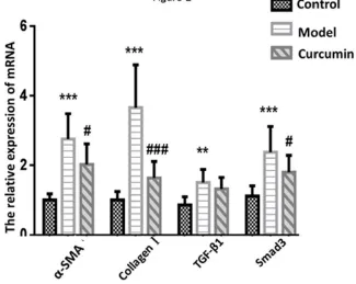
Curcumin decreased the mRNA expression levels of α-SMA, collagen I, TGF-β and Smad3 in rats with hepatic
fibrosis: The mRNA expression levels of α-SMA, collagen I,
TGF-β and Smad3 were significantly increased in the model group compared with those in the control group (P<0.05). However, the mRNA expression levels of α-SMA, collagen I and Smad3 were significantly lower in the control group than those in the model group (P < 0.05)(Fig. 2).
Curcumin decreased the protein expression levels of
α-SMA and Smad3 in rats with hepatic fibrosis: The protein
expression of α-SMA and Smad3 in the model group was significantly higher than that in the control group (P<0.05),
Table 1. Sequences for primers (forward primer; reverse
Gene name
Collagen TGGCCTTGGAGGAAACTTTG
α-SMA ACTGGGACGACATGGAAAAG
TGF-β1 TTGCCCTCTACAACCAACACAA
Smad3 AGGGCTTTGAGGCTGTCTACC
[image:3.595.121.490.182.251.2]β-actin ACGGCCAGGTCATCACTATTG
Table 2 Comparison of
Biochemical indicators ALT IU·L-1
AST IU·L-1
LN(μg·L-1)
P NP(μg·L-1)
HA(μg·L-1)
IV-C(μg·L-1)
Data are presented as mean ± SD.
the control group, #P < 0.05, significantly different from the values in the
Figure 1. Histological images of liver tissues stained with H & E, ×200, scale bar: 10
Figure 2. The mRNA levels of collagen I,
are presented as mean ± SD. **P < 0.01, ***
###
P < 0.001, significantly different fro
Sequences for primers (forward primer; reverse primer) used in qPCR
Forward primer sequence Reverse primer sequence
TGGCCTTGGAGGAAACTTTG CTTGGAAACCTTGTGGACCAG
ACTGGGACGACATGGAAAAG GTTCAGTGGTGCCTCTGTCA
TTGCCCTCTACAACCAACACAA GCTTGCGACCCACGTAGTA
AGGGCTTTGAGGCTGTCTACC GTCCAC GCTGGCATCTTCTG
[image:3.595.72.545.283.447.2]ACGGCCAGGTCATCACTATTG CAAGAAGGAAGGCTGGAAAAG
Table 2 Comparison of biochemical indicators in serum
Control Model
46.52±6.32 86.11±14.97* 62.63±12.52
114.19±8.59 217.63±21.16*** 159.87±11.55
64.02±4.58 174.04±18.32*** 132.44±16.71
35.95±3.96 85.81±12.52** 53.60±8.32
86.74±8.42 192.31±20.36*** 142.41±12.09
47.14±4.11 86.08±8.62** 68.14±7.51
Data are presented as mean ± SD. *P < 0.05, **P < 0.01, ***P < 0.001, significantly different from the values in
< 0.05, significantly different from the values in the model group.
Histological images of liver tissues stained with H & E, ×200, scale bar: 100 μm (control, model, curcumin)
collagen I, α-SMA, TGF-β, and Smad3 in the liver tissues were examined by q
***P < 0.001, significantly different from the values in the control group,
< 0.001, significantly different from the values in the model group
primer) used in qPCR
Reverse primer sequence
CTTGGAAACCTTGTGGACCAG GTTCAGTGGTGCCTCTGTCA GCTTGCGACCCACGTAGTA GTCCAC GCTGGCATCTTCTG CAAGAAGGAAGGCTGGAAAAG
Curcumin 62.63±12.52 159.87±11.55#
132.44±16.71 53.60±8.32#
142.41±12.09#
68.14±7.51 < 0.001, significantly different from the values in
0 μm (control, model, curcumin)
in the liver tissues were examined by q-PCR. Data
< 0.001, significantly different from the values in the control group, #P < 0.05,
[image:3.595.143.468.493.752.2]while the protein expression of α-SMA and Smad3 in the curcumin group decreased (P<0.05) (Fig. 3).
DISCUSSION
Liver fibrosis is a process of repairing persistent tissue damage caused by external stimuli such as viral hepatitis, alcohol, drugs, and toxic substances. In the process of liver fibrosis, α-SMA is a marker of hepatic stellate cell activation. Activated hepatic stellate cells can synthesize additional α-SMA and collagen I that are abnormally deposited in the inner cell, which can cause liver tissue morphology abnormalities and
dysfunction5).Therefore, reducing liver fibrosis indicators and
liver tissue pathological changes is considered to be an effective strategy for the treatment of liver fibrosis. The results of our study show that curcumin can significantly reduce the serum levels in rats with CCl4-induced liver fibrosis and improve the degree of steatosis and fibrosis of liver tissue. Our results suggest that curcumin has obvious protective effects on the liver in rats with hepatic fibrosis. In addition, curcumin can significantly down regulate the mRNA expression levels of collagen I, α-SMA, TGF-β, and Smad3 as well as the protein expression levels of α-SMA and Smad3.
Therefore, we speculate that the protective effect of curcumin on the liver of rats with hepatic fibrosis may be through the regulation of the TGF-β/Smad signaling pathway. Many cytokines and signaling pathways are involved in the development of liver fibrosis. Among them, TGF-β is one of the most effective profibrotic factors, and it plays a key role in promoting the development of liver fibrosis Caja, 2018. TGF-β can not only induce the expression of collagen in the liver but also activate hepatic stellate cells. The activated stellate cells can synthesize and secrete a large number of collagen I and α-SMA and can participate in the formation of liver fibrosis and the reconstruction of the liver structure. Smad is a downstream signaling molecule of TGF-β, and Smad plays a key role in the passing process of TGF-β signaling from cell surface receptors
to the nucleus Wang et al., 2017; Chen et al., 2017). Research
shows that the JianpiRuangan Recipe can significantly inhibit the expression of Smad3, Smad7, and TGF-β1 as well as receptors in the TGF-β/Smad signaling pathway, which can effectively alleviate the degree of liver fibrosis induced by
CCl4 in the rat model. In addition, ursolic acid extracted from
medicinal plants is a natural triterpenoid compound that can
inhibit the TGF-β signaling pathway and reduce the expression of collagen I, and it has a clear antifibrosis effect. The results of our study also confirmed that the expression levels of collagen I, α-SMA and Smadwere significantly increased while upregulating TGF-β, which is consistent with studies at home and abroad. The addition of curcumin simultaneously reduced these indicators, indicating that curcumin effectively alleviated CCl4-induced liver fibrosis in rats through the TGF-β/Smad signaling pathway. In conclusion, curcumin can reduce liver hepatic lesions and improve liver fibrosis by downregulating the TGF-β/Smad signaling pathway, and this result has potential therapeutic value for liver fibrosis treatment. The study of curcumin provides a new strategy for the study of liver fibrosis in Chinese medicine.
Acknowledgements
This work was supported by grants from the Natural Science Foundation of China (91729101 and 81573110) and Wenzhou Municipal Research Program (Y20160074, Y20160009 and ZS2017014).
Conflicts of interest: The authors declare that they have no
competing interests.
REFERENCES
Afrin R., Arumugam S., Soetikno V., Thandavarayan RA., Pitchaimani V., Karuppagounder V., Sreedhar R., Harima M., Suzuki H., Miyashita S., Nomoto M., Suzuki K., Watanabe K. 2015. Curcumin ameliorates streptozotocin-induced liver damage through modulation of endoplasmic
reticulum stress-mediated apoptosis in diabetic rats. Free
radical research,49, 279-289.
Badria FA., Ibrahim AS., Badria AF., Elmarakby AA. 2015. Curcumin Attenuates Iron Accumulation and Oxidative Stress in the Liver and Spleen of Chronic Iron-Overloaded
Rats. PloS one,10, e0134156.
Caja L., Dituri F., Mancarella S., Caballero-Diaz D., Moustakas A., Giannelli G., Fabregat I. 2018. TGF-beta and the Tissue Microenvironment: Relevance in Fibrosis
and Cancer. International journal of molecular sciences,19
[image:4.595.57.552.47.239.2]Chen Q., Zhang H., Cao Y., Li Y., Sun S., Zhang J., Zhang G. 2017. Schisandrin B attenuates CCl4-induced liver fibrosis in rats by regulation of Nrf2-ARE and TGF-beta/Smad
Figure 3: Expression of proteins in the liver tissues. The protein expressions of α-SMAand Smad3 were detected by western blots.
Quantitative analyses of the immunoblots were shown for α-SMA and Smad3. Data are presented as mean ± SD. *P < 0.05, ***P < 0.001,
significantly different from the values in the control group, #P < 0.05, significantly different from the values in the model group.
signaling pathways. Drug design, development and therapy,11, 2179-2191.
Lee HY., Kim SW., Lee GH., Choi MK., Jung HW., Kim YJ., Kwon HJ., Chae HJ. 2016. Turmeric extract and its active compound, curcumin, protect against chronic
CCl4-induced liver damage by enhancing antioxidation. BMC
complementary and alternative medicine,16, 316.
Lee YA., Wallace MC., Friedman SL. 2015. Pathobiology of
liver fibrosis: a translational success story. Gut,64,
830-841.
Wang Y., Shen RW., Han B., Li Z., Xiong L., Zhang FY., Cong BB., Zhang B. 2017. Notch signaling mediated by TGF-beta/Smad pathway in concanavalin A-induced liver
fibrosis in rats. World journal of gastroenterology,23,
2330-2336.
Yin C., Evason KJ., Asahina K., Stainier DY. 2013. Hepatic stellate cells in liver development, regeneration, and cancer.
The Journal of clinical investigation,123, 1902-1910.