ISSN Online: 2165-3410 ISSN Print: 2165-3402
DOI: 10.4236/aim.2019.912062 Dec. 6, 2019 971 Advances in Microbiology
Clinical Utility of Molecular Diagnosis of Blood
Stream Infections in Allogeneic Hematopoietic
Stem Cell Transplantation Recipients with
Hematologic Malignancies
Atsushi Fujieda
1, Kazunori Nakase
2, Akiko Nakamura
3, Kohshi Ohishi
4,
Yuka Sugimoto
1, Fumihiko Monma
1, Masahiro Masuya
1, Naoyuki Katayama
11Department of Hematology and Oncology, Mie University Hospital, Tsu, Japan 2Cancer Center, Mie University Hospital, Tsu, Japan
3Central Clinical Laboratories, Mie University Hospital, Tsu, Japan 4Transfusion Service, Mie University Hospital, Tsu, Japan
Abstract
Blood stream infections (BSIs) are a serious problem in patients with hema-tologic malignancies receiving allogeneic hematopoietic stem cell transplan-tation (ASCT). We evaluated the clinical utility of molecular diagnosis for the management of BSIs in such patients. We prospectively performed a polyme-rase chain reaction (PCR) analysis of microbial DNA in blood samples from 10 consecutive patients with hematological malignancies at least once a week for one month after ASCT. In total, 51 and 54 samples were analyzed by bac-terial and fungal PCR assays, respectively. Bacteria were detected in 24 sam-ples from 8 patients by PCR, but in only 2 samsam-ples from one patient by blood culture. Notably, the bacteria detected in at least half of the 24 samples were considered to have originated from the oral cavity. Fungi were detected in 5 samples from 3 patients by PCR, but not by blood culture. Most cases with positive PCR results were manageable with empirical antimicrobial therapy without disclosure of DNA data. Our DNA analyses did not directly contri-bute to management of BSIs, but did provide valuable microbiological evi-dence for the patients. Additionally, oral management appears to require a critical re-evaluation to reduce the occurrence of BSIs in ASCT recipients.
Keywords
Allogeneic Hematopoietic Stem Cell Transplantation, Blood Stream Infection, PCR Analysis
How to cite this paper: Fujieda, A., Na-kase, K., Nakamura, A., Ohishi, K., Sugimoto, Y., Monma, F., Masuya, M. and Katayama, N. (2019) Clinical Utility of Molecular Diagnosis of Blood Stream Infections in Allogeneic Hematopoietic Stem Cell Trans-plantation Recipients with Hematologic Malignancies. Advances in Microbiology, 9, 971-982.
https://doi.org/10.4236/aim.2019.912062
Received: November 6, 2019 Accepted: December 3, 2019 Published: December 6, 2019
Copyright © 2019 by author(s) and Scientific Research Publishing Inc. This work is licensed under the Creative Commons Attribution International License (CC BY 4.0).
DOI: 10.4236/aim.2019.912062 972 Advances in Microbiology
1. Introduction
Allogeneic hematopoietic stem cell transplantation (ASCT) offers the chance of cure for patients with hematologic malignancies. However, blood stream infec-tions (BSIs) are major causes of morbidity and mortality for patients undergoing ASCT [1]. Therefore, successful management of these infectious complications is essential to further improve the clinical outcome of such patients. BSIs com-monly develop early after ASCT despite the use of prophylactic anti-infective drugs [1] [2]. However, conventional blood culture (BC), which is the gold standard for identifying causative pathogens, has a low isolation rate and is very time-consuming [3]. At present, there is no solution for BSI management other than empirical antimicrobial therapy despite the lack of microbiological evi-dence in a majority of ASCT recipients. On the other hand, polymerase chain reaction (PCR)-based analysis of microbial DNA is a promising tool for the sen-sitive and rapid detection of etiological organisms [3] [4]. PCR assays are re-portedly useful in managing febrile neutropenia in patients with hematologic disorders [5] [6]. To our knowledge, however, these molecular techniques have not been well evaluated in hematologic patients undergoing ASCT.
In this study, we prospectively applied an original PCR assay to find causative organisms in peripheral blood (PB) samples of patients with hematologic ma-lignancies who had received ASCT, and we evaluated its clinical usefulness for managing BSIs. Our DNA data provide valuable microbiological information for the management to reduce the occurrence of BSIs in ASCT recipients.
2. Materials and Methods
2.1. Patients and Definitions
DOI: 10.4236/aim.2019.912062 973 Advances in Microbiology Table 1. Patient characteristics and conditioning regimen.
Patient
no. Age/Sex Underlying disease transplantation Status at Conditioning Stem cell source GVHD prophylaxis Prophylactic antibiotics
1 49/male ATLL CR1 TBI (12 Gy) + CY UR-BM Tacrolimus + sMTX LVFX
2 44/male AML CR1 Flu + BU + TBI (2 Gy) UR-BM Tacrolimus + sMTX -
3 20/male ALL CR1 TBI (12 Gy) + CY Rel-PB Ciclosporin + sMTX TFLX
4 45/female ALL CR2 TBI (12 Gy) + CA + CY UR-CB Tacrolimus + sMTX LVFX
5 44/male LBL PR1 TBI (12 Gy) + CA + CY UR-CB Tacrolimus + sMTX LVFX
6 44/male MDS CR1 TBI (12 Gy) + CA + CY UR-CB Tacrolimus + sMTX LVFX
7 62/male ALL CR2 Flu + Mel + TBI (4 Gy) UR-CB Tacrolimus + sMTX LVFX
8 24/male AML CR2 TBI (12 Gy) + CY UR-BM Tacrolimus + sMTX LVFX
9 46/male ATLL CR1 TBI (12 Gy) + CY Rel-PB Ciclosporin + sMTX CPFX
10 19/male ALCL primary refractory TBI (12 Gy) + CY UR-BM Tacrolimus + sMTX LVFX
ATLL adult T-cell leukemia/lymphoma, AML acute myeloid leukemia, ALL acute lymphoblastic leukemia, LBL lymphoblastic lymphoma, MDS myelodys-plastic syndrome, ALCL anamyelodys-plastic large cell lymphoma, CR complete remission, PR patial remission, TBI total body irradiation, CY cyclophosphamide, flu fludarabine, CA cytarabine, Mel melphalan, UR-BM unrelated bone marrow, Rel-PB related peripheral blood, CB cord blood, sMTX short methotrexate, LVFX levofloxacin, TFLX tosufloxacin, CPFX ciprofloxacin.
the European Organization of Research and Treatment of Cancer/Mycosis Study Group (EORTC/MSG) [7]. In neutropenic (≤500/μl) patients at the onset of fev-er (≥38˚C) and in afebrile patients with neutropenia but with signs or symptoms of infection, empirical antibacterial therapy was initiated with broad-spectrum β-lactam antibiotics [cefepime, meropenem (MEPM), or doripenem (DRPM)] with or without aminoglycoside (arbekacin), and when a substantial mucosal injury occurred, a glycopeptide [vancomycin (VCM) or teicoplanin] was added [8]. As a surrogate marker for mucosal injury, we adopted the severity of stoma-titis and diarrhea according to the National Cancer Institute Common Termi-nology Criteria for Adverse Events Scales (NCI-CTCAE) [9]. PCR results were not shared with the physicians, but were disclosed if fever continued despite em-pirical antimicrobial treatment and if the physician requested them. For febrile ep-isodes, BC was performed as necessary. Blood samples were cultured in an auto-mated system (BacT/Alert 3D, BioMerieux, France). Serological (1-3)-β-D-glucan (BDG) and galactomannan antigen (GM) assays were performed at least once weekly. BDG and GM were measured in each sample using the Beta-glucan test (Wako Pure Chemical Industries, Ltd., Osaka, Japan) and the Platelia™ Aspergillus enzyme immunoassay(Bio-Rad, Marnes-la-Coquette, France), respectively. GM results were recorded as an index relative to the mean optical density of the thre-shold controls (GM index = optical density of sample/mean optical density of the threshold control sample). A positive GM result was defined as an index value of ≥ 0.5. A positive BDG level was defined as a serum level of ≥ 11 pg/ml.
2.2. Molecular Detection of Bacteria and Fungi
DOI: 10.4236/aim.2019.912062 974 Advances in Microbiology detect various pathogenic bacteria and fungi, and the broad range primers, which amplify a conserved region of the bacterial 16S rRNA gene and the fungal 18S rRNA gene, were used. Extraction and purification of DNA from bacteria and fungi were performed, as we previously described [6] [10]. EDTA-anticoagulated PB (1 ml) was centrifuged at 1000g for 10 min. The supernatant was used for detection of bacterial DNA, while the buffy coat was used for detection of fungal DNA. The supernatant was centrifuged at 13,000 g for 10 min, and the pellet was washed with phosphate-buffered saline (PBS). Extraction and purification of Bacterial DNA were performed using Mora Extract (Kyokuto Seiyaku Co., Ltd., Tokyo, Japan). For fungal DNA, the buffy coat was washed twice with PBS and centrifuged at 3000 g for 10 min [6] [10]. The supernatant was decanted off and the pellet was reacted with 50 μl of lysis buffer (COBAS® AMPLICOR S.E.T.S II Kit; Roche Diagnostics, Meylan, France) at room temperature for 2 min and then centrifuged at 1000 g for 1 min. The pellet was again incubated with lysis buffer at 90˚C for 20 min, and then centrifuged at 13,000 g for 10 min. Fungal DNA was extracted and purified from the pellet, using Mora Extract (Kyokuto Seiyaku). For amplification and detection of bacterial DNA the complete 16S rRNA gene was amplified by PCR using two oligonucleotide primers: UN-F: 5'-CAG CAG CCG CGC TAA TAC-3' and UN-R: 5'-CCG TCA ATT CCT TTG AGT TT-3'. PCR reactions were carried out in a DNA thermal cycler (GeneAmp PCR System 9600; Applied Biosystems, Foster City, CA, USA) with preliminary denaturation at 95˚C for 10 min, followed by 45 cycles of amplification consist-ing of denaturation at 94˚C, primer annealconsist-ing at 62˚C and elongation at 72˚C, each lasting for 1 min. For species identification, positive PCR products were sequenced using the ABI PRISM BigDye terminator cycle sequencing ready reaction kit and ABI PRISM 377 genetic analyzer (Applied Biosystems). For phylogenetic identification, sequences were compared with those of known bac-teria listed in official databases using the BLAST program available at the Na-tional Center for Biotechnology Information (http://ncbi.nlm.nih.gov). For fun-gal DNA, the complete 18S rRNA gene was amplified by PCR using two oligo-nucleotide primers: Fung-F: 5'-TTCGATGGTAGGATAGTGGCC-3 and B4R: 5'-TGA TCG TCT TCG ATC CCC TA-3'. This broad-range PCR system was able to detect a wide range of fungi other than Zycomycetes and Fusarium spe-cies. PCR reactions were conducted in a DNA thermal cycler with preliminary denaturation at 94˚C for 5 min, followed by 40 cycles of amplification consisting of 94˚C for 30 sec, 55˚C for 1 min, and 72˚C for 1 min. After amplification, all PCR products were precipitated by the addition of ethanol and amplified by nested PCR. The primers were designed to separate PCR products into groups based on their susceptibilities to antifungal agents. We used reacting systems for the group resistant to fluconazole (FLCZ) (Fung-F and n-Asp/Pen R:
DOI: 10.4236/aim.2019.912062 975 Advances in Microbiology and a wide-range fungal group (n-Fung-F: 5'-GAATAAGGGTTCGATTCCGG-3' and n-Fung-R: 5'-CCCCGACCGTCCCTATTAAT-3'). The wide-range fungal PCR was simultaneously performed during second-round PCR to detect the small amounts of fungal DNA that could not be detected by first-round PCR and the sequence identified. We validated that the gene products amplified by the susceptibility primers were indeed derived from fungal DNA and obtained the same level of sensitivity to detect fungal species other than Candida spp., Asper-gillus spp., and Penicillium spp. that were detectable by nested PCR with these susceptibility primers. The temperature conditions and number of cycles were the same as in the first PCR. Species were identified in any positive PCR prod-ucts using the same methods as those used to identify bacteria. The positive con-trols for bacterial PCR were 100 CFU/ml of Staphylococcus aureus ATCC29213, 500 CFU/ml of Candida albicans JCM1542, 500 CFU/ml of Candida glabrata JCM1539, and 500 CFU/ml of Aspergillus fumigatus JCM1617. The negative control was nuclease-free water. When these positive and negative controls did not work as expected, we considered the assay invalid. We validated the bacterial and fungal PCR using 18 strains of 9 bacteria and 26 strains of 13 fungi (data not shown). The sensitivity of detecting fungus was 100 CFU/ml while that for bac-teria was 50 CFU/ml.
3. Results
3.1. Molecular Detection of Bacteria
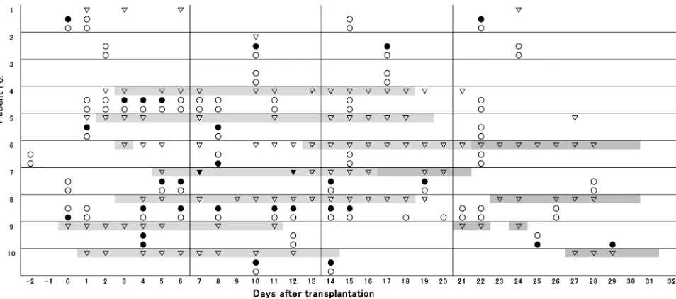
Fifty-one PB samples from 10 patients were analyzed by bacterial PCR assays during the first month after ASCT (Figure 1). Bacteria were detected in 24 samples
The upper row in each patient: ▼ Blood culture (+); ▽ Blood culture (−). The middle row in each patient: ● Bacterial PCR (+); ○ Bacterial PCR (−). The lower row in each patient: ● Fungal PCR (+); ○ Fungal PCR (−). Neutropenic fever; Non-neutropenic fever due
[image:5.595.62.539.449.660.2]to engraftment syndrome (all cases manifested regional skin eruption and a slight CRP elevation, which might include the symptom of GVHD in some cases).
DOI: 10.4236/aim.2019.912062 976 Advances in Microbiology from 8 patients (Table 2). The species were identified in 17 samples; and 14 (82%) were gram-positive cocci and 3 (18%) gam-negative rods. In 7 positive samples from 3 patients (day 0 in patient 1, days 8, 11, 12, 14 and 15 in patient 8, day 4 in patient 9), the bacterial species could not be identified because PCR products formed numerous bands. Multiple species in Table 2 indicates such cases, which were suspected of polymicrobial infections. Bacteria detected in 12 of 24 PCR-positive samples were considered to have originated from the oral cavity. Representative organisms identified were Streptococcus salivarius (days 3 - 5 in patient 4), Fusobacterium nucleatum (day 6 in patient 8), Abiotrophia spp. (days 10 and 17 in patient 2), and Streptococcus intermedius (day 8 in patient 5).
Table 2. Clinical characteristics of bacterial PCR positive cases.
Patient
no. Bacterial PCR Day of PCR positivity Blood culture Fever Stomatitis (grade) Diarrhea (grade) PCR(+) day (/μl) WBC counts on during PCR(+) days Antibiotics used 1st week (day 0 - 6)
1 multiple species day 0 − − 1 3 40 MEPM + ABK
4 Streptococcus salivarius* day 3, 4, 5 − + 2 3 20, 20, 40 CFPM + VCM
5 Enterococcus species** day 1 − − 2 3 20 MEPM + VCM
7 Enterococcus faecalis** day 5, 6 Enterococcus faecalis*** + 0 3 30, 20 MEPM + VCM
8 Streptococcus bovis** day 4 − + 0 1 20 DRPM + VCM
8 Fusobaterium nucleatum* day 6 − + 2 1 10 DRPM + VCM
9 multiple species day 4 − + 2 3 430 MEPM + VCM
2nd week (day 7 - 13)
2 Abiotrophia species* day 10 − − 1 0 90 MEPM + ABK
5 Streptococcus intermedius* day 8 − + 3 2 50 MEPM + VCM
8 multiple species* day 8, 11, 12 − + 2 0 20, 50, 90 PZFX + VCM +
CLDM
10 MRSA day 10 − + 2 2 110 DRPM + VCM
3rd week (day 14 - 20)
2 Abiotrophia species* day 17 − − 2 0 2870 DRPM + TEIC
7 Stenotrophomonas maltophilia day 14, 19 Stenotrophomonas maltophilia**** + 0 1 210, 4250 PZFX + VCM + MINO
7 Staphylococcus epidermidis day 19 − + 0 1 4250 PZFX + VCM +
MINO
8 multiple species* day 14, 15 − + 2 0 350, 570 PZFX + VCM +
CLDM + ABPC/SBT
10 MRSA day 14 − + 1 2 200 DRPM + VCM
4th week (day 21 - 27)
1 Enterococcus species** day 22 − − 0 1 4180 DRPM + TEIC
DOI: 10.4236/aim.2019.912062 977 Advances in Microbiology Multiple species detected in patient 8 seemed to also be of oral origin, as this pa-tient suffered from stomatitis but not diarrhea. On the other hand, Enterococcus spp. (day 1 in patient 5, and day 22 in patient 1), Enterococcusfaecalis (days 5 and 6 in patient 7) and Streptococcus bovis (day 4 in patient 8) were thought to have derived from the gut because of the presence of substantial diarrhea sug-gestive of intestinal mucosal injury. Concerning the other isolates: Stenotro-phomonas maltophilia (days 14 and 19 in patient 7) and methicillin-resistant Staphylococcus aureus (MRSA) (days 10 and 14 in patient 10), their source re-mains to be determined. BC was performed on 106 PB samples, and bacteria were isolated from only 2 samples from one patient (patient 7). The isolated bacteria, E. faecalis (day 7) and S. maltophilia (day 12), were the same species identified by the molecular methods, although the detection timings of BC and PCR were slightly different.
Seven of 10 patients developed fever during neutropenic status after ASCT (Figure 1). Bacteria were detected by PCR in samples taken from 6 of these pa-tients (papa-tients 4, 5, 7, 8, 9 and 10). However, the remaining patient (patient 6) showed no molecular evidence of bacterial infection despite the presence of fev-er. Five of the 6 febrile patients showing bacterial PCR-positive results were ma-nageable with empirical antibiotic therapies using broad-spectrum β-lactam plus glycopeptide without disclosure of the DNA data (Table 2). Among them, pa-tient 7 followed a complicated clinical course, but the PCR results still did not need to be disclosed. At first, S. maltophilia was isolated by BC on day 12 during an empirical treatment with MEPM and VCM; thus, MEPM was changed to pa-zufloxacin (PZFX) plus minocycline. PCR detection of this bacterium continued (days 14 and 19) despite its disappearance from BC (day 13), and Staphylococcus epidermidis was additionally identified by PCR on day 19. However, the anti-bacterial therapy eventually contributed to clinical improvement. These repeated PCR detections could be explained by the levels of bacteria undetectable by BC or by already dead bacteria, and the detection of S. epidermidis may represent contamination due to normal skin commensals introduced during blood collec-tion. In the case for which the PCR results were disclosed (patient 8), a high fever and CRP elevation developed on day 11 during empiric DRPM and VCM therapy, and we changed DRPM to PZFX plus clindamycin to cover anaerobes. But, as the fever continued, DNA data were at last disclosed to the physicians. PCR analyses on days 8, 11 and 12 showed multiple species, but F. nucleatum had been identi-fied on day 6; thus, ampicillin sulbactam was added on day 13 with a successful outcome. F. nucleatum may have been among the multiple species detected by PCR. Even in afebrile cases, bacteria were also detected by PCR in 5 samples from 3 patients (days 0 and 22 in patient 1, days 10 and 17 in patient 2, and day 1 in patient 5). These cases did not present any clinical problems, but still needed empirical antibiotic therapy for the symptoms of suspected infections.
3.2. Molecular Detection of Fungi
DOI: 10.4236/aim.2019.912062 978 Advances in Microbiology and positive results were obtained in 5 samples from 3 patients (patients 6, 8 and 9) (Table 3). No fungi were isolated from BC of any PB samples. The fungal PCR-positive cases were manageable without disclosure of the molecular data. CT scans of patient 8 revealed several cavitary nodules in both lungs 12 days be-fore ASCT. VRCZ had been given on suspicion of fungal infection, and these le-sions disappeared just before preconditioning. The PCR data on day 0 identified Aspergillus fumigatus DNA, but at this time we were able to confirm the causal pathogen by PCR and to justify our VRCZ therapy. In patient 9, Candida crusei along with many other bacterial species were detected in a febrile situation on day 4. These organisms may have been derived from the gut, because severe di-arrhea persisted at that time. This yeast was only temporarily detected and dis-appeared while we used prophylactic ITCZ. In this patient, Aspergillus niger was also identified by PCR during the afebrile period on days 25 and 29. Since the CT scan on day 17 had already shown an infiltration shadow in the right upper lung, we changed the antifungal from ITCZ (prophylactic use) to liposomal amphotericin B, which overcame the infection. The molecular information later corroborated the correctness of our antifungal management. In patient 6, Peli-omyces lilacinus was transiently detected during an afebrile period and became undetectable during continuous prophylaxis with ITCZ.
4. Discussion
[image:8.595.57.538.538.688.2]In this study, we performed broad-range PCR assays to detect a wide spectrum of bacteria and fungi to compliment the drawbacks of BC and evaluated the clinical usefulness of this system for planning BSI treatment during the first month after ASCT. Although PCR analysis proved far more sensitive than BC for the detection of both bacteria and fungi, most cases with PCR-positive results were manageable with empirical antimicrobial therapy alone and without the DNA da-ta. This indicates that our PCR assay did not directly contribute to BSIs manage-ment early after ASCT. However, the PCR data seemed to provide etiologically
Table 3. Clinical characteristics of fungal PCR positive cases.
Patient
no. Fungal PCR Day of PCR positivity culture Blood Fever BDG level index GM WBC (/μl) criteria IFD Antifungal used on PCR (+) day 1st week (day 0 - 6)
8 Aspergillus fumigatus day 0 - - negative negative 30 No category VCZ
9 Candida crusei day 4 - + negative negative 430 No category ITCZ (prophylaxis)
2nd week (day 7 - 13)
6 Peliomyces lilacinus day 8 - - negative negative 20 No category ITCZ (prophylaxis)
4th week (day 21 - 27)
9 Aspergillus niger day 25, 29 - - negative negative 2980, 5670 Possible L-AmB
DOI: 10.4236/aim.2019.912062 979 Advances in Microbiology valuable microbiological evidence.
As for the origin of bacteria identified by PCR, it is noteworthy that more than half of the organisms were considered to have originated from the oral cavity. In patients receiving intensive chemotherapy, not only neutropenia, but also treat-ment-induced mucosal injury appears to be crucial for the development of BSIs, and causative pathogens can originate from the endogenous microbiota inhabit-ing the mouth and the gut of the patients [11] [12]. Furthermore, severe oral mucositis occurs quite often in ASCT recipients due to the adverse effects of preconditioning with TBI [13] and cytotoxic drugs, such as high-dose cyclo-phosphamide and/or cytarabin [11], and the use of methotrexate containing GVHD prophylaxis [14]. Accordingly, BSIs due to orally derived organisms may develop more frequently in ASCT recipients than in patients receiving conven-tional chemotherapies. In addition, fluoroquinolone prophylaxis has been asso-ciated with an increased incidence of BSIs due to gram-positive bacteria, and oral viridans streptococci is one of among the most common blood stream iso-lates [15] [16], consistently with our observations. Orally derived bacteria appear to break into systemic circulation easily, because they do not pass through the major phagocytic system, such as the liver [17], and tend to be detected in PB with unexpected frequency compared to intestinal bacteria, as shown in our study. Since oral management is reported to effectively decrease the severity of stomatitis during ASCT [18] [19] [20], the same techniques should be evaluated for control of BSIs in ASCT recipients.
The other bacteria identified, E. faecalis [21], S. epidermidis [22], S. maltophi-lia [23] and MRSA [24], have also been described as orally derived pathogens in immunocompromised hosts. Since we performed DNA analyses only on PB samples and not on samples from mouth mucosa, we could not confirm their oral origin. However, such an origin seemed unlikely, as the former 3 species were detected in patients lacking stomatitis. In our study, contamination by ex-ogenous microbes was distinguishable in most cases by the identity of the spe-cies, frequency of detection, and clinical evaluation. Transitory appearance of S. epidermidis was likely to be originated from contamination from the skin [25] [26].
The present study also demonstrated that all cases showing gram-positive bacteria in PCR were effectively treated by empirical therapy with glycopeptides [8]. To our knowledge, there have been no reports in which the effectiveness of this antibacterial strategy was confirmed etiologically based on molecular data from ASCT recipients.
microor-DOI: 10.4236/aim.2019.912062 980 Advances in Microbiology ganisms in such cases.
In conclusion, although the molecular analyses did not directly contribute to manage BSI management early after ASCT, the DNA data were at least useful in elucidating the microbiological etiology of the BSIs. In addition, oral manage-ment appears to require a critical re-evaluation to reduce BSIs in ASCT reci-pients [18] [19] [20] [27]. However, as the main limitation of this study was the small number of cases examined, future large-scale studies will be required to validate our observations.
Acknowledgements
The authors thank all of the physicians and staff in the Department of Hematol-ogy and OncolHematol-ogy, Mie University Hospital, who assisted with the provision of data for this study.
Conflicts of Interest
The authors declare no conflicts of interest regarding the publication of this pa-per.
References
[1] Blennow, O., Ljungman, P., Sparrelid, E., Mattsson, J. and Remberger, M. (2014) Incidence, Risk Factors, and Outcome of Bloodstream Infections during the Pre-Engraftment Phase in 521 Allogeneic Hematopoietic Stem Cell Transplantation.
Transplant Infectious Disease,16, 106-114.https://doi.org/10.1111/tid.12175
[2] Kikuchi, M., Akahosh,i Y., Nakao, H., Ugai, T., Wada, H., Yamasaki, R., et al. (2015) Risk Factors for Pre- and Post-Engraftment Bloodstream Infections after Al-logeneic Hematopoietic Stem Cell Transplantation. Transplant Infectious Disease, 17, 56-65.https://doi.org/10.1111/tid.12345
[3] Book, M., Lehmann, L.E., Zhang, X. and Stuber, F. (2013) Monitoring Infection: From Blood Culture to Polymerase Chain Reaction (PCR). Best Practice & Re-search: Clinical Anaesthesiology, 27, 279-288.
https://doi.org/10.1016/j.bpa.2013.06.010
[4] Khot, P.D. and Fredricks, D.N. (2009) PCR-Based Diagnosis of Human Fungal In-fections. Expert Review of Anti-Infective Therapy, 7, 1201-1221.
https://doi.org/10.1586/eri.09.104
[5] von Lilienfeld-Toal, M., Lehmann, L.E., Raadts, A.D., Hahn-Ast, C., Orlopp, K.S., Marklcin, G., et al. (2009) Utility of a Commercially Available Multiplex Real-Time PCR Assay to Detect Bacterial and Fungal Pathogens in Febrile Neutropenia. Jour-nal of Clinical Microbiology, 47, 2405-2410.https://doi.org/10.1128/JCM.00491-09
[6] Nakamura, A., Sugimoto, Y., Ohishi, K., Sugawara, Y., Fujieda, A., Monma, F., et al. (2010) Diagnostic Value of PCR Analysis of Bacteria and Fungi from Blood in Em-piric-Therapy-Resistant Febrile Neutropenia. Journal of Clinical Microbiology,48, 2030-2036.https://doi.org/10.1128/JCM.01700-09
[7] De Pauw, B., Walsh, T.J., Donnelly, J.P., Stevens, D.A., Edwards, J.E., Calandra, T.,
DOI: 10.4236/aim.2019.912062 981 Advances in Microbiology
Mycoses Study Group (EORTC/MSG) Consensus Group. Clinical Infectious Dis-eases, 46, 1813-1821.https://doi.org/10.1086/588660
[8] Freifeld, A.G., Bow, E.J., Sepkowitz, K.A., Boeckh, M.J., Ito, J.I., Mullen, C.A., et al. (2011) Clinical Practice Guideline for the Use of Antimicrobial Agents in Neutro-penic Patients with Cancer: 2010 Update by the Infectious Diseases Society of America. Clinical Infectious Diseases, 52, e56-e93.
https://doi.org/10.1093/cid/cir073
[9] National Cancer Institute (2009) Common Terminology Criteria for Adverse Events v4.0. NIH Publication #09-7473.
[10] Sugawara, Y., Nakase, K., Nakamura, A., Ohishi, K., Sugimoto, Y., Fujieda, A., et al. (2013) Clinical Utility of a Panfungal Polymerase Chain Reaction Assay for Invasive Fungal Diseases in Patients with Haematologic Disorders. European Journal of Haematology, 90, 331-339.https://doi.org/10.1111/ejh.12078
[11] Wardley, A.M., Jayson, G.C., Swindell, R., Morgenstern, G.R., Chang, J., Bloor, R.,
et al. (2000) Prospective Evaluation of Oral Mucositis in Patients Receiving Mye-loablative Conditioning Regimens and Haemopoietic Progenitor Rescue. British Journal of Haematology, 110, 292-299.
https://doi.org/10.1046/j.1365-2141.2000.02202.x
[12] Blijlevens, N.M.A., Donnelly, J.P. and De Pauw, B. (2005) Prospective Evaluation of Gut Mucosal Barrier Injury Following Various Myeloablative Regimens for Hae-matopoietic Stem Cell Transplant. Bone Marrow Transplant, 35, 707-711.
https://doi.org/10.1038/sj.bmt.1704863
[13] Robien, K., Schubert, M.M., Bruemmer, B., Lloid, M.E., Potter, J.D. and Ulrich, C.M. (2004) Predictors of Oral Mucositis in Patients Receiving Hematopoietic Cell Transplants for Chronic Myeloid Leukemia. Journal of Clinical Oncology, 22, 1268-1275.https://doi.org/10.1200/JCO.2004.05.147
[14] Cutler, C., Li, S., Kim, H.T., Laglenne, P., Szeto, K.C., Hoffmeister, L., et al. (2005) Mucositis after Allogeneic Hematopoietic Stem Cell Transplantation: A Cohort Study of Methotrexate- and Non-Methotrexate-Containing Graft-versus-Host Dis-ease Prophylaxis Regimens. Biology of Blood and Marrow Transplantation, 11, 383-388.https://doi.org/10.1016/j.bbmt.2005.02.006
[15] Busca, A., Cavecchia, I., Locatelli, F., D’Ardia, S., De Rosa, F.G., Marmont, F., et al. (2012) Blood Stream Infections after Allogeneic Stem Cell Transplantation: A Sin-gle-Center Experience with the Use of Levofloxacin Prophylaxis. Transplant Infec-tious Disease, 14, 40-48.https://doi.org/10.1111/j.1399-3062.2011.00650.x
[16] Kimura, M., Araoka, H., Yoshida, A., Yamamoto, H., Abe, M., Okamot, Y., et al. (2016) Breakthrough Viridans Streptococcal Bacteremia in Allogeneic Hematopoie-tic Stem Cell Transplant Recipients Receiving Levofloxacin Prophylaxis in a Japa-nese Hospital. BMC Infectious Diseases, 16, Article No. 372.
https://doi.org/10.1186/s12879-016-1692-y
[17] Nakase, K., Tsuji, K., Miyanishi, E. and Shirakawa, S. (1990) Pathogenesis of Fever of Unknown Origin in Patients with Acute Leukemia and Granulocytopenia. Medi-cal Hypotheses, 33, 235-237.https://doi.org/10.1016/0306-9877(90)90133-Y
[18] Mori, T., Hasegawa, K., Okabe, A., Tsujimura, N., Kawata, Y., Yashima, T., et al. (2008) Efficacy of Mouth Rinse in Preventing Oral Mucositis in Patients High-Dose Cytarabine for Allogeneic Hematopoietic Stem Cell Transplantation. International Journal of Hematology, 88, 583-587.https://doi.org/10.1007/s12185-008-0181-5
DOI: 10.4236/aim.2019.912062 982 Advances in Microbiology
Clinical Outcomes of Oral Mucositis. Clinical Transplantation, 25, 325-328.
https://doi.org/10.1111/j.1399-0012.2010.01283.x
[20] Yamagata, K., Arai, C., Sasaki, H., Takeuchi, Y., Onizawa, K., Yanagawa, T., et al. (2012) The Effect of Oral Management on the Severity of Oral Mucositis during Hematopoietic SCT. Bone Marrow Transplant,47, 725-730.
https://doi.org/10.1038/bmt.2011.171
[21] Baik, J.E., Choe, H.-I., Hong, S.W., Kang, S.-S., Ahn, K.B., Cho, K., et al. (2016) Human Salivary Proteins with Affinity to Lipoteichoic Acid of Enterococcus faeca-lis. Molecular Immunology, 7, 52-59.https://doi.org/10.1016/j.molimm.2016.07.013
[22] Soga, Y., Maeda, Y., Ishimaru, F., Tanimoto, M., Maeda, H., Nishimura, F., et al. (2011) Bacterial Substitution of Coagulase-Negative Staphylococci for Streptococci on the Oral Mucosa after Hematopoietic Cell Transplantation. Support Care Can-cer, 19, 995-1000.https://doi.org/10.1007/s00520-010-0923-9
[23] Soga, Y., Saito, T., Nishimura, F., Ishimaru, F., Mineshiba, J., Mineshiba, F., et al. (2008) Appearance of Multidrug-Resistant Opportunistic Bacteria on the Gingiva during Leukemia Treatment. Journal of Periodontology,79, 181-186.
https://doi.org/10.1902/jop.2008.070205
[24] Ebinuma, T., Soga, Y., Sato, T., Matsunaga, K., Kudo, C., Maeda, H., et al. (2014) Distribution of Oral Mucosal Bacteria with mec4 in Patients Undergoing Hemato-poietic Cell Transplantation. Support Care Cancer,22, 1679-1683.
https://doi.org/10.1007/s00520-014-2151-1
[25] Costa, S.F., Micell, M.H. and Anaissie, E.J. (2004) Mucosa or Skin as Source of Coag Ulate-Negative Staphylococcal Bacteremia? The Lancet Infectious Diseases, 4, 278-286.https://doi.org/10.1016/S1473-3099(04)01003-5
[26] Kleinschmidt, S., Huygens, F., Faoagali, J., Rathnayake, I.U. and Hafner, L.M. (2015) Staphylococcus epidermidis as a Cause of Bacteremia. Future Microbiology, 10, 1859-1879.https://doi.org/10.2217/fmb.15.98
[27] Yamagata, K., Onizawa, K., Yanagawa, T., Hasegawa, Y., Kojima, H., Nagasawa, T.,