From attention to memory along the dorsal-ventral axis of
the medial prefrontal cortex: some methodological
considerations
Helen J. Cassaday1*, Andrew J. D. Nelson1,2and Marie A. Pezze1 1
School of Psychology, University of Nottingham, Nottingham, UK 2
School of Psychology, Cardiff University, Cardiff, UK
Edited by:
Chris John Tinsley, Nottingham Trent University, UK
Reviewed by:
Marco Atzori, University of Texas at Dallas, USA
Nandakumar Narayanan, Yale University, USA
*Correspondence: Helen J. Cassaday, School of Psychology, University of Nottingham, University Park, Nottingham NG7 2RD, UK e-mail: helen.cassaday@ nottingham.ac.uk
Distinctions along the dorsal-ventral axis of medial prefrontal cortex (mPFC), between anterior cingulate (AC), prelimbic (PL), and infralimbic (IL) sub-regions, have been proposed on a variety of neuroanatomical and neurophysiological grounds. Conventional lesion approaches (as well as some electrophysiological studies) have shown that these distinctions relate to function in that a number behavioral dissociations have been demonstrated, particularly using rodent models of attention, learning, and memory. For example, there is evidence to suggest that AC has a relatively greater role in attention, whereas IL is more involved in executive function. However, the well-established methods of behavioral neuroscience have the limitation that neuromodulation is not addressed. The neurotoxin 6-hydroxydopamine has been used to deplete dopamine (DA) in mPFC sub-regions, but these lesions are not selective anatomically and noradrenalin is typically also depleted. Microinfusion of drugs through indwelling cannulae provides an alternative approach, to address the role of neuromodulation and moreover that of specific receptor subtypes within mPFC sub-regions, but the effects of such treatments cannot be assumed to be anatomically restricted either. New methodological approaches to the functional delineation of the role of mPFC in attention, learning and memory will also be considered. Taken in isolation, the conventional lesion methods which have been a first line of approach may suggest that a particular mPFC sub-region is not necessary for a particular aspect of function. However, this does not exclude a neuromodulatory role and more neuropsychopharmacological approaches are needed to explain some of the apparent inconsistencies in the results.
Keywords: attention, associative leaning, object recognition memory, anterior cingulate, prelimbic, infralimbic, dopamine
INTRODUCTION
The interconnectivity of medial prefrontal cortex (mPFC) and hippocampus is well-established (Gabbott et al., 2005) and although there are differences between rat and primate mPFC a number of functional homologies have been documented. Indeed data from rat models has confirmed the importance of functional interactions between hippocampus and mPFC (Aggleton et al., 1995; Chudasama et al., 2004; Vertes, 2006; Ragozzino, 2007; Bizon et al., 2012). The signature effect of mPFC lesions is gen-erally viewed as one of cognitive inflexibility (Ragozzino, 2007), in particular when animals need to respond adaptively based on changed reinforcement contingencies (de Bruin et al., 1994; Aggleton et al., 1995; Bussey et al., 1997; Ragozzino et al., 1999; Birrell and Brown, 2000; Killcross and Coutureau, 2003). Such deficits in executive function tend to manifest as perseverative behavior—animals continue to respond according to previously acquired contingencies. Importantly, the delineation of the role of mPFC in executive function has high translational relevance given that mPFC dysfunction results in neuropsychiatric dysfunctions
such as schizophrenia (Andreasen et al., 1992; Barch et al., 2001; Spieker et al., 2012).
However, the mPFC is not a uniform entity. Evidence from neuroanatomical studies, including cytoarchitectonics, suggests a distinct heterogeneity to the structure comprising sub-regions such as anterior cingulate cortex (AC) as well as the prelimbic (PL) and infralimbic (IL) cortices. These sub-regions are also distinguishable on the basis of their differing patterns of inter-connectivity (Fisk and Wyss, 1999; Heidbreder and Groenewegen, 2003; Vertes, 2004). Thus, hodologically, there are good grounds to expect that the mPFC sub-regions should be dissociable in relation to distinct aspects of cognitive function (Kesner and Churchwell, 2011).
of underlying mechanisms (Bizon et al., 2012). The successful dissociation of these mechanisms is likely to require consider-ation of the functional heterogenity of mPFC as well as the adoption of behavioral tests suitable to tap component pro-cesses. Indeed, behavioral dissociations have been demonstrated, particularly with respect to the distinction between AC, PL, and IL, predominantly using tasks which tax attention, learn-ing and/or memory. However, until recently, the majority of studies have employed mechanical, electrolytic or excitotoxic lesions to explore these functional subdivisions (Heidbreder and Groenewegen, 2003). Whilst reinforcing the role of intercon-nected structures within functional brain circuits, the neuropsy-chological evidence obtained by studying the effects of lesions which damage fibers and/or cell bodies indiscriminately does not speak to the role of specific neurotransmitter systems in modulat-ing function. Similarly, whilst they provide an important advance to distinguish effects on encoding vs. retrieval, reversible inacti-vation studies do not specifically address the role of DA function and the border of the temporary lesion is typically ill-defined (Ragozzino, 2007). And, whilst providing important information about functional dissociations between sub-regions with excel-lent anatomical resolution, the majority of electrophysiological studies do not speak to the role of particular neurotransmitters either.
Evidence from psychopharmacological studies is necessary in order to develop neuropsychopharmacological theories as to the function of mPFC and interconnected brain structures. Dopamine (DA) is a likely neuromodulator of mPFC function in that the region is richly innervated by DA fibers originating in the ventral tegemental area (Lindvall et al., 1974). Consistent with this proposition, the modulation of DA functioning within mPFC has been shown to play an important role in regulat-ing learnregulat-ing and memory, as well as other cognitive functions (Goldman-Rakic, 1998; Robbins, 2000; Robbins and Roberts, 2007).
Microinfusion studies too are not without their limita-tions. Although they provide a workable method to activate or inactivate particular receptor subtypes within brain regions of interest, their anatomical resolution is less precise and rarely verified. Indeed, studies microinjecting agents selective for dif-ferent DA receptor subtypes have typically targeted mPFC as an entity rather than attempting any systematic comparison between mPFC sub-regions (Ragozzino, 2002; Floresco et al., 2006b).
For the purposes of the present review, four tasks which have been systematically examined in relation to their dependence on mPFC sub-regions will be considered in some detail, particu-larly in relation to the available evidence pertaining to the role of DA. The serial reaction time task measures early attentional processing and a key role for mPFC has been established using excitotoxic lesion approaches (Passetti et al., 2002; Pezze et al., 2009). Studies using excitotoxic lesions have also been conducted to examine the role of mPFC in latent inhibition (Lacroix et al., 1998, 2000b; Schiller and Weiner, 2004; George et al., 2010). Earlier studies of trace conditioning used aspiration ( Kronforst-Collins and Disterhoft, 1998; Weible et al., 2000) and electrolytic lesions (McLaughlin et al., 2002; Runyan et al., 2004), both of which methods also destroy fibers of passage. However, the role of
mPFC has since been confirmed by a variety of other approaches including excitotoxic lesions (Table 1).
Both latent inhibition and trace conditioning are simple Pavlovian conditioning procedures in which the associability of the conditioning cue is manipulated, by stimulus pre-exposure in the case of latent inhibition or by the introduction of a time inter-val after cue offset in the case of trace conditioning. There are a variety of theoretical interpretations of latent inhibition, some of which are attentional, but its psychological mechanisms are still disputed (Lubow and Weiner, 2010). Trace conditioning similarly results in reduced stimulus associability but has been argued to tap working memory because of the need to bridge the time gap introduced by the trace interval (Sweatt, 2004). Similarly, there is good evidence that mPFC is involved in recognition memory as measured by tasks exploiting rats’ preferences to explore novelty as presented by previously unseen objects, less recently encoun-tered objects, or familiar objects in novel locations. Some such studies have been driven by the theoretical views on the function of mPFC, some by the known effects of hippocampal lesions on working memory. However, the role of different neuromodulators within these mPFC sub-regions is still poorly understood.
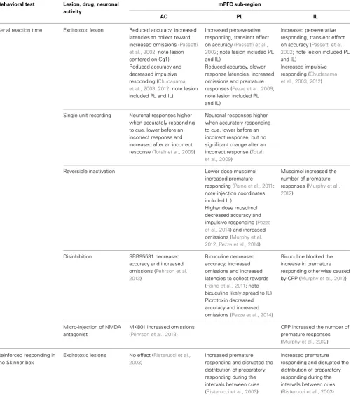
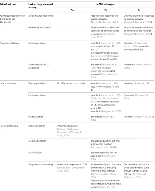
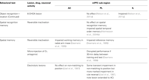
The present review will therefore address the functional dis-tinction between AC, PL, and IL mPFC sub-regions with respect to the mechanisms of attention, learning, and memory.Table 1 summarizes the behavioral effects associated with changes in neuronal activity in AC, PL, and IL mPFC sub-regions in tasks measuring serial reaction time, latent inhibition, trace condi-tioning and object recognition, as well as some related tests of learning and memory (for which the effects of similar interven-tions in different mPFC sub-regions have been examined). The advantages and disadvantages of the available neurophamacolog-ical techniques—for example in neurotoxic lesion, microinfusion and pharmacogenetic studies—to establish the role of DA will also be considered.
EARLY ATTENTION: THE SERIAL REACTION TIME TASK In rodents, the five choice serial reaction time task (5-CSRT) test, which resembles the human continuous performance test, is a commonly used test of attention (Robbins, 2002; Chudasama and Robbins, 2006; Lustig et al., 2013). In this task rodents are trained to detect a brief visual target presented randomly in one out of an array of five apertures in order to obtain food rewards. The proportion of nose pokes into the correct aperture, as opposed to incorrect apertures, provides a measure of attention. Other mea-sures such as correct response latency (for example) may also reflect attentional deficits; whereas magazine latency to collect food reward provides useful information about levels of activ-ity or motivation. Additionally, this task indexes response control or behavioral inhibition, as reflected in the number of premature responses (when the animal responds before the presentation of the light) and perseverative responses (when the animal carries on nose poking in the same aperture after a correct or an incorrect response).
EXCITOTOXIC mPFC LESIONS IMPAIR 5-CSRT PERFORMANCE
Table 1 | The behavioral effects associated with changes in neuronal activity in anterior cingulate (AC), prelimbic (PL), and infralimbic (IL) sub-regions of medial prefrontal cortex (mPFC) in tasks measuring serial reaction time, latent inhibition, trace conditioning and object
recognition, as well as related benchmark tests of learning and memory (for which the effects of similar interventions in different mPFC sub-regions have been examined).
Behavioral test Lesion, drug, neuronal activity
mPFC sub-region
AC PL IL
Serial reaction time Excitotoxic lesion Reduced accuracy, increased latencies to collect reward, increased omissions (Passetti et al., 2002; note lesion centered on Cg1) Reduced accuracy and decreased impulsive responding (Chudasama et al., 2003, 2012; note lesion included PL and IL)
Increased perseverative responding, transient effect on accuracy (Passetti et al., 2002; note lesion included PL and IL)
Reduced accuracy, slower response latencies, increased omissions and premature responses (Pezze et al., 2009; note lesion included PL and IL)
Increased perseverative responding, transient effect on accuracy (Passetti et al., 2002; note lesion included PL and IL)
Increased impulsive responding (Chudasama et al., 2003, 2012)
Single unit recording Neuronal responses higher when accurately responding to cue, lower before an incorrect response and increased after an incorrect response (Totah et al., 2009)
Neuronal responses higher when accurately responding to cue, lower before an incorrect response, but no significant change after an incorrect response (Totah et al., 2009)
Reversible inactivation Lower dose muscimol
increased premature responding (Paine et al., 2011; note injection coordinates included IL)
Higher dose muscimol decreased accuracy and impulsive responding (Pezze et al., 2014) and increased omissions (Murphy et al., 2012; Pezze et al., 2014)
Muscimol increased the number of premature responses (Murphy et al., 2012)
Disinhibition SRB95531 decreased accuracy and increased omissions (Pehrson et al., 2013)
Bicuculine decreased accuracy, increased omissions and increased latencies to collect rewards (Paine et al., 2011; note bicuculine likely spread to IL) Picrotoxin decreased accuracy and increased omissions (Pezze et al., 2014)
Bicuculine blocked the increase in premature responding otherwise caused by CPP (Murphy et al., 2012)
Micro-injection of NMDA antagonist
MK801 increased omissions (Pehrson et al., 2013)
CPP increased the number of premature responses (Murphy et al., 2012)
Reinforced responding in the Skinner box
Excitotoxic lesions No effect (Risterucci et al., 2003)
Increased premature responding and disrupted the distribution of preparatory responding during the intervals between cues (Risterucci et al., 2003)
Increased premature responding and disrupted the distribution of preparatory responding during the intervals between cues (Risterucci et al., 2003)
Table 1 | Continued
Behavioral test Lesion, drug, neuronal activity
mPFC sub-region
AC PL IL
Reinforced responding in the Skinner box (Continued)
Single neuron recording Fast transient responses to sucrose delivery
(Burgos-Robles et al., 2013)
Delayed prolonged responses to sucrose delivery
(Burgos-Robles et al., 2013)
Reversible inactivation Muscimol without effect on collection of earned sucrose rewards (Burgos-Robles et al., 2013)
Muscimol delayed collection of earned sucrose rewards (Burgos-Robles et al., 2013)
Pre-pulse inhibition Excitotoxic lesion No effect (Lacroix et al., 1998; note lesion included AC and IL)
Increased by larger lesions (Lacroix et al., 2000b; note lesion included AC and IL)
No effect (Sullivan and Gratton, 2002; note lesion encroached on PL)
Micro-injection of D1
antagonist
Impaired (Ellenbroek et al., 1996; note injection coordinates included IL) Impaired (Shoemaker et al., 2005)
Impaired (Shoemaker et al., 2005)
Latent inhibition Electrolytic lesion No effect (Joel et al., 1997) No effect (Joel et al., 1997; note lesion included AC and IL)
No effect (Joel et al., 1997)
Excitotoxic lesion No effect (Lacroix et al., 1998, 2000b; Schiller and Weiner, 2004; note lesions centered on PL, encroached on IL and/or AC)
No effect (George et al., 2010)
Enhanced (George et al., 2010)
6-OHDA lesion Enhanced (Nelson et al.,
2010)
No effect (Nelson et al., 2010)
Trace conditioning Aspiration lesion Impaired acquisition (Kronforst-Collins and Disterhoft, 1998; Weible et al., 2000)
Electrolytic lesion Impaired acquisition but only at longer CS duration (McLaughlin et al., 2002)
Erk inhibition Impaired retention but not
acquisition (Runyan et al., 2004)
Single neuron recording Attentional responses to CSs (Weible et al., 2003; Hattori et al., 2014)
Increased activity to the trace conditioned CS, including within the trace interval (Gilmartin and McEchron, 2005)
Persistent activity within the trace interval during retention tests (Hattori et al., 2014)
Decreased activity to the trace conditioned CS, no change in trace interval activity (Gilmartin and McEchron, 2005)
Table 1 | Continued
Behavioral test Lesion, drug, neuronal activity
mPFC sub-region
AC PL IL
Trace conditioning (Continued)
Reversible inactivation Impaired acquisition (Kalmbach et al., 2009; note “caudal” placement included PL)
Excitotoxic lesionc-fos
expression
Impaired by lesion and associated with increased neuronal activity in Cg1 but not Cg2 (Han et al., 2003)
Excitotoxic lesion Impaired by
immediate-post-training lesions (Oswald et al., 2010)
Impaired by
1-week-post-training lesions (Oswald et al., 2008, 2010)
Fear conditioning Microstimulation No effect on the expression or extinction of conditioned fear (Vidal-Gonzalez et al., 2006)
Increased the expression of conditioned fear and prevented extinction (Vidal-Gonzalez et al., 2006)
Decreased the expression of conditioned fear and facilitated extinction (Vidal-Gonzalez et al., 2006)
Single neuron recording Decreased activity during extinction and extinction memory in male rats; females showed increased activity during extinction (Fenton et al., 2014)
Increased activity during extinction and extinction memory (no sex difference;
Fenton et al., 2014)
Reversible inactivation Muscimol impaired fear
expression, but had no effect on extinction memory (Sierra-Mercado et al., 2011)
Muscimol had no effect of fear expression, but impaired extinction memory
(Sierra-Mercado et al., 2011)
Object
recognition—identity
Excitotoxic lesion No effect (Ennaceur et al., 1997)
No effect (Ennaceur et al., 1997; Barker et al., 2007)
Reversible inactivation No effect (Hannesson et al., 2004a)
6-OHDA lesion No effect (Nelson et al.,
2011a)
No effect (Nelson et al., 2011a)
Object
recognition—recency
Excitotoxic lesion Impaired (Barker et al., 2007; note lesion encroached on IL)
Reversible inactivation Impaired (Hannesson et al., 2004a)
6-OHDA lesion Impaired (Nelson et al.,
2011a; note PL lesion anatomically selective)
Impaired (Nelson et al., 2011a; note IL lesion encroached on PL)
Single neuron recording Increased activity when in location of a “missing” object at 6 h or 30 day delay (Weible et al., 2012)
Object
recognition—location
Excitotoxic lesion No effect unless lesion extended to include retrosplenial cortex (Ennaceur et al., 1997)
No effect (Ennaceur et al., 1997; Barker et al., 2007)
Table 1 | Continued
Behavioral test Lesion, drug, neuronal activity
mPFC sub-region
AC PL IL
Object recognition— location (Continued)
6-OHDA lesion No effect (Nelson et al.,
2011a)
Impaired (Nelson et al., 2011a)
Spatial recognition memory
Reversible inactivation No effect on spatial recognition memory, impaired spatial temporal order memory (Hannesson et al., 2004b)
Spatial memory Reversible inactivation Impaired working memory in radial arm maze (Seamans et al., 1995)
Impaired reference memory (Seamans et al., 1995)
Micro-injection of D1
antagonist
Disrupted performance if 30 min delay between training and test (Seamans et al., 1998)
Electrolytic lesions No effect on non-matching to position (Joel et al., 1997)
Some transient impairment in non-matching to position but more marked impairment in rule reversal (Joel et al., 1997; note lesion extended to AC)
shown that complete mPFC lesions impair choice accuracy and increase both omissions and perseverative responses, as well as latency to respond correctly (Passetti et al., 2002; Pezze et al., 2009). More discrete lesions have helped to clarify the contribu-tion of specific mPFC sub-regions. Indeed,Passetti et al. (2002)
also compared the effects of complete mPFC excitotoxic lesions with those of AC and more ventral PL plus IL mPFC lesions. When more restricted lesions were employed, those with the AC placements resulted in reduced accuracy in the absence of any effect on perseverative responding, although there was some tran-sient increase in the number of omissions (Passetti et al., 2002). In contrast, animals with complete mPFC lesions showed impaired accuracy combined with increased perseverative responding in the absence of any significant change in omissions or latencies (Passetti et al., 2002).
In a later study from the same laboratory, excitotoxic lesions using the same mPFC coordinates as those used by Passetti et al. (2002), reduced accuracy was seen in conjunction with slower response latencies, increased omissions and premature responses (Pezze et al., 2009). It has also been shown that whilst both combined lesions of AC and PL (Chudasama et al., 2003; Chudasama and Robbins, 2006) and lesions restricted to AC (Passetti et al., 2002) impair attentional performance measured as accuracy, lesions restricted to IL increase impulsive responding (Chudasama et al., 2003, 2012).
TEMPORARY INACTIVATION OF mPFC IMPAIRS 5-CSRT PERFORMANCE Complementary to the study of permanent excitotoxic lesions, temporary inactivation studies have also helped to clarify the differential engagement of mPFC sub-regions in attention and
response control. Paine and co-workers reported that low doses of muscimol (6.25 to 50 ng/side), a GABA(A) receptor agonist, selectively increased premature responding when infused into the PL plus IL mPFC without affecting accuracy (Paine et al., 2011). In contrast, a high dose of muscimol (500 ng/side) decreased response accuracy and increased the percentage of omissions reflecting an attentional deficit when infused into the PL or IL (Murphy et al., 2012). Increased reward-collection latency how-ever indicated that the high dose may have caused motivational impairments. Lower doses of muscimol (62.5 to 250 ng/side) in this case injected into PL have since been found to decrease atten-tional performance without causing nonspecific impairments in reward-collection latency, consistent with the importance of PL mPFC in attention (Pezze et al., 2014). Moreover, also consis-tent with the results of the excitotoxic lesion studies reported above, infusion of the N-methyl-D-aspartate (NMDA) receptor antagonist MK801 into the AC selectively impaired attentional parameters of performance (Pehrson et al., 2013). Further con-firming the role of IL mPFC in impulsivity, both the NMDA receptor antagonist CPP and muscimol increased the number of premature responses when infused into the IL (Murphy et al., 2012).
DISINHIBITION IN mPFC IMPAIRS 5-CSRT PERFORMANCE
of non-specific impairments (Pezze et al., 2014). Yet another GABA(A) receptor antagonist SRB95531 also selectively reduced attentional aspects of performance after AC infusion (Pehrson et al., 2013). On the other hand when infused in IL the GABA(A) receptor antagonist bicuculine blocked the increase in premature responding otherwise caused by the NMDA receptor antagonist CPP (Murphy et al., 2012). These data converge in suggesting that while the more dorsal parts of the mPFC (AC plus PL) may drive attentional functions, the IL region rather controls inhibitory behavior.
ELECTROPHYSIOLOGICAL CORRELATES OF 5-CSRT PERFORMANCE This dissociation based on attentional vs. inhibitory behaviors has been further confirmed by electrophysiological studies of the dif-ferential involvement of mPFC sub-regions, in this case in the ability to allocate preparatory attention to improve subsequent stimulus processing and response selection. Totah and co-workers have used single unit recording in a three choice serial reaction time task to show that levels of activity in both PL and AC are related to preparatory attention in the period before the presen-tation of the light cue (Totah et al., 2009). In this study, neuronal responses were higher when the rat was accurately responding to the cue, lower before an incorrect response and not different from baseline before an omission. The authors suggested that both PL and AC may be involved in signaling reward expectation of the upcoming goal-relevant stimuli. However, in contrast to the neu-ral activity in PL, neuneu-ral activity in the AC increased after an incorrect response, suggesting that this structure also codes for error related events.
DA MODULATES 5-CSRT PERFORMANCE
Consistent with the importance of DA as a modulator of early attentional processes, a history of amphetamine exposure also appears to alter performance on the 5-CSRT test. For example, rats which have been trained to self-administer amphetamine showed a transient deficit in attention reflected by decreased accu-racy and increased omissions when tested on the 5-CSRT (Dalley et al., 2005). More recently, it has been reported that repeated and intermittent exposure to escalating doses of amphetamine in rats increased the number of omissions during the amphetamine treatment or the withdrawal phase (Fletcher et al., 2007). When the attentional load of the task was increased by shortening the stimulus duration, the level of accurate responding was also decreased in amphetamine pre-treated rats. The similarity of the impairments to those caused by lesion of the dorsal mPFC sug-gested that the effect of amphetamine treatment on attention may have been mediated by the dorsal mPFC.
A few more anatomically specific neurochemical stud-ies have investigated the role of DA release in the mPFC in attention and/or response control (Robbins, 2002). Using post-mortem neurochemistry a positive correlation between 3,4-dihydroxyphenylacetic acid (DOPAC)/DA ratio and choice accu-racy was observed in the right hemisphere (Puumala and Sirvio, 1998). In freely moving rats,in vivomicrodialysis has confirmed increased extracellular levels of DA in the PL and IL sub-regions of animals performing a simplified one choice version of the task (Dalley et al., 2002b). In the same study, it was demonstrated
that increased DA levels in the mPFC of rats treated with an amphetamine challenge paralleled increased numbers of prema-ture responses; and post-mortem neurochemistry revealed that impulsive rats had a higher level of DA turnover in both PL and IL sub-regions of the mPFC (Dalley et al., 2002a). Thus, these neu-rochemical studies point to the conclusion that whilst attention clearly requires DA in the mPFC, too much may trigger impulsive responding.
While profound catecholamine depletions of the mPFC did not alter baseline performance on the 5-CSRT, deficits emerged when the rats were behaviorally challenged. For example, the introduction of a variable inter-trial-interval (ITI) to make the target stimulus less predictable induced a deficit in accuracy in rats with 6-OHDA lesion of the mPFC (Robbins et al., 1998; Robbins, 2002). Moreover, microinfusion studies have confirmed the importance of DA D1 receptor transmission, in attentional
processes in particular (Dalley et al., 2004; Boulougouris and Tsaltas, 2008). Granon and co-workers showed an enhancement in attentional processes after infusion into PL mPFC of the DA D1 receptor agonist SKF38393, though only at low levels
of attentional performance (Granon et al., 2000). This effect was subsequently replicated using a more selective D1
recep-tor agonist SKF81297, in a variation of the task in which rats were trained to detect a visual target and then to remember the location of this visual stimulus (Chudasama and Robbins, 2004). Moreover, the decline in accurate responding and the increase in omissions induced by amphetamine withdrawal could be prevented by infusion of the D1 receptor agonist SKF38393
into the PL mPFC (Fletcher et al., 2007). In contrast, the D1
receptor antagonist SCH23390 impaired the accuracy of atten-tional performance in rats that performed at high baseline levels (Granon et al., 2000). This set of data may reflect an inverted U-shaped function relating attentional processes to DA-receptor stimulation in the mPFC. Similarly, working memory (Sawaguchi and Goldman-Rakic, 1991; Williams and Goldman-Rakic, 1995; Goldman-Rakic, 1995; Zahrt et al., 1997) and the expression of conditioned fear (Pezze et al., 2003) have also been suggested to require an optimal level of prefrontal dopamine-receptor stimulation.
5-CSRT CONCLUSIONS
Thus, neurochemical evidence converges to suggest that mPFC DA is involved in early attentional processes, as well as in response inhibition (seeExecutive Functions in mPFCbelow). In common with other functions mediated in mPFC, as well as the more established interconnectivity with hippocampus (Gabbott et al., 2005), there is evidence pointing to the importance of its inter-connections with other brain regions such as NAc (Pezze et al., 2007, 2009). For example, the attentional dysfunction demon-strated in the serial reaction time task after mPFC lesions was ameliorated by treatment with the DA D2 antagonist sulpiride
administered intra-NAc (Pezze et al., 2009). This finding points to the importance of neuromodulation, specifically by DA D2
FIGURE 1 | The connections of rat medial prefrontal cortex (mPFC) sub-regions with other key brain regions; based onHeidbreder and Groenewegen (2003).The different arrow line formats (use of dashed and gray lines) denote connections to (and from) different target structures.
Effective early attention or vigilance of the kind measured by the 5-CSRT is necessary but not sufficient for the efficient functioning of subsequent learning and memory processes. For example, early attention is a likely modulator of associability or animals’ capacity to learn about environmental cues. However, this subsequent capacity is not directly assessed in the 5-CSRT.
ATTENTIONAL LEARNING: LATENT INHIBITION
(Lubow and Moore, 1959). The reduction in later learning is demonstrated when the pre-exposed CS subsequently comes to predict an outcome (unconditioned stimulus, US). In attentional terms, such a pre-exposed cue can be seen to be irrelevant. Consistent with a likely role for mPFC in latent inhibition, NAc and other interconnected regions such as entorhinal cortex, hip-pocampus, orbitofrontal cortex and basolateral amygdala (BLA), are known to modulate the expression of latent inhibition (Honey and Good, 1993; Tai et al., 1995; Coutureau et al., 1999; Schiller and Weiner, 2004; Gal et al., 2005). Consequently, a number of studies have been conducted to examine the role of mPFC in latent inhibition.
EXCITOTOXIC mPFC LESIONS CAN ENHANCE LATENT INHIBITION Contrary to expectation, a number of studies examining the effects of electrolytic lesions to the mPFC and its sub-regions as well as excitotoxic lesions (typically spanning two to three mPFC sub-regions) reported no effect on latent inhibition (Joel et al., 1997; Lacroix et al., 1998, 2000b; Schiller and Weiner, 2004). More restricted lesions have also been reported to be without effect (Joel et al., 1997).
However, a number of neuromodulatory effects are known to depend on the strength of the behavioral effect under examina-tion in that ceiling or floor effects can preclude the demonstraexamina-tion of enhanced or impaired performance, respectively. This has cer-tainly proven to be an important consideration in studies of latent inhibition. For example, whilst abolition of latent inhibition is most readily demonstrated when the effect is clear in the control group, the demonstration of potentiated latent inhibition may require a weakened level of latent inhibition in the control group. Weakened latent inhibition is produced experimentally either by using a low number of stimulus pre-exposures or a high num-ber of conditioning trials (Weiner and Arad, 2009). Indeed, such parametric manipulations have been found to be critical to the demonstration of the effects of lesions in BLA or core NAc and which do not disrupt latent inhibition under conditions which reliably produce latent inhibition in control (in this case sham-operated) animals, but which enhance latent inhibition under behavioral conditions known to be insufficient to produce latent inhibition in controls (Gal et al., 2005; Schiller and Weiner, 2005). In one previous study which similarly examined the effects of lesions to mPFC under conditions which did not produce latent inhibition in controls (in this case using a high number of condi-tioning trials), still no effects of mPFC lesions were demonstrated (Schiller and Weiner, 2004).
The functional heterogeneity of mPFC also needs to be taken into account and lesions restricted to IL mPFC have more recently been reported to potentiate latent inhibition (George et al., 2010). This study used 30 stimulus pre-exposures in a within-subject aversive on-the-baseline procedure, under which experimental conditions enhanced latent inhibition was demonstrated in a group with excitotoxic lesions restricted to IL mPFC but not PL mPFC. Since the demonstration of the potentiating effects of IL mPFC lesions did not in this case require the use of any experimental manipulation to weaken latent inhibition, the on-the-baseline procedure used by George et al may be particularly sensitive to small variations in the magnitude of latent inhibition.
The previous study which reported no effect of IL lesions used the standard off-the-baseline procedure (Joel et al., 1997).
There has been little further investigation—using experimen-tal parameters that do result in latent inhibition in untreated animals—of the involvement of the PL and IL mPFC sub-regions in relation to the expression of latent inhibition. However, with respect to sub-region there is evidence that the critical IL vs. PL lesion site to produce this effect depends on whether the lesion is made excitotoxically (Schiller and Weiner, 2004; George et al., 2010) or using more neurochemically selective neurotoxins (Nelson et al., 2010).
DA AND LATENT INHIBITION
The abolition of latent inhibition after treatment with systemic amphetamine has been demonstrated many times (Weiner et al., 1981, 1984, 1988; Weiner, 1990, 2003; Weiner and Arad, 2009) and this effect of amphetamine has been one of the key argu-ments used to support the use of latent inhibition as a behavioral marker for schizophrenia (Weiner, 1990, 2003; Weiner and Arad, 2009). Unsurprisingly therefore, further studies have gone on to examine the role of mPFC DA. When 6-OHDA was used to lesion DA terminals within PL vs. IL mPFC, it was the injection in PL which lead to the emergence of latent inhibition under weak pre-exposure conditions insufficient to produce latent inhibition in controls (Nelson et al., 2010). These results are contrary to those reported with excitotoxic lesions made with ibotenic acid, in that in this case the effective lesion was the ventral place-ment corresponding with IL mPFC (George et al., 2010). The fact that opposing patterns of outcome have been demonstrated depending on whether the lesion method has greater or lesser neurochemical selectivity clearly points to the importance of neu-romodulation by DA within PL vs. IL mPFC. This would be consistent with other evidence that DA within mPFC modulates cognitive control (Goldman-Rakic, 1998; Robbins, 2000; Robbins and Roberts, 2007). However, 6-OHDA lesions are not selec-tive to DA and typically also deplete noradrenalin (NA; see also Methodological Considerationsbelow).
On balance the available evidence point to the importance of mPFC sub-region, selectivity of the lesioning method to cat-echolamines and the level of latent inhibition generated by the behavioral parameters in use. For example, the results obtained with 6-OHDA IL lesions, which in fact depleted DA in both the IL and PL and were without effect on latent inhibition (Nelson et al., 2010), can be seen as consistent with the earlier reported finding that larger electrolytic or excitotoxic mPFC lesions do not affect latent inhibition (Joel et al., 1997; Lacroix et al., 1998, 2000b; Schiller and Weiner, 2004). Such dissociable effects of anatom-ically complete vs. partial mPFC lesions mirror the observation that total (shell plus core) and sub-regionally-selective (shell vs. core) NAc lesions have opposing effects on latent inhibition (Gal et al., 2005).
Further direct evidence on the neuromodulatory role of DA is lacking in that within mPFC local infusions of either D1 or D2
were conducted using standard latent inhibition procedures more suited to test for the abolition of latent inhibition rather than any potential enhancement; neither did they involve any com-parison of effects in relation to PL vs. IL placement. Thus, given the importance of both the precise behavioral parameters in use and the placement with respect to sub-region, although consis-tently negative to date, the available evidence to date suggests that the role of DA within PL may yet be confirmed by microinfusion studies.
LATENT INHIBITION CONCLUSIONS
Overall, the evidence base for the role of DA in mPFC sub-regions is more limited in the case of latent inhibition compared with that available in the case of the 5-CSRT. A particular role for DA in more dorsal mPFC regions would be broadly consistent with the importance of AC and PL sub-regions in 5-CSRT accu-racy as distinct from impulsive responding (Early Attention: the Serial Reaction Time Task;Table 1). However, to our knowledge there has to date been only one study to examine the role of AC in latent inhibition and this was an electrolytic lesion study. The latent inhibition procedure nonetheless adds to our understand-ing in that it allows for examination of the effects of attention on associative learning. However, the assessment of such effects is inevitably indirect and potentially confounded by other cogni-tive demands introduced by the latent inhibition task, such as the need to switch flexibly between stimulus-reinforcement contin-gencies. The interface between attention and executive function is discussed below (Executive Functions in mPFC).
WORKING MEMORY: TRACE CONDITIONING
Trace conditioning is the reduction in learning produced by the introduction of a trace interval between CS offset and US onset. Like latent inhibition, trace conditioning can be demonstrated in a variety of Pavlovian conditioning procedures. Also in common with latent inhibition, attentional processes have been identi-fied as essential to the normal acquisition of trace conditioning. However, unlike latent inhibition, trace conditioning has been argued to represent an essential aspect of working memory. The ability to bridge time delays to show associative learning in a trace conditioning procedure allows animals to associate what goes with what, when potentially causally-related events are sep-arated in time. With respect to anatomical substrates it is very well-established that conventional hippocampal lesions impair trace conditioning, and this effect is reproducible in a variety of conditioning procedures (though typically aversively moti-vated:McEchron et al., 2000; Beylin et al., 2001; Quinn et al., 2002).
Consistent with known projections from hippocampus, mPFC has also been shown to be part of the trace conditioning net-work. Following a number of lesion studies of eye blink con-ditioning in the rabbit which showed the importance of mPFC sub-regions including the caudal aspect of AC (Kronforst-Collins and Disterhoft, 1998; Weible et al., 2000) single neuron record-ing studies indicated an attentional role for AC, manifest par-ticularly in the early acquisition of trace conditioning (Weible et al., 2003; Oswald et al., 2008; Hattori et al., 2014). A sim-ilar effect, in this case of electrolytic lesions to PL mPFC in
the same eye blink conditioning preparation, has also been reported; however—consistent with some functional difference— the demonstration of PL lesion effects on acquisition was found to require the use of a relatively long duration CS (McLaughlin et al., 2002).
EXCITOTOXIC mPFC LESION EFFECTS ON TRACE CONDITIONING Further studies using the eye blink conditioning preparation have been conducted with more anatomically selective excito-toxic lesions. For example, the effects of post-training PL lesions point to the importance of PL in regulating retrieval: PL lesions made 1 week after training resulted in reduced expression of prior learning, however animals showed normal relearning (Oswald et al., 2008, 2010). Again using the same behavioral procedures, reduced expression of prior learning was found to result from excitotoxic AC lesions but in this case only if the lesions were made immediately post-training (Oswald et al., 2010). This dis-sociation between the effects of PL and AC lesions based on the timing of the lesions with respect to behavioral training points to a functional difference in relation to the retrieval of consolidated material in PL vs. processes related to consolidation which require AC (Oswald et al., 2010). Since initial acquisition was not exam-ined in the Oswald et al. studies (only relearning), the importance of AC for early attentional processing in trace conditioning is not excluded.
CONVERGENT EVIDENCE FOR DISSOCIATIONS BY mPFC SUB-REGION Consistent with the above studies byOswald et al. (2008, 2010), in a rat CER procedure, inhibition of extracellular signal-regulated (Erk) cascade to disrupt long term memory storage within PL impaired the retention butnot the acquisition of trace condi-tioning (Runyan et al., 2004). Thus, there is convergent evidence consistent with the importance of PL in retrieval-related pro-cesses. Moreover, excitoxic AC lesions have been reported to reduce trace conditioning in a rat CER procedure in the same way as an experimental distractor stimulus (based on the putative attentional role of AC:Han et al., 2003).
conditioning with a 20 s trace interval (Gilmartin and McEchron, 2005).
DA AND TRACE CONDITIONING
With respect to the role of DA, systemic amphetamines increased conditioning over a 30 s trace interval measured in a CER proce-dure (Norman and Cassaday, 2003; Horsley and Cassaday, 2007). The amphetamines are indirect catecholamine agonists so these results suggest a role for DA or NA in the neuromodulation of trace conditioning. However, particularly in the case of trace con-ditioning, an additional consideration arises in that lesion effects have typically been investigated in aversively motivated proce-dures (eye blink or CER). Lesions and drug treatments affecting the DA system have been found to have different effects in aver-sive (Norman and Cassaday, 2003; Horsley and Cassaday, 2007) and appetitive trace conditioning procedures (Kantini et al., 2004; Cassaday et al., 2005). This difference may relate to the different motivational systems engaged by aversive and appetitive condi-tioning procedures or inevitable differences in the salience of the aversive and appetitive UCSs used in such studies. Consistent with the latter interpretation, systematic comparison of the effects of mPFC lesions in two aversive trace conditioning variants, moti-vated by relatively mild corneal air puff vs. relatively aversive peri-orbital eye shock UCS showed greater trace conditioning deficits with the air puff UCS (Oswald et al., 2010). Therefore, it will be important to establish the role of mPFC DA using appetitive as well as aversive task variants, particularly given the evidence that the relationship between mPFC DA and cogni-tion follows an inverted U-funccogni-tion (Zahrt et al., 1997; Arnsten, 1998).
Reduced trace conditioning can be viewed as a cognitive impairment. Conversely, enhanced trace conditioning can be taken to reflect cognitive enhancement because of improved working memory function (Sweatt, 2004), as demonstrated after both treatment with amphetamines (Norman and Cassaday, 2003; Horsley and Cassaday, 2007) and catecholaminergic deple-tion within NAc (Nelson et al., 2011d). Connections of the AC (caudal aspect) point to the importance also of the choliner-gic system at least in the attentional processes necessary to trace conditioning (Weible et al., 2007). However, the role of other key neuromodulators within mPFC and the extent to which its sub-regions are differentially involved in trace conditioning are not yet established. A role for mPFC DA is to be expected, and the interconnectivity with NAc is consistent with the pattern of effects observed in latent inhibition procedures (Nelson et al., 2010, 2011b,c). Indeed, we have presented some preliminary evi-dence for differential effects of PL and IL dopamine depletion on trace conditioning measured in a CER procedure (Thur et al., 2010). However, statistically this result was marginal and needs replication. DA depletion in core rather than shell NAc provided the most likely explanation of the increased trace condition-ing demonstrated in our earlier study (Nelson et al., 2011d). Accordingly, based on the known interconnectivity between NAc core and shell with PL and IL mPFC, respectively, this effect would be predicted to be reproduced by DA depletion in PL. We have recently found that the early acquisition of appetitive trace con-ditioning is modulated by the DA D1agonist SKF81297 but we
have so far been unable to distinguish the role of different mPFC sub-regions (Pezze et al., unpublished data).
TRACE CONDITIONING CONCLUSIONS
In summary, there is some strong evidence for the functional significance of mPFC sub-regions from studies of trace con-ditioning, including some intriguing parallels with conclusions emerging from studies with the 5-CSRT. Together with evi-dence from systemic drug studies this points to the role of DA within mPFC as a likely modulator of trace conditioning also. However, these lines of enquiry have not yet led to any compelling neuropsychopharmacological studies of trace conditioning.
WHAT, WHERE, AND WHEN: OBJECT RECOGNITION MEMORY
Recognition memory relies on the ability to discriminate familiar “known” stimuli from novel stimuli which have not previously been encountered, or which have been seen in a different location or in the same location but less recently. The fact that rats have a natural tendency to preferentially explore relatively novel cues, provided by the intrinsic features of objects (“what”), new spa-tial context (“where”), or mere forgetting over time (“when”), has been exploited to devise tests of object recognition which do not require any additional motivation or training. Such familiarity-based discrimination tests have been widely used to investigate the neurobiological basis of recognition memory (Ennaceur and Delacour, 1988; Dere et al., 2007; Winters et al., 2008; Warburton and Brown, 2010).
As in the case of serial reaction time processing, latent inhi-bition and trace conditioning, structures interconnected with hippocampus have been identified as key. As might be expected, hippocampus is essential for recognition discriminations based on the spatial location of items, the where aspect of familiarity (Wan et al., 1999; Hardt et al., 2009). Perirhinal cortex has been found to be necessary for both object recognition (Aggleton et al., 1997; Bussey et al., 1999; Brown and Aggleton, 2001) and the processing of temporal information, to allow the performance of familiarity discriminations based on the timing or recency of the encounter (Barker et al., 2007). Thus, perirhinal cortex plays an important role in mediating both what and when aspects of famil-iarity. There is also good evidence that the mPFC sub-regions modulate different aspects of recognition memory.
ELECTROPHYSIOLOGICAL STUDIES OF OBJECT RECOGNITION
method to distinguish unconditioned effects, this experimental design cannot dissociate memory for features inextricably related to object identity from memory for object location. However, some lesion studies of mPFC allow comparison across different object recognition variants.
INACTIVATION AND LESION STUDIES OF OBJECT RECOGNITION Specifically, although mPFC does not seem to be necessary for object recognition as measured by the standard test variant based on object identity (Ennaceur et al., 1997; Hannesson et al., 2004a), performance on object recency variants has been shown to be disrupted by either reversible inactivation ofHannesson et al. (2004a)or excitotoxic lesions toBarker et al. (2007)PL mPFC. Thus, the available evidence suggests that although—to the extent that mPFC lesions are without effect in standard tests of object recognition—mPFC may not be necessary for familiarity judg-mentsper se, it nonetheless plays an important role in familiarity based on when an object has been presented. The role of mPFC in familiarity based on where an object has been presented is less well-established. Performance in object location variants has been reported to be disrupted by excitotoxic lesions to AC plus retros-plenial cortex. However, excitotoxic lesions restricted to AC were without effect under the same test conditions and neither was there any effect of relatively selective PL lesions on object location recognition (Ennaceur et al., 1997).
DA AND OBJECT RECOGNITION
To delineate the role of neuromodulators such as DA within mPFC requires the use of neuropharmacological approaches. For example, the effects of 6-OHDA lesions to the PL and IL mPFC sub-regions have been systematically compared across novel object recognition variants requiring judgments about object identity, recency, and location. Consistent with earlier reports that mPFC is not involved in simple novel object recognition based on intrinsic features of the object, DA depletion in PL/IL left perfor-mance based on object identity intact. In contrast, in the recency variant of the task, lesions to both PL and IL interfered with animals’ ability to differentiate between earlier presented (“old”) and more recently presented (“familiar”) objects (Nelson et al., 2011a). These findings point to a role of mPFC DA in familiarity based on when an object has been presented.
With respect to the neuroanatomical locus of neuromodula-tion by DA within mPFC, the lesion study with 6-OHDA points to a role for PL DA (and/or NA). The PL lesion which was sufficient to impair object recognition based on recency, was anatomi-cally highly selective and spared catecholamine content in the more ventral IL (Nelson et al., 2011a). This selectivity of the behaviorally effective PL lesion in turn suggests that the same behavioral deficit seen also in the IL-lesioned group was due to catecholamine loss produced by the lesion in the PL rather than the IL or entire mPFC (Nelson et al., 2011a). The importance of PL mPFC is consistent with neuroanatomical data. There are reciprocal interconnections between the perirhinal cortex and the mPFC, as well as behavioral evidence to suggest that functional interactions between these regions mediate recognition mem-ory performance requiring recency judgments (Hannesson et al., 2004a; Barker et al., 2007). Notably, connections are particularly
strong between perirhinal cortex and PL rather than IL mPFC (Vertes, 2004; Hoover and Vertes, 2007).
Neurotoxic 6-OHDA lesions which depleted DA in IL mPFC also partially disrupted recognition memory for object location; however in the case of the location variant DA depletion in PL was without effect (Nelson et al., 2011a). The 6-OHDA IL lesions in use significantly depleted catecholamines also in the PL, but since at the same time animals with selective PL lesions performed at comparable levels to shams, the impairment in object recogni-tion based on locarecogni-tion was attributed to IL (Nelson et al., 2011a). Thus, the results of this study suggest that DA within IL may con-tribute to the ability to detect a change in the spatial location of a familiar object. Previous studies which showed intact object location memory used excitoxic lesions rather than DA depletion, were targeted at the PL or entire mPFC rather than the IL and were conducted using the basic variant of object displacement (Ennaceur et al., 1997; Barker et al., 2007).
UNDERLYING MEMORY MECHANISMS
A variety of further object location recognition variants have been developed, some of which introduce contextual associa-tions (by introducing new distinctive environments) in order to manipulate familiarity in relation to the test environment (Dere et al., 2007). One further aspect of context is the spatial relations between the objects themselves: in object-in-place variants, for example, of an array of four objects two swap positions (Dix and Aggleton, 1999). These contextual variants are likely to tap differ-ent underlying memory mechanisms. Indeed, excitotoxic mPFC lesions and inactivation with lidocaine are known to depend on the contextual vs. spatial object recognition variant in use in that they can disrupt memory for object-in-place whilst leav-ing discriminations based on object location intact (Ennaceur et al., 1997; Hannesson et al., 2004a; Barker et al., 2007). In other words, the effects of mPFC lesions depend on whether suc-cessful discrimination requires recognition of the topographical relationship between sets of objects in a particular context or the detection of some alteration in the spatial location of a familiar object. To our knowledge the effects of DA depletion or treatment with DA agonists have not been examined in the object-in-place task variant.
temporal information (Kesner and Holbrook, 1987; Kesner, 1989) and such findings are consistent with the hypothesis that the tem-poral sequencing of behavior is a key aspect of mPFC function (Fuster, 2001; Dalley et al., 2004; see also Temporal Control of Respondingbelow). Notwithstanding these other data, it is diffi-cult in effect to differentiate between the two alternative accounts of performance on the recency task empirically (Ennaceur, 2010; Barker and Warburton, 2011). Moreover, impairments on the recency task manifest behaviorally as equivalent exploration of two objects presented at the test stage of the procedure. Therefore, lesion effects could arise because both objects were identified as equally novel or because both were recognized as equally familiar (Ennaceur, 2010).
OBJECT RECOGNITION CONCLUSIONS
On balance, the evidence from object recognition lesion (includ-ing 6-OHDA and reversible inactivation) studies points to a particular role for mPFC DA (Nelson et al., 2011a) in the regu-lation of aspects of recognition memory, particularly if the task conflates the need to make recency judgments (Hannesson et al., 2004b) or spatial aspects of contextual processing (Ennaceur et al., 1997; Hannesson et al., 2004a; Barker et al., 2007).
However, a full consideration of the role of mPFC DA requires an additional level of analysis to examine the role of the dif-ferent receptor subtypes. Although lesion methods suggest that mPFC is not necessary for object recognition as measured by the standard test variant based on object identity (Ennaceur et al., 1997; Hannesson et al., 2004a; Nelson et al., 2011a), this does not exclude a modulatory role for mPFC. Indeed, evidence that the role of DA D1-like receptors in the consolidation of novel object
recognition memory (de Lima et al., 2011) is mediated in mPFC has been provided by evidence for DA D1 regulation of protein
synthesis after novel object exposure (Nagai et al., 2007) as well by micro-injection experiments comparing the effects of DA D1–
like receptor agents with DA D2receptor agents in mPFC (Nagai
et al., 2007; Rossato et al., 2013). Similarly, we have recently found that novel object recognition is impaired by the DA D1 agonist
SKF81297, both after systemic injections and after microinfusion directly into PL mPFC (Pezze et al., unpublished data).
With respect to the wider circuitry, the IL mPFC receives input from the hippocampus (Hoover and Vertes, 2007) and hippocam-pus is similarly necessary for object recognition discriminations based on the spatial location (Wan et al., 1999; Hardt et al., 2009). Moreover, the IL projects to NAc shell (Berendse et al., 1992) and NAc shell DA similarly plays a critical role in object location mem-ory (Nelson et al., 2010). Thus, object location familiarity is likely to depend on a circuit of which IL mPFC is just one component.
Similarly within the general domain of recognition memory, “what, where, and when” components have been particularly attributed to perirhinal cortex, hippocampus and mPFC, respec-tively. However, these interconnected areas are functionally inter-dependent so functional dissociations are not absolute (Figure 1). For example, perirhinal cortex plays a role in processing when as well as what has been presented (Hannesson et al., 2004a; Barker et al., 2007); mPFC plays a role in identifying where as well as when an object was presented, in this case with some evidence for dissociation by sub-region (Nelson et al., 2011a).
RELATED BENCHMARK TESTS OF LEARNING AND MEMORY The literature on mPFC function is vast and we have primar-ily focused on four behavioral tasks for which there has been some research effort to distinguish the relative roles of mPFC sub-regions and for which there is evidence for neuromodula-tion by DA. We should also consider the extent to which related benchmark tests of learning and memory (for which the effects of similar interventions in different mPFC sub-regions have been examined) may rely on the same underlying psychological processes.
MOTIVATED RESPONDING
Excitotoxic lesions to mPFC have been reported to disrupt preparatory responding in a reaction time task requiring lever manipulation, in this case release, in response to presentation of a cue light. IL and PL lesions were compared but in this study there was no difference between these more restricted lesions and whole mPFC lesions, only lesions restricted to AC were without effect. All lesions involving IL and/or PL increased pre-mature responding and disrupted the distribution of preparatory responding during the intervals between the cues (Risterucci et al., 2003). Electrophysiological and pharmacological evidence has recently come from a multichannel single unit recording study which systematically compared the responses of neurons in IL and PL sub-regions whilst rats were lever pressing for sucrose pellets on a variable interval schedule. Whilst a similar propor-tion of neurons were activated during reinforced lever pressing, the time course of responses in IL and PL was clearly different, as was the response to pharmacological inactivation with muscimol (Burgos-Robles et al., 2013). Lever pressing is a very simple oper-ant response which nonetheless requires attention, learning and memory, as well as motivation, for optimal performance at dif-ferent stages of training. The rats tested in the above studies were pre-trained and so the findings do not address such distinctions in relation to the component psychological processes. Importantly, 5-CSRT test procedures include a number of parameters suitable to distinguish motivational from attentional effects on accu-racy. Motivational impairments can be distinguished as increased latencies to collect food reward and increased omissions.
PRE-PULSE INHIBITION
As was the case for latent inhibition, initial studies of pre-pulse inhibition reported no effect of excitotoxic mPFC lesions, either non-selective with respect to sub-region (Lacroix et al., 1998) or targeted to the IL sub-region (Sullivan and Gratton, 2002). However, larger excitotoxic mPFC lesions have since been shown to increase pre-pulse inhibition (Lacroix et al., 2000b). Conversely, prepulse inhibition is dose-dependently reduced by micro-injection of DA D1 or DA D2 antagonists in mPFC
(Ellenbroek et al., 1996). Moreover, the impairment follow-ing injection of a D1 antagonist can be reproduced by
FEAR CONDITIONING
Fear conditioning is an important and influential area of research in its own right and largely beyond the scope of the current review. However, it must be acknowledged that the majority of the latent inhibition and trace conditioning studies conducted to examine the role of mPFC sub-regions have been conducted in CER procedures (Han et al., 2003; Schiller and Weiner, 2004; Nelson et al., 2010) or other aversively motivated procedures such as eyeblink conditioning (Kronforst-Collins and Disterhoft, 1998; Weible et al., 2000). Naturally such studies are routinely run with control groups so that effects on basic conditioning can be distinguished from those specific to latent inhibition or trace conditioning.
Micro-injection studies have identified differential modula-tion of fear condimodula-tioning by mPFC DA D4receptors vs. D1-like
receptors). For example, DA D4activation by PD168077 increased
conditioning to low (0.4 mA) intensity foot shock UCS but blocked conditioning to higher intensity (0.8 mA) foot shock UCS when injected in the PL region of mPFC. This finding combined with the fact that there was no effect on the recall of previously learned emotionally salient memories is consistent with a role for DA D4 receptors in the encoding of nonsalient (but not salient)
aversive outcomes (Lauzon et al., 2009).
There is also evidence for distinct roles of mPFC sub-regions in different aspects of fear conditioning. One highly influential study used reversible inactivation (produced by injecting musci-mol) to identify dissociable roles of IL vs. PL in the extinction vs. the expression of conditioned fear (Sierra-Mercado et al., 2011). Moreover, this proposed distinction is consistent with electro-physiological recording and microstimulation studies which have identified a role for PL in the expression vs. the extinction of conditioned fear responses (Vidal-Gonzalez et al., 2006) albeit there is also evidence for sex differences (Fenton et al., 2014). A role of mPFC in the initial acquisition as well as the expres-sion of fear has also been identified, particularly when the task demands for contextual or temporal processing are increased, for example by the introduction of a trace interval (Gilmartin et al., 2014).
SPATIAL RECOGNITION MEMORY
Rats’ tendency to explore novel object locations has been exploited to devise analogous tests of recognition memory in the radial arm maze (Hannesson et al., 2004b).Hannesson et al. (2004b) conducted an analogous series of experiments in the radial arm maze. This study similarly relied on rats’ tendency to spontaneously explore but in this case to measure spatial recog-nition memory (measured as the tendency to explore a novel arm not previously visited) and spatial temporal order memory (mea-sured as preference for the least recently visited arm, based on the order in which arms have been visited). Lidocaine inactiva-tion of mPFC (with the injecinactiva-tion targeted in dorsal PL) had no effect on the basic recognition memory variant but impaired task performance based on recency (Hannesson et al., 2004b).
Similarly, although PL and IL placements were not systemati-cally compared, both 6-OHDA lesions to PL mPFC (Bubser and Schmidt, 1990) and infusions of DA D1 antagonists (Seamans
et al., 1998) have been shown to disrupt spatial memory, as
measured in T-maze delayed alternation or in radial arm maze tasks with a delay between training and testing, respectively.
SPATIAL WORKING AND REFERENCE MEMORY
In more standard radial arm maze tests, reversible inactivation produced using lidocaine impaired working memory when tar-geted in AC whereas impaired reference memory resulted from inactivation targeted in PL (Seamans et al., 1995). A role for DA was established in a later study from the same laboratory in that performance was impaired by micro-injection of a DA D1
antag-onist targeted on PL mPFC provided there was a delay between training and test (Seamans et al., 1998).
Similarly, delayed-non-matching to sample (using lever posi-tion in the Skinner box) has been reported to show some impair-ment after PL plus AC mPFC lesions. Delayed-non-matching to sample is also a classic test of working memory. However, reduced performance was transient (in that depressed accuracy improved after the introduction of each delay) and was moreover not shown to be delay-dependent and—in the absence of any effect on initial acquisition—rule reversal learning from non-matching to match-ing variants of the task showed a more marked impairment (Joel et al., 1997). There was no effect of lesions restricted to AC in this study. Thus, the PL may be the critical site for rule shifting in this task, reflecting impairment in executive function rather than working memory (Joel et al., 1997).
Although a few studies have shown little or no spatial deficits after mPFC lesions (Kesner et al., 1989; Granon et al., 1996), on balance it is well-established that the mPFC (Kolb et al., 1983; Aggleton et al., 1995) and in particular its DA innervation is important for aspects of spatial memory (Brozoski et al., 1979; Bubser and Schmidt, 1990; Ragozzino et al., 1998; Seamans et al., 1998).
TEMPORAL CONTROL OF RESPONDING
Last but not least, the ability to time responses to environmen-tal contingencies is critical to performance in the 5-CSRT as well as other reaction time tasks, and in the various delayed response tasks used to assess working memory in object recognition as well as other standard procedures. Moreover, impaired temporal con-trol can also be expected to disrupt the expression of learning in the Pavlovian procedures considered in this review.
brain activity to control response times (Narayanan et al., 2013). Thus, there is compelling evidence that DA signaling in mPFC modulates the temporal control of responding which underlies performance across the range of tasks considered here. Evidence suggesting that timing may be a mPFC-mediated aspect of exec-utive function has been fully reviewed elsewhere, and in relation to the cognitive symptoms of Parkinson’s disease which include timing deficits (Parker et al., 2013b).
EXECUTIVE FUNCTIONS IN mPFC
As discussed above, aspects of executive function such as tim-ing may be a key factor across the spectrum of tasks examined. Even attention is not a unitary construct and subsumes vigilance as measured by the serial reaction time task as well as atten-tional learning in so far as this is measured by latent inhibition procedures. In any event, neither latent inhibition task nor serial reaction time procedures provide pure measures of attentional processes (Weiner, 1990, 2003; Muir, 1996; Lubow and Weiner, 2010). For example, a role for mPFC DA in mediating latent inhibition enhancement through failure to switch attentional resources to the now relevant previously pre-exposed stimulus (Weiner, 1990, 2003) would be consistent with a wider role in integrating information about different stimulus-reinforcement contingencies and adjusting behavior in relation to past vs. present contingencies (Kehagia et al., 2010; Nelson et al., 2010).
Serial reaction time task performance has received almost as much prominence as a potential test of impulsivity or executive function (Chudasama et al., 2003, 2012). Overall, the available data from serial reaction time studies suggest that while the PL and AC control attentional processes, IL mPFC is more involved in generating response inhibition (i.e., in impulsivity or executive function). There is also independent evidence that DA signaling in PL mPFC modulates attentional functions. Interestingly, atten-tional processes appear to require an optimal level of prelimbic neural activity (Pezze et al., 2014) and DA D1receptor stimulation
(Granon et al., 2000). It is known that DA D1receptor stimulation
increases the excitability of inhibitory interneurons ( Goldman-Rakic, 1998; Seamans et al., 2001; Tseng and O’Donnell, 2007; Trantham-Davidson et al., 2008). Also the appropriate level of neuronal activation known to underlie attentional processes may depend of DA D1receptor stimulation in the PL.
Of course the majority of behavioral tasks involve basic aspects of attention, learning, and memory as well as the higher order executive functions necessary to show behavioral flexibility. Accordingly experimental interventions affecting the DA system may do so at a number of levels and executive function may be a common underlying factor. For example, both NA deaf-ferentation (McGaughy et al., 2008) and direct blockade of DA D1
receptors (Ragozzino, 2002) within the mPFC have been shown to disrupt rule shifting; conversely, DA activity in mPFC is increased during reversal learning (van der Meulen et al., 2007).
METHODOLOGICAL CONSIDERATIONS
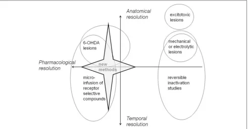
All methods have their limitations. Most important for present purposes is the trade-off between neuroanatomical and neu-rochemical resolution which is a feature of the approaches most commonly adopted to date. Excitotoxic lesion and
electrophysiological methods offer excellent neuroanatomical res-olution but tell us little about the role specifically of DA within mPFC. However, the standard method to deplete DA using 6-OHDA makes lesions which may be less anatomically cir-cumscribed than those made by standard excitotoxic methods. However, this difference may be more apparent than real in that neurochemical lesions are more typically verified by HPLC rather than methods which allow visualization of the boundaries of the lesion. Quantitative immunocytochemistry can be used to determine the final boundaries of such lesions (using for exam-ple antibodies against the rate limiting biosynthetic enzymes). However, delineation of lesion boundaries is known to depend on the method in use. For example, excitotoxic lesions show differ-ent boundaries using an immunohistochemical marker compared with those identified using conventional Nissl staining for his-tological assessment (Jongen-Relo and Feldon, 2002). Moreover, HPLC remains the method of choice to quantify changes in other neurotransmitters as well as secondary changes in neurotransmit-ter function in inneurotransmit-terconnected brain regions. Functional recovery might be seen to be a particular issue in the case of neurotoxic lesions made with 6-OHDA but the delineation of all lesions is confounded by secondary compensatory changes in response to the damage.
SELECTIVITY OF 6-OHDA LESIONS
A further constraint on the selectivity of 6-OHDA lesions arises in that the pre-treatments routinely used to protect NA neu-rons are not reliably successful and changes in NA as well as DA typically result from 6-OHDA lesions in mPFC (Nelson et al., 2010, 2011a) as well as in other brain regions such as NAc (Nelson et al., 2011b,c,d). However, neurotoxic 6-OHDA lesions do not result in non-specific neuronal damage in that they do not—for example—result in significant changes in 5-HT levels in target regions such as mPFC (Nelson et al., 2010, 2011a). Primary and secondary effects of neurotoxic lesions should also be distinguished, but 6-OHDA lesions do not necessarily result in secondary changes in control regions selected on the basis of their known connectivity with the mPFC (Sesack et al., 1989; Takagishi and Chiba, 1991).
Anatomically, the dominant view is that 6-OHDA lesions are typically less well-circumscribed. Infusion of 6-OHDA into the PL can nonetheless produce selective depletion within this target structure and spares catecholamine content in the more ventral IL cortex (Nelson et al., 2010, 2011a). However, infusion of 6-OHDA into the IL has a less selective effect in that it depletes cate-cholamines in both the IL and more dorsally in the PL (Naneix et al., 2009; Nelson et al., 2010, 2011a). On the other hand, in contrast to the evidence that mPFC lesions can enhance the responsiveness of DA neurons in NAc (Deutch et al., 1990), nei-ther PL nor IL 6-OHDA lesions have any effect on striatal DA (Rosin et al., 1992; Nelson et al., 2010, 2011a). Thus, these lesions are nonetheless in some respects more selective.
DOSE-RELATED EFFECTS OF DRUGS ADMINISTERED BY MICROINFUSION