2-Acetylpyridinium bromanilate
Lynne H. Thomas,a* Bryan Boyle,aLesley A. Clive,bAnna Collins,aLynsey D. Currie,aMalgorzata Gogol,bClaire Hastings,bAndrew O. F. Jones,aJennifer L. Kennedy,b Graham B. Kerr,bAlastair Kidd,bLorreta M. Lawton,a Susan J. Macintyre,b Niall M. MacLean,bAlan R. G. Martin,b Kate McGonagle,bSamantha Melrose,aGaius A. Rew,bColin W. Robinson,aMarc Schmidtmann,a
Felicity B. Turnbull,b Lewis G. Williams,aAlan Y.
Wiseman,bMalgorzata H. Wocialband Chick C. Wilsona
a
WestCHEM, Department of Chemistry, University of Glasgow, University Avenue, Glasgow G12 8QQ, Scotland, andbDepartment of Chemistry, University of Glasgow, University Avenue, Glasgow G12 8QQ, Scotland
Correspondence e-mail: [email protected] Received 8 April 2009; accepted 1 May 2009
Key indicators: single-crystal X-ray study;T= 100 K; mean(C–C) = 0.003 A˚;
Rfactor = 0.022;wRfactor = 0.050; data-to-parameter ratio = 14.4.
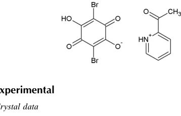
In the crystal of the title molecular salt (systematic name: 2-acetylpyridinium 2,5-dibromo-4-hydroxy-3,6-dioxocyclo-hexa-1,4-dienolate), C7H8NO
+
C6HBr2O4
, centrosymmetric rings consisting of two cations and two anions are formed, with the components linked by alternating O—H O and N— H O hydrogen bonds. Short O Br contacts [3.243 (2) and 3.359 (2) A˚ ] may help to consolidate the packing.
Related literature
For the structure of bromanilic acid, see: Robl (1987). For related structures, see: Tomura & Yamashita (2000); Zamanet al.(2001, 2004); Horiuchiet al.(2005).
Experimental
Crystal data
C7H8NO+C6HBr2O4
Mr= 419.03
a= 9.1323 (5) A˚ b= 13.3821 (7) A˚ c= 12.2287 (7) A˚ = 112.396 (2)
V= 1381.74 (13) A˚3
MoKradiation = 5.89 mm1 T= 100 K
0.250.20.1 mm
Data collection
Rigaku R-AXIS RAPID IP diffractometer
Absorption correction: empirical (using intensity measurements) (CrystalClear; Rigaku/MSC, 2008)
Tmin= 0.561,Tmax= 1.000 (expected range = 0.311–0.555) 17193 measured reflections 3156 independent reflections 2793 reflections withI> 2(I) Rint= 0.036
Refinement
R[F2> 2(F2)] = 0.022 wR(F2) = 0.050 S= 1.04 3156 reflections 219 parameters
H atoms treated by a mixture of independent and constrained refinement
max= 0.43 e A˚
3
min=0.31 e A˚
[image:1.610.51.230.568.683.2]3
Table 1
Hydrogen-bond geometry (A˚ ,).
D—H A D—H H A D A D—H A
O4—H1 O5 0.78 (3) 2.20 (3) 2.798 (2) 134 (3)
N1—H6 O2i
0.91 (3) 1.83 (3) 2.673 (2) 154 (3)
Symmetry code: (i)xþ2;yþ1;zþ1.
Data collection:CrystalClear(Rigaku/MSC, 2008); cell refinement:
CrystalClear; data reduction:CrystalClear; program(s) used to solve structure:SHELXS97(Sheldrick, 2008); program(s) used to refine structure: SHELXL97 (Sheldrick, 2008); molecular graphics:
ORTEP-3(Farrugia, 1997) andMercury(Macraeet al., 2006); soft-ware used to prepare material for publication: WinGX (Farrugia, 1999).
Supplementary data and figures for this paper are available from the IUCr electronic archives (Reference: HB2948).
References
Allen, F. H., Kennard, O., Watson, D. G., Brammer, L., Orpen, A. G. & Taylor, R. (1995).International Tables for Crystallography, Vol. C, edited by A. J. C. Wilson, pp. 685–706. Dordrecht: Kluwer Academic Publishers.
Farrugia, L. J. (1997).J. Appl. Cryst.30, 565. Farrugia, L. J. (1999).J. Appl. Cryst.32, 837–838.
Horiuchi, S., Kumai, R. & Tokura, Y. (2005).J. Am. Chem. Soc.127, 5010– 5011.
Macrae, C. F., Edgington, P. R., McCabe, P., Pidcock, E., Shields, G. P., Taylor, R., Towler, M. & van de Streek, J. (2006).J. Appl. Cryst.39, 453–457. Rigaku/MSC (2008).CrystalClear. Rigaku/MSC, The Woodlands, Texas, USA. Robl, C. (1987).Z. Kristallogr.180, 249–253.
Sheldrick, G. M. (2008).Acta Cryst.A64, 112–122.
Tomura, M. & Yamashita, Y. (2000).CrystEngComm,2, 92–95.
Zaman, Md. B., Tomura, M. & Yamashita, Y. (2001).J. Org. Chem.66, 5987– 5995.
Zaman, Md. B., Udachin, K. A. & Ripmeester, J. A. (2004).Cryst. Growth Des. 4, 585–589.
Structure Reports
Online
supporting information
Acta Cryst. (2009). E65, o1218 [doi:10.1107/S1600536809016456]
2-Acetylpyridinium bromanilate
Lynne H. Thomas, Bryan Boyle, Lesley A. Clive, Anna Collins, Lynsey D. Currie, Malgorzata
Gogol, Claire Hastings, Andrew O. F. Jones, Jennifer L. Kennedy, Graham B. Kerr, Alastair Kidd,
Lorreta M. Lawton, Susan J. Macintyre, Niall M. MacLean, Alan R. G. Martin, Kate McGonagle,
Samantha Melrose, Gaius A. Rew, Colin W. Robinson, Marc Schmidtmann, Felicity B. Turnbull,
Lewis G. Williams, Alan Y. Wiseman, Malgorzata H. Wocial and Chick C. Wilson
S1. Comment
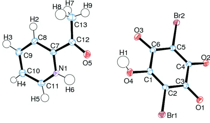
The stucture of the molecular proton-transfer salt of bromanilic acid with 2-acetylpyridine at 100 K is reported (Fig. 1). A
proton is transferred from the bromanilic acid molecule to the N atom on the acetylpyridine (Fig. 1). All previously
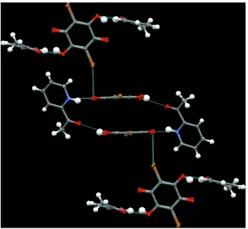
reported structures containing bromanilic acid have shown the tendency for extended chains of molecules to form. In this
case, hydrogen-bonded rings are formed between alternating cations and anions (Fig. 2) and these rings are held together
to form a three-dimensional structure by one Br···O close contact of 3.243 (2)Å (cf the sum of the van der Waals radii for
Br and O of 3.37Å) and one on the limit of the sum of the van der Waals radii of of 3.359 (2)Å (Fig. 3). The deprotonated
hydroxyl group on the bromanilic acid molecule is stabilized by forming a moderate hydrogen bond [2.673 (2)Å] with the
N atom on the 2-acetylpyridine molecule to which the proton has been transferred, and a short O···Br contact with
another bromanilic acid molecule. The C—O bond length to the deprotonated oxygen is notably shortened compared to
that to the protonated hydroxyl group [1.253 (2)Å versus 1.322 (2)Å]. The longer of the two O···Br close contacts is to
the C═O group on the bromanilic acid [C═O bond length 1.221 (2)Å].
S2. Experimental
Red blocks of (I) were grown by slow evaporation of solvent from a 1:1 solution of bromanilic acid and 2-acetylpyridine
in methanol.
S3. Refinement
The H atoms were identified in the difference map, and their positions were freely refined. The O- and N-bonded species
Figure 1
The molecular structure of (I) with displacement ellipsoids drawn at the 50% probability level.
Figure 2
The hydrogen bonded ring between alternating bromanilic acid and acetylpyridine molecules. The hydrogen bonds are
[image:3.610.130.483.312.444.2]Figure 3
The short bromine-oxygen close contacts connecting the hydrogen bonded rings. The short contacts and hydrogen bonds
are indicated by dashed lines.
2-Acetylpyridinium bromanilate
Crystal data
C7H8NO+·C6HBr2O4− Mr = 419.03
Monoclinic, P21/c Hall symbol: -P 2ybc
a = 9.1323 (5) Å
b = 13.3821 (7) Å
c = 12.2287 (7) Å
β = 112.396 (2)°
V = 1381.74 (13) Å3 Z = 4
F(000) = 816
Dx = 2.014 Mg m−3
Mo Kα radiation, λ = 0.71073 Å Cell parameters from 13698 reflections
θ = 6.1–55.2°
µ = 5.89 mm−1 T = 100 K Block, red
0.25 × 0.2 × 0.1 mm
Data collection
Rigaku R-AXIS RAPID IP diffractometer
Graphite monochromator
ω scans
Absorption correction: empirical (using intensity measurements)
(CrystalClear; Rigaku/MSC, 2008)
Rint = 0.036
θmax = 27.5°, θmin = 3.0°
k = −17→17
l = −15→15
Refinement
Refinement on F2 Least-squares matrix: full
R[F2 > 2σ(F2)] = 0.022 wR(F2) = 0.050 S = 1.04 3156 reflections 219 parameters 0 restraints
Primary atom site location: structure-invariant direct methods
Secondary atom site location: difference Fourier map
Hydrogen site location: difference Fourier map H atoms treated by a mixture of independent
and constrained refinement
w = 1/[σ2(F
o2) + (0.0229P)2 + 0.7894P] where P = (Fo2 + 2Fc2)/3
(Δ/σ)max = 0.001 Δρmax = 0.43 e Å−3 Δρmin = −0.31 e Å−3
Special details
Geometry. All s.u.'s (except the s.u. in the dihedral angle between two l.s. planes) are estimated using the full covariance matrix. The cell s.u.'s are taken into account individually in the estimation of s.u.'s in distances, angles and torsion angles; correlations between s.u.'s in cell parameters are only used when they are defined by crystal symmetry. An approximate (isotropic) treatment of cell s.u.'s is used for estimating s.u.'s involving l.s. planes.
Refinement. Refinement of F2 against ALL reflections. The weighted R-factor wR and goodness of fit S are based on F2, conventional R-factors R are based on F, with F set to zero for negative F2. The threshold expression of F2 > 2σ(F2) is used only for calculating R-factors(gt) etc. and is not relevant to the choice of reflections for refinement. R-factors based on F2 are statistically about twice as large as those based on F, and R- factors based on ALL data will be even larger. The isotropic displacement parameters for the hydrogen atoms involved in hydrogenbonds are refined freely. All other hydrogen atoms are refined against the atoms to which they are bonded.
Fractional atomic coordinates and isotropic or equivalent isotropic displacement parameters (Å2)
x y z Uiso*/Ueq
H6 0.611 (3) 0.307 (2) 0.195 (3) 0.041 (8)*
O5 0.50649 (17) 0.44777 (11) 0.25644 (13) 0.0201 (3)
N1 0.5396 (2) 0.29814 (12) 0.11977 (16) 0.0152 (3)
C12 0.4282 (2) 0.45501 (15) 0.15172 (18) 0.0160 (4)
C11 0.5549 (2) 0.22016 (16) 0.05849 (19) 0.0200 (4)
H5 0.637 (3) 0.1735 (18) 0.102 (2) 0.024*
C8 0.3294 (2) 0.36207 (16) −0.04572 (18) 0.0173 (4)
H2 0.263 (3) 0.4122 (18) −0.076 (2) 0.021*
C10 0.4555 (3) 0.20854 (17) −0.0595 (2) 0.0223 (5)
H4 0.468 (3) 0.156 (2) −0.099 (2) 0.027*
C7 0.4298 (2) 0.37017 (14) 0.07151 (17) 0.0145 (4)
C9 0.3417 (3) 0.28035 (17) −0.11190 (19) 0.0217 (4)
H3 0.274 (3) 0.2759 (19) −0.195 (2) 0.026*
C13 0.3272 (3) 0.54328 (17) 0.0970 (2) 0.0232 (5)
H9 0.351 (3) 0.595 (2) 0.152 (2) 0.028*
H7 0.340 (3) 0.5618 (19) 0.027 (2) 0.028*
H8 0.222 (3) 0.5245 (19) 0.076 (2) 0.028*
H1 0.663 (4) 0.511 (2) 0.426 (3) 0.050 (10)*
Br1 0.92699 (2) 0.416206 (15) 0.738904 (17) 0.01764 (6)
O1 1.19471 (16) 0.56928 (11) 0.79086 (12) 0.0180 (3)
O2 1.22536 (16) 0.73058 (10) 0.67034 (12) 0.0186 (3)
O3 0.73311 (16) 0.64428 (11) 0.37219 (12) 0.0198 (3)
C5 0.9790 (2) 0.69726 (14) 0.51628 (17) 0.0150 (4)
O4 0.70848 (17) 0.48853 (11) 0.48976 (14) 0.0197 (3)
C1 0.8359 (2) 0.54445 (15) 0.54043 (18) 0.0152 (4)
C6 0.8462 (2) 0.63450 (15) 0.46801 (17) 0.0149 (4)
C4 1.1029 (2) 0.67882 (14) 0.62391 (18) 0.0139 (4)
C3 1.0879 (2) 0.58650 (14) 0.69581 (17) 0.0143 (4)
C2 0.9474 (2) 0.52459 (14) 0.64757 (17) 0.0145 (4)
Atomic displacement parameters (Å2)
U11 U22 U33 U12 U13 U23
O5 0.0225 (8) 0.0205 (7) 0.0161 (7) −0.0024 (6) 0.0059 (6) −0.0021 (6)
N1 0.0126 (8) 0.0165 (8) 0.0135 (8) 0.0001 (6) 0.0014 (7) 0.0008 (7)
C12 0.0157 (10) 0.0161 (10) 0.0184 (10) −0.0023 (7) 0.0088 (8) 0.0005 (8)
C11 0.0178 (10) 0.0179 (10) 0.0219 (11) 0.0044 (8) 0.0048 (9) 0.0002 (8)
C8 0.0129 (10) 0.0206 (10) 0.0158 (10) 0.0007 (8) 0.0024 (8) 0.0035 (8)
C10 0.0277 (12) 0.0212 (11) 0.0185 (11) 0.0005 (9) 0.0093 (9) −0.0056 (9)
C7 0.0138 (9) 0.0143 (9) 0.0158 (10) −0.0004 (7) 0.0062 (8) 0.0011 (8)
C9 0.0242 (11) 0.0246 (11) 0.0137 (10) −0.0027 (9) 0.0045 (9) −0.0002 (8)
C13 0.0271 (12) 0.0209 (11) 0.0249 (12) 0.0057 (9) 0.0135 (10) 0.0023 (9)
Br1 0.01649 (11) 0.01824 (11) 0.01617 (11) −0.00241 (7) 0.00395 (8) 0.00493 (7)
Br2 0.02145 (12) 0.01616 (11) 0.01540 (11) −0.00364 (7) 0.00360 (9) 0.00339 (7)
O1 0.0164 (7) 0.0188 (7) 0.0144 (7) −0.0007 (5) 0.0011 (6) 0.0018 (6)
O2 0.0172 (7) 0.0158 (7) 0.0185 (7) −0.0026 (5) 0.0020 (6) 0.0016 (6)
O3 0.0182 (7) 0.0229 (8) 0.0137 (7) −0.0020 (6) 0.0008 (6) 0.0032 (6)
C5 0.0184 (10) 0.0128 (9) 0.0129 (9) −0.0001 (7) 0.0051 (8) 0.0021 (7)
O4 0.0168 (8) 0.0208 (8) 0.0154 (7) −0.0058 (6) −0.0008 (6) 0.0026 (6)
C1 0.0155 (10) 0.0153 (9) 0.0154 (10) −0.0005 (7) 0.0065 (8) −0.0014 (8)
C6 0.0156 (10) 0.0165 (10) 0.0127 (9) 0.0016 (7) 0.0057 (8) 0.0005 (8)
C4 0.0147 (10) 0.0122 (9) 0.0152 (10) 0.0003 (7) 0.0061 (8) −0.0013 (7)
C3 0.0164 (10) 0.0136 (9) 0.0145 (10) 0.0013 (7) 0.0077 (8) −0.0015 (7)
C2 0.0167 (10) 0.0130 (9) 0.0151 (9) −0.0001 (7) 0.0075 (8) 0.0012 (7)
Geometric parameters (Å, º)
O5—C12 1.209 (2) C13—H7 0.94 (3)
N1—C11 1.323 (3) C13—H8 0.93 (3)
N1—C7 1.353 (2) Br1—C2 1.8826 (19)
N1—H6 0.91 (3) Br2—C5 1.8922 (19)
C12—C13 1.491 (3) O1—C3 1.221 (2)
C12—C7 1.504 (3) O2—C4 1.253 (2)
C11—C10 1.390 (3) O3—C6 1.239 (2)
C11—H5 0.96 (3) C5—C4 1.392 (3)
C8—C7 1.381 (3) C5—C6 1.407 (3)
C10—C9 1.381 (3) C1—C2 1.344 (3)
C10—H4 0.88 (3) C1—C6 1.520 (3)
C9—H3 0.97 (3) C4—C3 1.552 (3)
C13—H9 0.93 (3) C3—C2 1.451 (3)
C11—N1—C7 122.38 (18) H9—C13—H7 112 (2)
C11—N1—H6 119.2 (18) C12—C13—H8 107.8 (16)
C7—N1—H6 118.2 (18) H9—C13—H8 110 (2)
O5—C12—C13 123.61 (19) H7—C13—H8 107 (2)
O5—C12—C7 118.77 (18) C4—C5—C6 123.42 (18)
C13—C12—C7 117.62 (18) C4—C5—Br2 119.00 (14)
N1—C11—C10 120.53 (19) C6—C5—Br2 117.57 (14)
N1—C11—H5 115.3 (15) C1—O4—H1 107 (2)
C10—C11—H5 124.2 (15) O4—C1—C2 123.31 (19)
C7—C8—C9 119.83 (19) O4—C1—C6 114.51 (17)
C7—C8—H2 117.2 (16) C2—C1—C6 122.18 (17)
C9—C8—H2 122.9 (16) O3—C6—C5 127.49 (19)
C9—C10—C11 118.8 (2) O3—C6—C1 114.86 (17)
C9—C10—H4 122.0 (16) C5—C6—C1 117.65 (17)
C11—C10—H4 119.1 (16) O2—C4—C5 126.44 (18)
N1—C7—C8 119.03 (18) O2—C4—C3 116.07 (17)
N1—C7—C12 116.30 (17) C5—C4—C3 117.48 (17)
C8—C7—C12 124.67 (18) O1—C3—C2 122.78 (18)
C10—C9—C8 119.39 (19) O1—C3—C4 118.58 (17)
C10—C9—H3 120.6 (15) C2—C3—C4 118.64 (17)
C8—C9—H3 119.9 (15) C1—C2—C3 120.51 (18)
C12—C13—H9 109.4 (16) C1—C2—Br1 121.41 (15)
C12—C13—H7 110.4 (16) C3—C2—Br1 118.08 (14)
Hydrogen-bond geometry (Å, º)
D—H···A D—H H···A D···A D—H···A
O4—H1···O5 0.78 (3) 2.20 (3) 2.798 (2) 134 (3)
N1—H6···O2i 0.91 (3) 1.83 (3) 2.673 (2) 154 (3)