Spiro[1,3-dioxolane-2,3
000-indolin]-2
000-one
Yan Menga* and Yanqing Miaob
aSchool of Environmental Engineering, Chang’an University, South Second Cycle Road 368#, Xi’an 710054, Shannxi, People’s Republic of China, andbDepartment of Pharmacy, Xi’an Medical University, Hanguang Round No. 137, Xi’an 710021, Xi’an, People’s Republic of China
Correspondence e-mail: [email protected]
Received 28 March 2010; accepted 2 May 2010
Key indicators: single-crystal X-ray study;T= 273 K; mean(C–C) = 0.003 A˚; Rfactor = 0.048;wRfactor = 0.137; data-to-parameter ratio = 11.7.
The title compound, C10H9NO3, was synthesized by the
condensation reaction of isatin (systematic name 1H-indole-2,3-dione) with glycol in presence of p-toluenesulfonic acid. The indol-2-one ring system is essentially planar [N—C—C— C torsion angle = 3.1 (2)], and the 1,3-dioxolane ring is slightly distorted. The crystal structure exhibits intermolecular N—H O hydrogen bonds.
Related literature
For the synthesis of the title compound, see: Santos et al. (2008). For the bioactivity of the title compound, see: Demostheneset al.(1998); Rajopadhye & Popp (1988).
Experimental
Crystal data
C10H9NO3
Mr= 191.18
Monoclinic,P21=c
a= 7.484 (2) A˚
b= 5.650 (1) A˚
c= 20.942 (5) A˚
= 97.889 (8)
V= 877.1 (4) A˚3
Z= 4
MoKradiation
= 0.11 mm1
T= 273 K
0.360.270.21 mm
Data collection
Bruker SMART CCD diffractometer
Absorption correction: multi-scan (SADABS; Bruker, 2002)
Tmin= 0.963,Tmax= 0.989
4056 measured reflections 1534 independent reflections 1093 reflections withI> 2(I)
Rint= 0.070
Refinement
R[F2> 2(F2)] = 0.048
wR(F2) = 0.137
S= 1.09 1534 reflections 131 parameters
H atoms treated by a mixture of independent and constrained refinement
max= 0.23 e A˚
3
min=0.20 e A˚
3
Table 1
Hydrogen-bond geometry (A˚ ,).
D—H A D—H H A D A D—H A
N1—H1 O1i 0.87 (3) 2.07 (3) 2.941 (3) 174 (2)
Symmetry code: (i)xþ1;y;zþ1.
Data collection:SMART(Bruker, 2002); cell refinement: SAINT-Plus(Bruker, 2002); data reduction:SAINT-Plus; program(s) used to solve structure: SHELXS97(Sheldrick, 2008); program(s) used to refine structure:SHELXL97(Sheldrick, 2008); molecular graphics: SHELXTL(Sheldrick, 2008); software used to prepare material for publication:SHELXTL.
Supplementary data and figures for this paper are available from the IUCr electronic archives (Reference: LX2143).
References
Bruker (2002). SMART, SAINT-Plus and SADABS. Bruker AXS Inc, Madison, Wisconsin, USA.
Demosthenes, F., William, J. R., David, S. C. & David, L. C. (1998).
Tetrahedron Lett.39, 2235–2238.
Rajopadhye, M. & Popp, F. D. (1988).J. Med. Chem.31, 1001–1005. Santos, E. L., Gomes, W. A. Jr, Ribeiro, N. M. & Andrade, H. M. C. (2008).J.
Mol. Catal. A,295, 18–23.
Sheldrick, G. M. (2008).Acta Cryst.A64, 112–122. Acta Crystallographica Section E
Structure Reports
Online
supporting information
Acta Cryst. (2010). E66, o1305 [https://doi.org/10.1107/S1600536810016132]
Spiro[1,3-dioxolane-2,3
′
-indolin]-2
′
-one
Yan Meng and Yanqing Miao
S1. Comment
Isatin derivatives, has caught great attention of many researchers as a versatile lead molecule for designing of potential
drugs for the variety of biological activities, such as anti-bacteria, anti-virus, anti-tumor and neuroprotection. Among
these compounds, spiro-oxindol analogues have received considerable attention as potential anti-bacteria and
neuroprotection agents(Demosthenes et al. 1998; Rajopadhye et al. 1988; Santos et al. 2008).
The X-ray structural analysis confirmed the assignment of its structure from spectroscopic data. The molecular structure
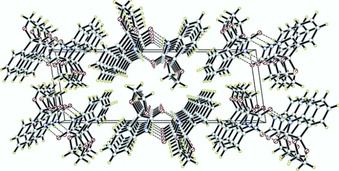
is depicted in Fig. 1, and a diagram of interactions between the title compounds is depicted in Fig. 2. Geometric
parameters of the title compound are in the usual ranges. The crystal packing (Fig. 2) is stabilized by intermolecular N—
H···O hydrogen bonds between the indoline H atom and the oxygen of the C═O unit, with a N1—H1···O1i (Table 1).
S2. Experimental
Isatin (1 mmol) and glycol (1 mmol) was dissolved in cyclohexane (20 ml), and 0.01 mmol TsOH was added. The
mixture was stirred under reflux. After completion of the reaction, it was evaporated to dryness, followed by
chromatography to the pure title compound. 1H-NMR (D
6-Acetone, 400 MHz) delta: 10.44 (1H, s), 7.33 (2H, m), 7.00
(1H, td, J = 7.2, 0.8 Hz), 6.82 (1H, d, J = 7.6 Hz), 4.33 (2H, m), 4.23 (2H, m); EI–MS, m/z (%): 233 (M+)
S3. Refinement
The H atom bound N atom was located from difference Fourier map and refined freely. All H atoms of C atoms were
positioned geometrically and refined using a riding model, with C–H = 0.93 Å for aryl and 0.97 Å for methylene H
Figure 1
The molecular structure of the title compound with the atom numbering scheme. Displacement ellipsoids are drawn at the
30% probability level. H atoms are presented as a small cycles of arbitrary radius.
Figure 2
N–H···O interactions (dotted lines) in the crystal structure of the title compound.
Spiro[1,3-dioxolane-2,3′-indolin]-2′-one
Crystal data
C10H9NO3
Mr = 191.18
Monoclinic, P21/c
Hall symbol: -P 2ybc
a = 7.484 (2) Å
b = 5.650 (1) Å
c = 20.942 (5) Å
β = 97.889 (8)°
V = 877.1 (4) Å3
Z = 4
F(000) = 400
Dx = 1.448 Mg m−3
Mo Kα radiation, λ = 0.71073 Å Cell parameters from 7304 reflections
θ = 1.5–25.0°
µ = 0.11 mm−1
[image:3.610.136.474.308.479.2]Data collection
Bruker SMART CCD diffractometer
Radiation source: fine-focus sealed tube Graphite monochromator
phi and ω scans
Absorption correction: multi-scan (SADABS; Bruker, 2002)
Tmin = 0.963, Tmax = 0.989
4056 measured reflections 1534 independent reflections 1093 reflections with I > 2σ(I)
Rint = 0.070
θmax = 25.0°, θmin = 2.0°
h = −8→8
k = −6→6
l = −21→24
Refinement
Refinement on F2
Least-squares matrix: full
R[F2 > 2σ(F2)] = 0.048
wR(F2) = 0.137
S = 1.09 1534 reflections 131 parameters 0 restraints
Primary atom site location: structure-invariant direct methods
Secondary atom site location: difference Fourier map
Hydrogen site location: difference Fourier map H atoms treated by a mixture of independent
and constrained refinement
w = 1/[σ2(F
o2) + (0.0648P)2 + 0.151P]
where P = (Fo2 + 2Fc2)/3
(Δ/σ)max < 0.001
Δρmax = 0.23 e Å−3
Δρmin = −0.20 e Å−3
Special details
Geometry. All esds (except the esd in the dihedral angle between two l.s. planes) are estimated using the full covariance matrix. The cell esds are taken into account individually in the estimation of esds in distances, angles and torsion angles; correlations between esds in cell parameters are only used when they are defined by crystal symmetry. An approximate (isotropic) treatment of cell esds is used for estimating esds involving l.s. planes.
Refinement. Refinement of F2 against ALL reflections. The weighted R-factor wR and goodness of fit S are based on F2,
conventional R-factors R are based on F, with F set to zero for negative F2. The threshold expression of F2 > 2sigma(F2) is
used only for calculating R-factors(gt) etc. and is not relevant to the choice of reflections for refinement. R-factors based on F2 are statistically about twice as large as those based on F, and R- factors based on ALL data will be even larger.
Fractional atomic coordinates and isotropic or equivalent isotropic displacement parameters (Å2)
x y z Uiso*/Ueq
O1 0.6983 (2) 0.2241 (3) 0.50491 (8) 0.0600 (5)
O3 0.6788 (2) 0.6884 (3) 0.43906 (8) 0.0551 (5)
O2 0.8104 (2) 0.4151 (3) 0.38133 (8) 0.0560 (5)
N1 0.4575 (3) 0.1613 (4) 0.42609 (9) 0.0469 (6)
H1 0.407 (4) 0.044 (5) 0.4439 (13) 0.070 (9)*
C7 0.3813 (3) 0.2722 (4) 0.36864 (10) 0.0400 (6)
C2 0.6494 (3) 0.4723 (4) 0.40675 (10) 0.0417 (6)
C8 0.4869 (3) 0.4639 (4) 0.35588 (10) 0.0402 (6)
C3 0.4343 (3) 0.6053 (4) 0.30311 (11) 0.0506 (6)
H3A 0.5032 0.7355 0.2945 0.061*
C6 0.2254 (3) 0.2139 (4) 0.32859 (11) 0.0512 (6)
H6A 0.1566 0.0834 0.3370 0.061*
C1 0.6076 (3) 0.2731 (4) 0.45352 (10) 0.0447 (6)
C5 0.1752 (3) 0.3571 (5) 0.27544 (11) 0.0550 (7)
H5A 0.0707 0.3216 0.2477 0.066*
H4A 0.2381 0.6443 0.2272 0.065*
C10 0.8530 (4) 0.7641 (6) 0.4348 (2) 0.0911 (11)
H10A 0.9164 0.7972 0.4774 0.109*
H10B 0.8504 0.9075 0.4092 0.109*
C9 0.9435 (4) 0.5760 (6) 0.40444 (16) 0.0809 (10)
H9B 1.0019 0.6383 0.3694 0.097*
H9C 1.0340 0.5011 0.4355 0.097*
Atomic displacement parameters (Å2)
U11 U22 U33 U12 U13 U23
O1 0.0568 (12) 0.0592 (12) 0.0595 (10) −0.0091 (9) −0.0079 (9) 0.0079 (8)
O3 0.0577 (11) 0.0369 (9) 0.0735 (11) −0.0087 (8) 0.0196 (9) −0.0187 (8)
O2 0.0441 (10) 0.0489 (10) 0.0777 (11) −0.0054 (8) 0.0176 (8) −0.0210 (8)
N1 0.0488 (13) 0.0362 (11) 0.0535 (11) −0.0084 (10) −0.0008 (9) 0.0106 (9)
C7 0.0435 (14) 0.0317 (12) 0.0452 (12) 0.0029 (10) 0.0081 (10) 0.0008 (9)
C2 0.0424 (13) 0.0305 (12) 0.0538 (12) −0.0037 (10) 0.0127 (10) −0.0069 (10)
C8 0.0435 (13) 0.0297 (12) 0.0490 (12) 0.0015 (10) 0.0122 (10) −0.0006 (9)
C3 0.0542 (15) 0.0397 (13) 0.0605 (14) 0.0000 (11) 0.0174 (12) 0.0095 (11)
C6 0.0495 (16) 0.0433 (14) 0.0596 (14) −0.0058 (11) 0.0032 (11) 0.0019 (11)
C1 0.0464 (14) 0.0368 (13) 0.0497 (12) 0.0005 (11) 0.0022 (11) −0.0019 (10)
C5 0.0483 (15) 0.0612 (17) 0.0540 (13) 0.0034 (13) 0.0014 (11) 0.0042 (12)
C4 0.0558 (16) 0.0569 (16) 0.0505 (13) 0.0139 (14) 0.0095 (11) 0.0114 (11)
C10 0.059 (2) 0.0579 (19) 0.160 (3) −0.0178 (16) 0.028 (2) −0.043 (2)
C9 0.0602 (18) 0.084 (2) 0.102 (2) −0.0233 (17) 0.0217 (16) −0.0377 (19)
Geometric parameters (Å, º)
O1—C1 1.223 (2) C3—C4 1.393 (3)
O3—C10 1.387 (4) C3—H3A 0.9300
O3—C2 1.399 (3) C6—C5 1.386 (3)
O2—C9 1.386 (3) C6—H6A 0.9300
O2—C2 1.420 (3) C5—C4 1.370 (3)
N1—C1 1.347 (3) C5—H5A 0.9300
N1—C7 1.406 (3) C4—H4A 0.9300
N1—H1 0.87 (3) C10—C9 1.452 (4)
C7—C6 1.380 (3) C10—H10A 0.9700
C7—C8 1.388 (3) C10—H10B 0.9700
C2—C8 1.503 (3) C9—H9B 0.9700
C2—C1 1.552 (3) C9—H9C 0.9700
C8—C3 1.377 (3)
C10—O3—C2 108.95 (19) C5—C6—H6A 121.2
C9—O2—C2 109.03 (19) O1—C1—N1 126.8 (2)
C1—N1—C7 111.8 (2) O1—C1—C2 125.8 (2)
C1—N1—H1 124.0 (17) N1—C1—C2 107.44 (18)
C7—N1—H1 123.8 (17) C4—C5—C6 121.5 (2)
C6—C7—N1 128.6 (2) C6—C5—H5A 119.2
C8—C7—N1 109.75 (19) C5—C4—C3 120.5 (2)
O3—C2—O2 107.13 (17) C5—C4—H4A 119.8
O3—C2—C8 115.39 (18) C3—C4—H4A 119.8
O2—C2—C8 111.88 (17) O3—C10—C9 107.6 (2)
O3—C2—C1 111.09 (17) O3—C10—H10A 110.2
O2—C2—C1 109.05 (18) C9—C10—H10A 110.2
C8—C2—C1 102.17 (17) O3—C10—H10B 110.2
C3—C8—C7 120.0 (2) C9—C10—H10B 110.2
C3—C8—C2 131.7 (2) H10A—C10—H10B 108.5
C7—C8—C2 108.34 (17) O2—C9—C10 106.1 (2)
C8—C3—C4 118.7 (2) O2—C9—H9B 110.5
C8—C3—H3A 120.6 C10—C9—H9B 110.5
C4—C3—H3A 120.6 O2—C9—H9C 110.5
C7—C6—C5 117.6 (2) C10—C9—H9C 110.5
C7—C6—H6A 121.2 H9B—C9—H9C 108.7
C1—N1—C7—C6 −177.7 (2) C7—C8—C3—C4 −0.9 (3)
C1—N1—C7—C8 1.5 (3) C2—C8—C3—C4 178.4 (2)
C10—O3—C2—O2 −0.6 (3) C8—C7—C6—C5 −1.3 (4)
C10—O3—C2—C8 −125.9 (3) N1—C7—C6—C5 177.8 (2)
C10—O3—C2—C1 118.4 (3) C7—N1—C1—O1 176.7 (2)
C9—O2—C2—O3 7.5 (3) C7—N1—C1—C2 −5.3 (3)
C9—O2—C2—C8 134.9 (2) O3—C2—C1—O1 −51.7 (3)
C9—O2—C2—C1 −112.8 (2) O2—C2—C1—O1 66.2 (3)
C6—C7—C8—C3 1.8 (3) C8—C2—C1—O1 −175.3 (2)
N1—C7—C8—C3 −177.5 (2) O3—C2—C1—N1 130.3 (2)
C6—C7—C8—C2 −177.7 (2) O2—C2—C1—N1 −111.9 (2)
N1—C7—C8—C2 3.1 (2) C8—C2—C1—N1 6.7 (2)
O3—C2—C8—C3 54.2 (3) C7—C6—C5—C4 −0.1 (4)
O2—C2—C8—C3 −68.6 (3) C6—C5—C4—C3 0.9 (4)
C1—C2—C8—C3 174.9 (2) C8—C3—C4—C5 −0.4 (4)
O3—C2—C8—C7 −126.4 (2) C2—O3—C10—C9 −6.2 (4)
O2—C2—C8—C7 110.7 (2) C2—O2—C9—C10 −11.1 (4)
C1—C2—C8—C7 −5.8 (2) O3—C10—C9—O2 10.7 (4)
Hydrogen-bond geometry (Å, º)
D—H···A D—H H···A D···A D—H···A
N1—H1···O1i 0.87 (3) 2.07 (3) 2.941 (3) 174 (2)