Partially supported by the Ministry of Agriculture of the Czech Republic (Grant No. MZE-0002716201), Grants No. FOOD-CT-2005-007081 (PathogenCombat) and Cost Action 929 ENVIRONET of Brussels, EC.
Hepatitis E virus: a review
P. Vasickova
1,2, I. Psikal
2, P. Kralik
2, F. Widen
3, Z. Hubalek
4,5, I. Pavlik
2 1University of Veterinary and Pharmaceutical Sciences Brno, Czech Republic2Veterinary Research Institute, Brno, Czech Republic 3The National Veterinary Institute,Uppsala, Sweden
4Faculty of Science, Masaryk University, Brno, Czech Republic
5Institute of Vertebrate Biology of the Academy of Sciences of the Czech Republic, Brno,
Czech Republic
ABSTRACT: The hepatitis E virus (HEV), the causative agent of hepatitis E, is a non-enveloped RNA virus. The HEV genome is formed by a non-segmented positive-sense RNA chain. The 3´end of the chain is polyadenylated and the 5´end is structurally characterised by the so called “capping”. According to currently accepted taxonomy, HEV is classified in the genus Hepevirus, the only member of the Hepeviridae family. HE is usually transmitted via the faecal-oral route due to the fact that drinking water or water for industrial purposes is contaminated due to poor sanitation. This spread of HEV has been reported in developing countries of Asia, Africa, South and Central America. However, cases in countries with the sporadic occurrence of HEV have been associated with travelling to countries with an increased risk of infection (developing countries in Asia, Africa and America). HEV infections have subsequently been described in people who have not travelled to endemic countries. Further studies of the HEV suggested other routes of transmission and a zoonotic potential of the virus (pigs and deer as the potential source of human infection).
Keywords: risk assessment; food safety; foodborne viral outbreaks; zoonoses; pigs
Contents
1. Introduction 2. HE in humans
2.1. Clinical manifestations
2.2. Immune response of an infected organism 2.3. Gross lesions in liver tissue
3. HE in animals
3.1. Domestic animals 3.1.1. Domestic pigs 3.1.2. Poultry 3.1.3. Dogs and cats
3.1.4. Other domestic animals 3.2. Wild animals
3.2.1. Rodents
3.2.2. Artiodactyla 3.2.3. Carnivores
4. Characteristic features and prevalence of HEV in various animal species
4.1. HEV genome
4.1.1. Open reading frame 1 (ORF1) 4.1.2. Open reading frame 2 (ORF2) 4.1.3. Open reading frame 3 (ORF3)
4.2. Genotypes and prevalence of human HEV strains
4.3. Swine HEV and its prevalence
1. Introduction
Hepatitis E virus (HEV), the etiological agent of hepatitis E (HE), was described for the first time using electron microscopy in 1983 as a spherical viral particle being 27 to 30 nm in size. The virus originated from the stool of a volunteer orally in-fected with faeces from suspect cases of non-A and non-B hepatitis (Balayan et al., 1983). The HEV genome comprises a non-segmented positive-sense RNA chain and the virus is non-enveloped (Acha and Szyfres, 2003). HEV was suggested to be clas-sified in the Picornaviridae family (Balayan et al., 1983). However, later studies showed that it does not belong to members of this family. Between 1988 and 1998, HEV was tentatively classified in the Caliciviridae family, based on virion morphology. This classification was also rejected after a phyl-ogeny analysis of the HEV genome, and HEV was newly classified as an independent genus HEV-like virus, unassigned to any family (Berke and Matson, 2000; Acha and Szyfres, 2003).
At present, HEV is the only member of the
Hepevirus genus, Hepeviridae family (Emerson et al., 2004; http://www.ncbi.nlm.nih.gov/ICTVdb/ Ictv/fs_hepev.htm). Phylogenetically, HEV shows the highest, but limited, similarity with the Rubella virus: Alphavirus genus, Togaviridae family and with the Necrotic yellow vein virus of sugar beet:
Furovirus genus, Togaviridae family (Berke and Matson, 2000).
HEV causes acute sporadic and epidemic viral hepatitis worldwide. HEV infections are spread mainly by the faecal-oral route and large epidem-ics due to this virus are often associated with con-taminated water (Ashbolt, 2004; Koopmans and
Duizer, 2004; Vasickova et al., 2005). There is also a possibility of zoonotic transmission of the virus. Seroepidemiological studies revealed that anti-HEV antibodies are present in numerous animal species including pigs, rodents, chickens, dogs, cows, sheep and goats from developing and in-dustrialised countries (Tien et al., 1997; Favorov et al., 1998; Arankalle et al., 2001). In addition, an isolate of HEV from a pig, designated as swine HEV, has been identified and shown to be closely related to two human isolates of HEV (US-1 and US-2) identified in the United States (Meng et al., 1997). Similar findings have also been reported in pigs from Taiwan with a different strain of swine HEV (Hsieh et al., 1999). Experimental cross spe-cies infection has been demonstrated: swine HEV has been shown to infect rhesus monkeys and chimpanzees and the US-2 strain of human HEV has been shown to infect pigs (Meng et al., 1998a; Halbur et al., 2001).
With regard to this knowledge, HEV may be viewed as a new emerging pathogen with a zoonotic potential. Accordingly, the purpose of the review article is to summarise current knowledge, which may serve for the identification and more detailed characterisation of the virus not only in the Czech Republic, but also in other member states of the European Union.
2. HE in humans
2.1. Clinical manifestations
HEV infections are spread by the faecal-oral route. However, the mechanism allowing the virus 5. Replication and expression of HEV
5.1. Hepatocytes – primary host target cells for HEV
5.2. The other HEV infected host tissues 6. HEV transmission
6.1. The most common HEV transmission route
6.2. Person to person contact and transplacental transmission
6.3. HEV transmission via foodstuffs 6.3.1. Pork and pig offal
6.3.2. Undercooked foodstuffs made from venison
6.3.3. Shellfish
6.4. Zoonotic potential of HEV
7. Risk groups in the population and HEV preven-tion
8. Diagnostic methods
8.1. Molecular detection of HEV 8.2. Immunologic diagnosis
8.1.1. Enzyme immunoassay (EIA)
8.1.2. Immune fluorescence microscopy (IFE)
8.3. Virus isolation
8.4. Immune electron microscopy (IEM) 9. Conclusions
to reach the site of primary multiplication has not been fully clarified yet. Replication of viral particles takes place in intestinal mucosa cells, but primary in the cytoplasm of hepatocytes. Virions are trans-ported with bile from liver tissue to the intestine (Williams et al., 2001). Aggarwal et al. (2000) ana-lysed faeces collected from 20 patients affected by acute HE, 35 days after appearance of the first signs of the disease. They did not detect HEV ribonu-cleic acid (RNA) in any of the samples, but HEV RNA was present in the blood serum 29 days after the appearance of the first symptoms. Viremia was detected in one patient after 45 days. The incuba-tion time ranges between 3 to 8 weeks; symptoms of the disease appeared in macaques two weeks after experimental intravenous infection (Li et al., 1994; Tsarev et al., 1994, as quoted by Anderson and Shrestha, 2002).
Various clinical manifestations of the disease have been observed, from more frequent subclin-ical forms to fulminant forms of hepatitis. HEV infection is most often seen in children, young to middle aged adults (15 to 40 years old) and might be serious in pregnant women. In most cases, the signs and symptoms of the disease include mod-erately severe hepatitis with concurrent signs of influenza-like symptoms, abdominal pain, tender-ness, nausea, vomiting and fever in the first (pre-icteric) phase of 1 to 10 days. The second ((pre-icteric) phase (15 to 40 days) with concurrent jaundice and dark urine is followed by viremia, liver enzyme el-evations, antibody seroconversion and clearing of the virus. Clinically, HE is typically a self-limiting disease without progression to chronic illness. HE is mostly asymptomatic in children but fulminant hepatitis occurs more frequently during pregnan-cy with a mortality rate of 20% among pregnant women in the thrird trimester, and can also cause premature births. The reason for the high mor-tality of pregnant women is not fully determined (Hussaini et al., 1997; Bednar et al., 1999; Worm et al., 2002; Emerson and Purcell, 2003).
2.2. Immune response of an infected organism
With the onset of symptoms, antibodies against HEV, IgM and subsequently IgG class appear in the titre sera of infected patients. The IgM sharply declines at the beginning of convalescence; these antibodies may be detected for the following two to
three months only (Chauhan et al., 1993). In contrast, IgG antibodies usually persist in the organisms of infected people for several years, in 47% of patients for more than 14 years (Clayson et al., 1995).
2.3. Gross lesions in liver tissue
Specific changes in the morphology of liver tissue have been observed in HE patients. Liver biopsy specimens have shown either non-specific inflam-matory or prominent canalicular bile stasis with a pseudoglandular arrangement of hepatocytes around distended bile canaliculi (cholestatic form). Histopathological changes gradually resolve over 3 to 6 months (Anderson and Shrestha, 2002).
3. HE in animals
Serological studies of HEV in endemic and non-endemic regions have shown that HEV anti-bodies are present in domestic and wild animal species. That gives evidence that these animals have been in contact with HEV or an antigenically re-lated agent. A reservoir may exist in these animals and constitute a risk for spreading to the human population (Arankalle et al., 1994; Favorov et al., 1998; Arankalle et al., 2001).
3.1. Domestic animals
3.1.1. Domestic pigs
3.1.2. Poultry
Anti-HEV antibodies have been detected in 44% of examined chicken in Vietnam (Tien et al., 1997) and in 20% of chicken in Brazil (Vitral et al., 2005). Among 70 tested flocks of hens in the USA, 54 (71%) gave seropositive responses for IgG to HEV (Huang et al., 2002a). Avian HEV was identified and characterised in bile samples from chickens showing hepatitis-splenomegaly syndrome (HS; Haqshenas et al., 2001). HEV in-fection was most likely a factor (however, not the only one) essential for the clinical development of HS syndrome. Regressive ovaries, red fluid in the abdomen, enlarged liver and spleen and up to 20% drop in egg production may be observed in infected poultry (Billam et al., 2005).
3.1.3. Dogs and cats
Anti-HEV antibodies were detected in 27% of tested dog serum samples in Vietnam (Tien et al., 1997) and 7% of animals in Brazil (Vitral et al., 2005). Among 135 domestic cats tested in Japan, HEV antibodies were detected in 44 (33%) of them (Okamoto et al., 2004).
3.1.4. Other domestic animals
[image:4.595.69.533.112.509.2]Serological screenings showed the presence of antibodies against HEV in 29% to 62% of cows in Somalia, Tajikistan and Turkmenistan (endem-ic regions), in 42% to 67% of sheep and goats in Turkmenistan and in about 12% of cows in the Table 1. Prevalence of the IgG antibody to hepatitis E virus in serum of domestic pigs (Sus scrofa domesticus) de-tected by enzyme immunoassay according to the geographic region
Country Number of References
tested positive %
Argentina 97 22 22.7 Munne et al., 2006
Brazil 357 227 63.6 Vitral et al., 2005
Canada 712 129 18.1 Meng et al., 1999
Canada 998 594 59.5 Yoo et al., 2001
China 82 22 25.0 Meng et al., 1999
China 419 330 78.8 Wang et al., 2002
Great Britain 256 219 85.5 Banks et al., 2004a
India 284 122 43.0 Arankalle et al., 2002
Indonesia 99 71 72.0 Wibawa et al., 2004
Korea 140 57 40.7 Meng et al., 1999
Mexico 125 8 6.0 Cooper et al., 2005
New Zealand 72 54 75.0 Garkavenko et al., 2001
Spain 60 15 25.0 Pina et al., 2000
Sweden 204 118 58.0 Banks et al., 2004a
Taiwan 275 102 37.1 Hsieh et al., 1999
Thailand 76 10 13.0 Cooper et al., 2005
Thailand 75 23 30.7 Meng et al., 1999
The Netherlands 34 8 23.5 Banks et al., 2004a
USA 293 202 68.9 Meng et al., 1997
USA 84 29 34.5 Withers et al., 2002
Ukraine in non-endemic geographic areas (Favorov et al., 1998). Among calf serum samples from vari-ous regions in China, only 6.3% (12/160) were positive against HEV; specific antibodies were not detected in goats from the same areas (Wang et al., 2002). Vitral et al. (2005) detected antibodies against HEV in Brazil in one of 70 (1.4%) tested cows; IgG was not found in any of the 12 sheep and five goats tested.
3.2. Wild animals
3.2.1. Rodents
Serological surveys showed the presence of antibodies against HEV in several species of ro-dents (Table 2), especially rats (Rattus norvegi-cus, R. rattus) in Brazil (Vitral et al., 2005), Japan (Hirano et al., 2003) and the USA (Kabrane-Lazizi et al., 1999a; Favorov et al., 2000).
3.2.2. Artiodactyla
Among 117 serum samples from Sika deer (Cervus nippon yesoensis, C. n. centralis and C. n. nippon) and 35 serum samples from wild boars (S. scrofa leucomystax) tested in Japan, antibodies to HEV were detected in 2% and 9%, respectively (Sonoda et al., 2004). Takahashi et al. (2004), who examined wild boar (S. scrofa leukomystax) and Sika deer (C. n. nippon) in Japan, detected HEV RNA in both species.
3.2.3. Carnivores
[image:5.595.63.534.113.433.2]There is an interesting report on the detection of antibodies against HEV in wild mongooses (Herpestes javanicus; Li et al., 2006b) and HEV infection in wild mongooses (Herpestes sp.) in Japan (Nakamura et al., 2006). Possible virological and epidemiological consequences are still unclear.
Table 2. Prevalence of the IgG antibody to hepatitis E virus detected by enzyme immunoassay in serum of rodents according to the species and to the geographic region
Country Animal species Number of References
tested positive %
Brazil Nectomys sp. 4 2 50.0 Vitral et al., 2005
India Rattus rattus rufescens 58 9 18.0 Arankalle et al., 2001 India Bandicota bengalensis 22 12 54.5 Arankalle et al., 2001
India Rattus norvegicus 32 0 0 Arankalle et al., 2001
India Rattus rattus andamanensis 55 2 3.6 Arankalle et al., 2001
India Mus musculus 12 0 0 Arankalle et al., 2001
India Mus spp. 25 0 0 Arankalle et al., 2001
Japan Rattus norvegicus 362 114 31.5 Hirano et al., 2003
Japan Rattus rattus 90 12 13.3 Hirano et al., 2003
Japan Mus caroli1 41 0 0 Hirano et al., 2003
Japan Apodemus sp. 12 0 0 Hirano et al., 2003
Japan Mus musculus 2 0 0 Hirano et al., 2003
USA Rattus rattus 113 102 90.3 Kabrane-Lazizi et al., 1999a USA Rattus norvegicus 108 83 76.9 Kabrane-Lazizi et al., 1999a USA Rattus exulans 18 15 83.3 Kabrane-Lazizi et al., 1999a
Total 954 351 36.8
4. Characteristic features and prevalence of HEV in various animal species
Anti-HEV antibodies were detected in many animal species. However, the HEV genome has been detected only in humans, pigs, rodents, Sika deer and mongoose. Fully or partly sequenced ge-nomes of these animal strains have been compared and assigned into four genotypes: I, II, III and IV (Schlauder and Mushahwar, 2001; Takahashi et al., 2004; Nakamura et al., 2006). Newly discovered avian HEV is genetically related to, but distinct from, other known HEV strains. Whether avian HEV represents a fifth of the HEV genotype or be-longs to a separate genus remains to be determined (Huang et al., 2002a).
4.1. HEV genome
HEV has a single-stranded positive-sense RNA genome of 7.2 kb. It consists of a short 5´ non-translated region (27 to 35 nucleotides in length) followed by three partially overlapping forward open reading frames (ORFs, from 5´end ORF1, ORF3, ORF2). The 3´ nontranslated region is 65 to 74 nucleotides in length, terminated with a poly(A) end with 150 to 200 nucleotides in length. The 5´ end of the RNA is modified by m7G capping (Kabrane-Lazizi et al., 1999b). The
HEV genome structure in all human and animal strains is comparable. The following data con-cerning numbers of nucleotides in respective ORFs HEV correspond to the Burma HEV strain: genotype I, GeneBank accession number M73218 (Tam et al., 1991, as quoted by Schlauder and Mushahwar, 2001).
4.1.1. Open reading frame 1 (ORF1)
ORF1 begins at the 5´end of the viral genome following the 27 to 35 nucleotide long non-coding region. This gene consists of 5 079 nucleotides and codes for a non-structural protein (polyprotein) with a length of 1 693 amino acids. ORF1 poly-protein participates in the replication of viral par-ticles and the modification of structural protein(s) (Worm et al., 2002; Emerson and Purcell, 2003). Computer analysis of ORF1 according to Koonin et al. (1992) revealed a few putative amino acid domains (from N to C end):
(i) methyltransferase
(ii) domain of an unknown role, designated Y ( Ru-bella virus-like)
(iii) papain-like protease (found in alphaviruses and
Rubella virus)
(iv) “proline-rich hinge” domain
(v) X domain of an unknown role (Rubella virus -like)
(vi) NTP-binding sequence associated with helicase activity (that supports RNA unfolding essential for genome replication and transcription) (vii) RNA-dependent RNA polymerase (identified
in clone ET1.1)
It is not clear whether this gene-encoded poly-protein is split following translation, which would result in the formation of two or more independ-ent enzymes, or whether it exists as an integrat-ed unit. Due to the fact that the 5´ non-coding genome region is only 27 to 35 nucleotides long; translation is obviously initiated by capping. The cap on the 5´ end was detected by both direct (by binding of anti-cap antibodies) and indirect (py-rophosphatase-dependent elongation reactions) methods (Kabrane-Lazizi et al., 1999b; Zhang et al., 2001). Capping enzyme (that might be present at the sites of putative protease) activity was dem-onstratively confirmed by the expression of the first 979 amino acids in ORF1 (Magden et al., 2001). A hypervariable proline-rich hinge domain was found between the X domain and the domain of papain-like protease. A very low similarity of nucleotides and amino acids between geographically different strains exists (Worm et al., 2002).
4.1.2. Open reading frame 2 (ORF2)
vi-ruses than for capsid proteins of non-enveloped viruses (Zafrullah et al., 1999).
HEV capsid protein was used to form virus-like particles (VLPs) by means of baculovirus expres-sion of the ORF2 gene inside insect cells (Li et al., 1997, as quoted by Xing et al., 1999). These particles have been characterised by means of cryo-electron microscopy and by three-dimensional reconstruc-tion of the structure. ORF2 protein (between 112 to 607 amino acids) formed 30 homodimers in a form of icosahedra; smaller in size in comparison with fresh virions (Xing et al., 1999). It should be empha-sised that the size and modification of infectious viral particle proteins has not been determined yet. Phylogenetic affinity of HEV to the Rubella virus (genus Alphavirus), with characteristic glycopro-tein protrusions on the envelope (Emerson and Purcell, 2003) is noteworthy.
4.1.3. Open reading frame 3 (ORF3)
The last and smallest open reading frame, ORF3, is found between ORF1 and ORF2. ORF3 overlaps the ORF1 segment by one nucleotide near its 5´end and at the 3´end overlaps ORF2 by 328 nucleotides (Wang et al., 2000). It should be noted that ORF3 does not overlap ORF1 in HEV-T1. The start codon is 28 nucleotides behind the ORF1 stop codon. The genotype IV forms a protein of 114 amino acids; the protein size in the other HEV types is 123 amino acids. The genotype group IV lacks the first 9 of 32 amino acids, which code for the hydrophobic domain associated with cytoskeleton binding. It is not known whether the ORF3 protein is a part of a virion, or whether it is a non-structural pro-tein. The protein is non-glycosylated, but phospho-rylated ex vivo in serin-80, which is a residuum, conserved in genotype groups I and III, but not in groups II and IV. Function of this protein re-mains obscure (Zafrullah et al., 1997; Emerson and Purcell, 2003).
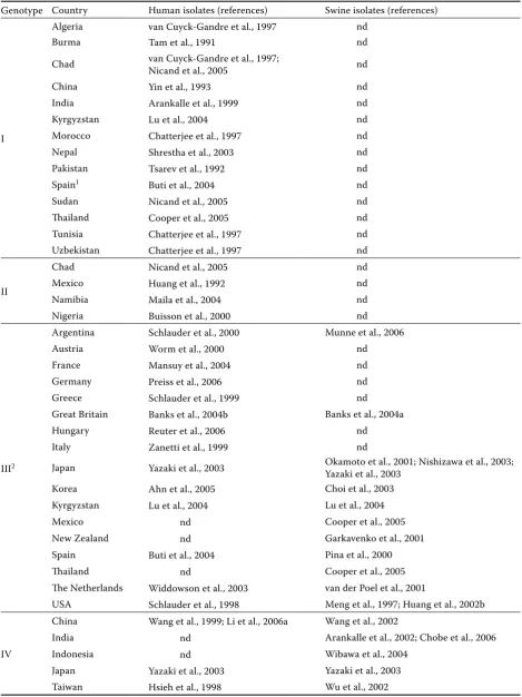
4.2. Genotypes and prevalence of human HEV strains
The HEV genome has only been complete-ly mapped in a few HEV strains, while most of them have been sequenced only partly. Based on all available data, phylogenetic analysis and com-parison of sequences have been performed: these
have revealed that there are four major genotypes of HEV: I, II, III and IV (Table 3). Homology of members of the same genotype is presumed not to be less than 81% (Schlauder and Mushahwar, 2001). The phylogenetic analysis divided HEV genotype I into five subtypes, genotype II into 2 subtypes, whereas genotypes III and IV were divided into 10 and 7 subtypes, respectively (Lu et al., 2006). A consensus for HEV classification does not yet exist.
4.3. Swine HEV and its prevalence
HEV from clinical samples of pigs kept in the USA was identified and described for the first time in 1997 (Meng et al., 1997). Subsequently, HEV strains have been detected in other countries with a high production of pork all over the world (Table 3).
A comparison of segments of sequenced ge-nomes and subsequent classification of these strains in HEV genotypes showed their affiliation with genotype III (Meng et al., 1997; Garkavenko et al., 2001; Okamoto et al., 2001; Huang et al., 2002b; Yazaki et al., 2003; Banks et al., 2004a; Lu et al., 2004; Cooper et al., 2005) and genotype IV (Arankalle et al., 2002; Wang et al., 2002; Wu et al., 2002; Yazaki et al., 2003). Swine HEV strains are usually most homologous with human HEV from the same geographic areas (Hsieh et al., 1999; Pina et al., 2000; Wang et al., 2002; Huang et al., 2002b; Yazaki et al., 2003), with the exceptions of swine HEV from Mexico and Thailand where hu-man HEV has been classified as genotype I and II, swine HEV as genotype III (Cooper et al., 2005). Human strains of HEV from Kyrgyzstan were clas-sified as genotype I and swine strains as genotype III (Lu et al., 2004), whereas human HEV from India as genotype I and that of swine as genotype IV (Arankalle et al., 2002).
4.4. Avian HEV and “big liver and spleen disease virus” (BLSV)
commer-Table 3. Genotypic designation for isolates of human and swine hepatitis E virus strains according to the countries
Genotype Country Human isolates (references) Swine isolates (references)
I
Algeria van Cuyck-Gandre et al., 1997 nd
Burma Tam et al., 1991 nd
Chad van Cuyck-Gandre et al., 1997; Nicand et al., 2005 nd
China Yin et al., 1993 nd
India Arankalle et al., 1999 nd
Kyrgyzstan Lu et al., 2004 nd
Morocco Chatterjee et al., 1997 nd
Nepal Shrestha et al., 2003 nd
Pakistan Tsarev et al., 1992 nd
Spain1 Buti et al., 2004 nd
Sudan Nicand et al., 2005 nd
Thailand Cooper et al., 2005 nd
Tunisia Chatterjee et al., 1997 nd
Uzbekistan Chatterjee et al., 1997 nd
II
Chad Nicand et al., 2005 nd
Mexico Huang et al., 1992 nd
Namibia Maila et al., 2004 nd
Nigeria Buisson et al., 2000 nd
III2
Argentina Schlauder et al., 2000 Munne et al., 2006
Austria Worm et al., 2000 nd
France Mansuy et al., 2004 nd
Germany Preiss et al., 2006 nd
Greece Schlauder et al., 1999 nd
Great Britain Banks et al., 2004b Banks et al., 2004a
Hungary Reuter et al., 2006 nd
Italy Zanetti et al., 1999 nd
Japan Yazaki et al., 2003 Okamoto et al., 2001; Nishizawa et al., 2003; Yazaki et al., 2003
Korea Ahn et al., 2005 Choi et al., 2003
Kyrgyzstan Lu et al., 2004 Lu et al., 2004
Mexico nd Cooper et al., 2005
New Zealand nd Garkavenko et al., 2001
Spain Buti et al., 2004 Pina et al., 2000
Thailand nd Cooper et al., 2005
The Netherlands Widdowson et al., 2003 van der Poel et al., 2001
USA Schlauder et al., 1998 Meng et al., 1997; Huang et al., 2002b
IV
China Wang et al., 1999; Li et al., 2006a Wang et al., 2002
India nd Arankalle et al., 2002; Chobe et al., 2006
Indonesia nd Wibawa et al., 2004
Japan Yazaki et al., 2003 Yazaki et al., 2003
Taiwan Hsieh et al., 1998 Wu et al., 2002
1a patient with a travel history to Ethiopia; 2comprises hepatitis E virus strains originating from countries with occasional
[image:8.595.63.533.101.728.2]cial breeds of hens in Australia. Comparison of sequenced genome segments of avian HEV and BLSV revealed a similarity of about 80% (Payne et al., 1999; Haqshenas et al., 2001).
Sequence analysis of the genome of avian HEV re-vealed 50% to 60% similarity in nucleotide sequence with human and swine HEV strains. Organisation of the genome of avian HEV is similar to the ge-nome arrangement found in other HEV strains. Dissimilarity in the localisation of the ORF3 was also observed: ORF3 does not overlap with ORF1, similarly to HEV-T1. It is not clear whether avian HEV has a novel genotype (V) of HEV or whether it is another member of the Hepevirus genus (Wang et al., 2000; Haqshenas et al., 2001).
5. Replication and expression of HEV
The exact mode of replication and expression of HEV has not been recognised yet. The assumed course of events has been mostly based on analogy with other viruses (where the genome is formed by a positive-sense RNA) and on the knowledge of conservative segments of non-structural HEV domains (Worm et al., 2002).
5.1. Hepatocytes – primary host target cells for HEV
The primary target cells for HEV are hepato-cytes. The positive-sense chain of viral RNA in their cytoplasm is translated into a non-structural protein necessary for viral replication. Negative-sense RNA serves as a template. It is not clear whether a non-structural polyprotein is split; if it was, the viral or host protease would be responsi-ble for this process. The product of a non-struc-tural gene comprises of RNA-dependent RNA polymerase, which evidently participates in the formation of both template sense and positive sense chains. This polymerase is only detectable at the beginning of replication (Panda et al., 2000). Translation is likely initiated by capping (Zhang et al., 2001). Structural protein ORF2 is posttrans-lationally glycosylated, which is usual for surface proteins of encapsulated viruses (Zafrullah et al., 1999). The ORF3 phosphorylated protein func-tion, encapsulation and excretion of virions from hepatocytes remains obscure (Worm et al., 2002; Emerson and Purcell, 2003).
5.2. The other HEV infected host tissues
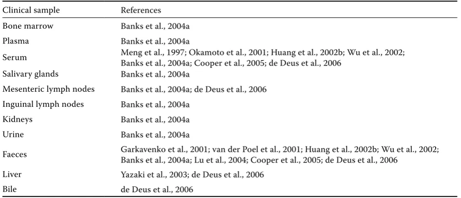
Using in situ hybridisation, HEV RNA was also detected on the luminal surface of epithelial cells in the biliary duct; which indicates a transient high HEV RNA concentration in bile. Other studies demonstrated HEV transfer from hepatocytes into epithelial cells and to biliary duct cells in an acute stage of the disease (Kawai et al., 1999; Williams et al., 2001). The template RNA was detected in the small intestine, spleen, lymph nodes, tonsils (Williams et al., 2001) and the HEV RNA was de-tected in bone marrow, salivary glands, mesenteric and inguinal lymphatic nodes, kidneys and urine (Banks et al., 2004a; de Deus et al., 2006). Possible replication of swine HEV outside the liver increased the fear of pathogen transmission with xenotrans-plantations (Williams et al., 2001).
6. HEV transmission
HEV is classified as one of the foodborne and waterborne viruses and it could be regarded as both an emerging anthroponosis and zoonosis. Developing countries of Asia, Africa, South and Central America (countries with endemic occur-rence of HE) are considered as risk areas (Hubalek, 2003; Ashbolt, 2004; Koopmans and Duizer, 2004; Vasickova et al., 2005). Sporadic occurrence of HE has been described in industrialised countries where affected people have been associated with a travel history to countries with an increased risk of infection (Skidmore et al., 1991; Dawson et al., 1992). Later studies reported HEV infections in people without a travel history to these countries (Schlauder et al., 1998; Smith, 2001). Further stud-ies of HEV suggested other routes of transmission and the zoonotic potential of HEV has been dis-cussed.
6.1. The most common HEV transmission route
Close genetic similarity of human and swine HEV viruses indicates that semi-liquid manure from pigs may also be a source of water contamination (Smith, 2001).
The first HE outbreak was recorded in Delhi, India, after the faecal contamination of a drinking water pump chamber. The outbreak, during which 29,300 people fell ill, arose in December, 1955: it culminated in two weeks and subsided within two months (Vishwanathan, 1957, as quoted by Worm et al., 2002).
6.2. Person to person contact and transplacental transmission
Person-to-person transmission of HE between family members has been documented in only 1% to 2% of cases, in contrast to 15% person-to-per-son transmission of hepatitis A (Khuroo, 1980). Transplacental transmission of HEV in the third trimester of pregnancy has been described; it is associated with a high perinatal mortality of the af-fected newborns (Khuroo et al., 1995; Bednar et al., 1999). Preterm delivery was recorded in two thirds of women infected with HE viremia included in the study. This group was reported to have a fatality rate approaching 26.9%; vertical transmission of HEV was described in 33.3% of cases (Kumar et al., 2004).
6.3. HEV transmission via foodstuffs 6.3.1. Pork and pig offal
Yazaki et al. (2003) and Tamada et al. (2004) re-ferred to an infection caused by the consumption of undercooked liver or meat from domestic pigs and wild boars in Japan. In nine out of ten cases, patients had consumed grilled or raw pig liver, two months to 19 days prior to the onset of the first symptoms. Subsequently, packages with raw liver from grocery stores in the area where the infected people lived (Hokkaido) were tested. Of a total of 363 samples HEV RNA (ORF2) was detected in seven packages (1.9%). After ORF2 gene sequenc-ing, the strains were classified as genotype groups III and IV. By comparison with human HEV geno-types detected in Japanese patients, the sample (swJL145) from genotype group IV showed 97.7 to 100% similarity of nucleotide sequences with HEV
from ten HE patients. Two isolates (swJV234 and swJV235) belonging to genotype group III had 96.6 to 100% homology with HEV from five peo-ple. Finally, 3 people from the investigated group admitted to the consumption of undercooked pig intestine.
Due to the fact that viral particles are secreted in the intestine together with bile and that replication of both swine and human HEV in the intestine has been demonstrated (Williams et al., 2001, as quoted by Yazaki et al., 2003), this foodstuff might also be a potential source of infection (Yazaki et al., 2003).
A case of HE was recorded in the UK, where the causative agent had 98% homology with a local swine HEV strain. The patient admitted to the con-sumption of raw pork in the past. However, it was not demonstrated whether this foodstuff was the source of the viral infection (Banks et al., 2004a). In Bali, IgG class antibodies to HEV were detected in 20% (54/276) of the tested population, in re-markable contrast with 4% (17/446) in Lombok and 0.5% (2/393) in Surabaya (Indonesia). Although the majority of the population in Indonesia is Muslim, Balinese people are Hindu and can consume pork. Therefore, serum samples were obtained from the 99 farm pigs in Bali and tested for HEV anti-bodies. The sera from 71 pigs (72%) were positive for anti-HEV antibodies and a 2-month-old pigs had detectable HEV RNA (Wibawa et al., 2004). This accumulating evidence suggests that eating undercooked liver or meat from domestic pigs and wild boars is associated with a high risk of acquir-ing HEV infection.
6.3.2. Undercooked foodstuffs made from venison
identical to a previous isolate from wild Sika deer (C. n. nippon) hunted in the same forest and to those isolates from four patients who contracted HE after eating raw meat form the deer. These findings suggest an interspecies HEV transmission between boar and deer in the wild and that both animal species might serve as a source of infection for humans (Takahashi et al., 2004).
6.3.3. Shellfish
The primary cause of HE outbreaks in develop-ing countries is poor sanitation. Particularly durdevelop-ing heavy rainfall, contamination of both drinking water supplies and coastal waters with human and animal faeces occurs (Balayan et al., 1983, 1997; Tsega et al., 1991). The propensity for waterborne transmission suggests that shellfish could also become contami-nated and thus acts as a vector for transmission of HEV. Although direct evidence is not currently avail-able, the association of shellfish with transmission of HEV is highly suggestive (Lees, 2000). Cacopardo et al. (1997) consider a stay in a tropical zone and consumption of raw or undercooked shellfish as risk factor for HEV transmission.
6.4. Zoonotic potential of HEV
The hypothesis of the zoonotic potential of HEV came first from a study by Balayan et al. (1990) who reported an experimental infection of domes-tic swine with human strains of HEV. Meng et al. (1998a) documented that swine HEV has the abil-ity to cross the species barrier and to infect rhe-sus monkeys, chimpanzees and vice versa. Human HEV (US-2) has also been shown to infect SPF pigs. Viraemia and HEV antibodies immediately appeared in the infected pigs, showing that hu-man HEV replication occurred in the pig organism. Identification of nucleotide sequences of HEV from pigs and rodents and their close relationship to human HEV from the same geographical regions (Meng et al., 1997; Tsarev et al., 1998; Yazaki et al., 2003) supports the hypothesis that a HEV reservoir may exist in animals.
The number of studies indicating that HE can be a zoonotic disease is now overwhelming (Favorov et al., 1998; van der Poel et al., 2001; Wang et al., 2002; Withers et al., 2002; Meng, 2003; Nishizawa et al., 2003; Tei et al., 2003, 2004; Banks et al., 2004b;
Takahashi et al., 2004; Tamada et al., 2004; Li et al., 2005; Masuda et al., 2005; Mizuo et al., 2005; Lu et al., 2006).
7. Risk groups in the population and HEV prevention
The WHO has specified groups of people exposed to HEV infection risk (http://www.who.int/disease/ hepatitis/HepatitisE_whocdscsredc2001_12.pdf): (i) people residing in areas where community
out-breaks occur
(ii) people travelling to areas with an endemic oc-currence of HE
(iii) people in overcrowded refugee camps, such as in Sudan, Somalia, Kenya and Ethiopia
(iv) people with chronic liver disease
(v) people handling non-human primates, pigs, cows, sheep and goats
Due to the fact that almost all HEV infections are spread by the faecal-oral route, good personal hygiene, high quality of standards for public water supplies and proper disposal of sanitary waste in developing countries have been recommended.
For travellers to highly endemic areas, the usual el-ementary local food hygiene precautions are recom-mended. These include avoiding drinking water and/or ice of unknown purity as well as eating uncooked shellfish and uncooked fruits or vegetables that are not peeled or prepared by the travellers themselves.
8. Diagnostic methods
8.1. Molecular detection of HEV
Nucleic acid-based techniques, especially nested RT-PCR and real-time RT-PCR, have emerged rap-idly as the method of first choice for sensitive and specific detection of RNA viruses. This method is very useful in research for the characterisation of divergent HEV strains whose serological responses have not been detected by some assays, especially in countries where infection is not endemic (Hsieh et al., 1998; Schlauder et al., 1999; Wang et al., 1999; Worm et al., 2000, 2002).
wild boar (S. scrofa leucomystax; Yazaki et al., 2003; Tamada et al., 2004), Sika deer(C. n. nippon; Tei at al., 2003; Takahashi et al., 2004; Li et al., 2005), mongoose (Herpestes sp.; Nakamura et al., 2006) and chickens (Wang et al., 2000; Haqshenas et al., 2001), but also in contaminated water (Pina et al., 2000; Grimm and Fout, 2002).
This molecular method consists of two or three steps (nested RT-PCR). The first step of RT-PCR, reverse transcription, uses specific primers, ran-dom hexamers or Oligo dT to rewrite viral RNA into cDNA. In the second or third step, PCR or nested PCR uses specific primers to amplify spe-cific segments of viral RNA. Primer binding sites can be spread over the whole genome. However, nested RT-PCR is prone to contamination and virus quantification cannot be undertaken. To overcome these difficulties, rapid and sensitive real-time RT-PCR assays have been developed for the detec-tion of HEV RNA in clinical samples (Ahn et al., 2006; Enouf et al., 2006; Jothikumar et al., 2006).
It should be noted that the choosing a suitable method for RNA extraction to ensure an adequate recovery of intact viral RNA and the elimination of inhibitory substances, is very important for the successful detection of viral genomes.
8.2. Immunologic diagnosis 8.2.1. Enzyme immunoassay (EIA)
EIA is a practical, highly sensitive and inexpensive diagnostic method for detection of HEV
anti-bodies. Antigenic domains have been found in all ORFs proteins of HEV (Khudyakov et al., 1999): (i) 12 antigenic domains in ORF1 (particularly in
the domain of the putative RNA-dependent RNA polymerase)
(ii) six antigenic domains in the ORF2 protein (iii) three antigenic domains within the ORF3
pro-tein
Recombinant proteins, originating from the ORF2 and ORF3 C-end domain or from a larger ORF2 seg-ment and complete ORF3, are used for the detection of IgG and IgM anti-HEV. A wider range of antigens expressed from a larger part of ORF2 or “capsid-like” particles are more effective in the detection of anti-bodies in the convalescent stage of the disease rather than rare antigens from the ORF2 and 3 C-ends or the whole of ORF3 (Ghabrah et al., 1998). Synthetic peptides may also be used as antigens; however, an-tibodies cannot be reliably detected in the convales-cence stage due to a low sensitivity. These peptide antigens are usually used for the confirmation of the EIA result with recombinant proteins and for the exclusion of non-specific reactions. Their use might increase the reaction specificity and determine the genotype group of HEV in the acute stage of hepatitis (Worm et al., 2002).
It should be noted that pair-wise comparison of 12 different EIA tests showed a concordance in blood donor sera ranging from 41% to 94% (mean, 68%), also a concordance among reactive sera from 0% to 89%: mean, 32% (Mast et al., 1998).
[image:12.595.61.535.101.305.2]At present, three commercial EIA tests for the detection of anti-HEV antibodies are available. The Table 4. Detection of hepatitis E virus RNA in domestic pigs using RT-PCR
Clinical sample References Bone marrow Banks et al., 2004a
Plasma Banks et al., 2004a
Serum Meng et al., 1997; Okamoto et al., 2001; Huang et al., 2002b; Wu et al., 2002; Banks et al., 2004a; Cooper et al., 2005; de Deus et al., 2006 Salivary glands Banks et al., 2004a
Mesenteric lymph nodes Banks et al., 2004a; de Deus et al., 2006 Inguinal lymph nodes Banks et al., 2004a
Kidneys Banks et al., 2004a
Urine Banks et al., 2004a
Faeces Garkavenko et al., 2001; van der Poel et al., 2001; Huang et al., 2002b; Wu et al., 2002; Banks et al., 2004a; Lu et al., 2004; Cooper et al., 2005; de Deus et al., 2006
Liver Yazaki et al., 2003; de Deus et al., 2006
first two are Genelabs-EIA; using four short
re-combinant proteins derived from the 3´end of ORF2 (42 amino acids) and ORF3 (33 amino acids) of the Burmese (genotype I) and Mexican (genotype II) strains for the detection of IgG or IgM antibodies.
The third commercially available EIA test (Abbott-EIA) is based on the detection of
anti-HEV IgG antibodies using two recombinant proteins obtained from the complete ORF3 (123 amino acids) of the Burmese (genotype I) strain. All tests may be used for routine diagnosis or epidemiological stud-ies; however, because sensitivity and specificity have not been precisely established, their usefulness for seroepidemiological studies is limited (Worm et al., 2000, 2002). Evaluation of the sensitivity, specificity, predictive positive and predictive negative values of the serology is difficult as it is not clear what the true positive is. Waar et al. (2005) defined the sera that were positive in the HEV PCR as true positive and ruled out the serum that was not available for PCR. The values for a combination of all three commer-cially available EIA’s were: sensitivity, 100%; specifi-city, 99.5%; positive predictive value, 75%; negative predictive value, 100%.
8.2.2. Immune fluorescence microscopy (IFE)
A few specialised laboratories use this technique for the detection of antibodies. IFE detects antibod-ies that react against the HEV antigen semiquan-titatively. Anti-HEV antibodies block the binding of fluorescein-conjugated anti-HEV IgG to HEV antigen in frozen liver tissue. The concentration of anti-HEV antibodies is estimated semiquantitative-ly (Krawczynski and Bradley, 1989). This method is laborious and expensive and thus not useful for routine diagnosis.
8.3. Virus isolation
Establishment of a practical cell culture system to allow the propagation of HEV in vitro is vital for virological characterisation as well as for diag-nosis and prevention of HEV infection. Several in vitro culture systems, such as human lung, kidney or liver (2BS, A549, Hep-G2) and macaques hepa-tocytes for HEV replication have been reported. Most of these, however, cannot provide authentic HEV particles or a high titre of VLPs and have poor
reproducibility (Tam et al., 1997; Wei et al., 2000; Worm et al., 2002). Currently there is no reliable cell culture system for HEV.
8.4. Immune electron microscopy (IEM)
IEM detects VLPs in clinical specimens (Balayan et al., 1983). HEV particles are precipitated with the native antibody to HEV derived from acute- or convalescent-phase sera. Anti-HEV antibodies con-centrations can be determined semiquantitatively by rating the antibody coating. Although IEM is a superior technique for specificity, the sensitiv-ity of the assay is insufficient for routine analy-sis. IEM is difficult to perform and most clinical specimens do not contain sufficient VLPs to be detected (Yarbough, 1999). Other antigen detec-tion methods have not been reported (Anderson and Shrestha, 2002).
9. Conclusions
HEV was first observed and described in 1983 (Balayan et al., 1983). The virus has been extensive-ly investigated since that time. After a long-term effort to assign this virus to a taxonomic class, the virus has been classified in the Hepevirus genus, Hepeviridae family in 2004 (Emerson et al., 2004; http://www.ncbi.nlm.nih.gov/ICTVdb/Ictv/fs_hep-ev.htm). Most of the information concerning HEV is based on molecular biology.
More thorough studies may be expected in this field, which would better clarify the virus-encod-ed protein types, their structures, functions and consequential biological characteristics presented inside the host organism and its cells.
The ecology of the virus has been insufficient-ly studied so far. The point which should be ad-dressed is the presence and circulation of HEV in populations of wild mammals, largely artiodactyls (wild boar, Sika deer), rodents (Rattus spp.) and carnivores. There is also a very urgent need for epizootiological studies contributing to a better understanding of the relationship between rats and domestic pigs in keeping the HEV circulation in the rural foci.
re-search is desirable. Above all direct confirma-tion of HEV transmission via foodstuffs is still needed. Viral RNA has been detected in the above mentioned foodstuffs; however, it does not mean that the virus present is infectious. HE patients with a history of undercooked meat consumption have been noted. During subsequent inspection of sources of these foodstuffs, HEV RNA was de-tected. Unfortunately, it could not be confirmed that the foodstuffs consumed by these people contained the infectious agent although there are significant epidemiological links. The fact that viral RNA detected in some patients showed a relatively high homology with HEV RNA found in meat intended for consumption supported the hypothesis that undercooked meat was the source of infection.
Anti-HEV antibodies (or viral RNA) have not only been found in people, but also detected in various animal species (Arankalle et al., 1994, 2001; Favorov et al., 1998). HEV capability to cross the species barrier was confirmed experimentally (Meng et al., 1998a). Knowledge of inter-species transmission supports the hypothesis about the zoonotic potential of HEV. However, further re-search of HEV incidence into other animal spe-cies and the possibility of spespe-cies barrier crossing should be performed. More detailed information obtained by this investigation might extend the knowledge of potential risk factors of the HEV transmission and facilitate the adoption of pre-ventive measures.
In general, the research concerning HEV is far from finished and various aspects of this disease remain obscure. Accordingly, it is necessary to ob-tain more information in this field.
10. Acknowledgments
Special thanks are expressed to Dr. Nigel Cook (Central Science Laboratory, Sand Hutton, York, UK) for critical reading of the manuscript. The au-thors are indebted to Anna Maslanova and Zdenka Gregorova (Veterinary Research Institute, Brno) for the help with cited references, Ing. Ludmila Faldikova, CSc. (Veterinary Research Institute, Brno) for English translation, and Mgr. Maria Vass (Swinburne University of Technology, Victoria, Australia) and Catherine Murdoch (Aberdeen University in Scotland, UK) for grammatical cor-rections.
11. REFERENCES
Acha P.N., Szyfres B. (2003): Zoonoses and Communi-cable Diseases Common to Man and Animals. 3rd ed.
Pan American Health Organization, Washington. Aggarwal R., Kini D., Sofat S., Naik S.R., Krawczynski
K. (2000): Duration of viremia and faecal viral excre-tion in acute hepatitis. Lancet, 356, 1081–1082. Ahn J.M., Kang S.G., Lee D.Y., Shin S.J., Yoo H.S. (2005):
Identification of novel human hepatitis E virus (HEV) isolates and determination of the seroprevalence of HEV in Korea. Journal of Clinical Microbiology, 43, 3042–3048.
Ahn J.M., Rayamajhi N., Kang S.G., Yoo H.S. (2006): Comparison of real-time reverse transcriptase-polymerase chain reaction and nested or commercial reverse transcriptase-polymerase chain reaction for the detection of hepatitis E virus particle in human serum. Diagnostic Microbiology and Infectious Dis-ease, 56, 269–274.
Anderson D.A., Shrestha I.L. (2002): Clinical Virology, Hepatitis E virus. 2nd ed. ASM Press, American
Soci-ety for Microbiology, Washington, DC, USA.
Arankalle V.A., Goverdhan M.K., Banerjee K. (1994): Antibodies against hepatitis E virus in Old World mon-keys. Journal of Viral Hepatitis, 1, 125–129.
Arankalle V.A., Paranjape S., Emerson S.U., Purcell R.H., Walimbe A.M. (1999): Phylogenetic analysis of hepa-titis E virus isolates from India (1976–1993). Journal of General Virology, 80, 1691–1700.
Arankalle V.A., Joshi M.V., Kulkarni A.M., Gandhe S.S., Chobe L.P., Rautmare S.S., Mishra A.C., Padbidri V.S. (2001): Prevalence of anti-HEV antibodies in different Indian animal species. Journal of Viral Hepatitis, 8, 223–227.
Arankalle V.A., Chobe L.P., Joshi M.V., Chadha M.S., Kundu B., Walimbe A.M. (2002): Human and swine hepatitis E viruses from Western India belong to dif-ferent genotypes. Journal of Hepatology, 36, 417–425. Ashbolt N.J. (2004): Microbial contamination of drink-ing water and disease outcomes in developdrink-ing regions. Toxicology, 198, 229–238.
Balayan M.S. (1997): Epidemiology of hepatitis E virus infection. Journal of Viral Hepatitis, 4, 155–165. Balayan M.S., Andzhaparidze A.G., Savinskaya S.S.,
Keti-ladze E.S., Braginsky D.M., Savinov A.P., Poleschuk V.F. (1983): Evidence for virus in non-A/non-B hepa-titis transmitted via the faecal-oral route. Intervirol-ogy, 20, 23–31.
Banks M., Heath G.S., Grierson S.S., King D.P., Gresham A., Girones R., Widen F., Harrison T.J. (2004a): Evi-dence for the presence of hepatitis E virus in pigs in the United Kingdom. Veterinary Record, 154, 223– 227.
Banks M., Bendall R., Grierson S., Heath G., Mitchell J., Dalton H. (2004b): Human and porcine hepatitis E virus strains, United Kingdom. Emerging Infectious Diseases, 10, 953–955.
Bednar M., Frankova V., Schindler J., Soucek A., Vavra J. (1999): Medical Microbiology (in Czech). 1st ed.
(Re-issue), Nakladatelstvi Marvil, Prague. 558 pp.
Berke T., Matson D.O. (2000): Reclassification of the Caliciviridae into distinct genera and exclusion of hepatitis E virus from the family on the basis of com-parative phylogenetic analysis. Archives of Virology, 150, 1421–1436.
Billam P., Huang F.F., Sun Z.F., Pierson F.W., Duncan R.B., Elvinger F., Guenette D.K., Toth T.E., Meng X.J. (2005): Systematic pathogenesis and replication of avian hepatitis E virus in specific-pathogen-free adult chickens. Journal of Virology, 79, 3429–3437.
Buisson Y., Grandadam M., Nicand E., Cheval P., van Cuyck-Gandre H., Innis B., Rehel P., Coursaget P., Tey-ssou R., Tsarev S. (2000): Identification of novel hep-atitis E virus in Nigeria. Journal of General Virology, 81, 903–909.
Buti M., Clemente-Casares P., Jardi R., Formiga-Cruz M., Schaper M., Valdes A., Rodriguez-Frias F., Esteban R., Girones R. (2004): Sporadic cases of acute autoch-thonous hepatitis E in Spain. Journal of Hepatology, 41, 126–131.
Cacopardo B., Russo R., Preiser W., Benanti F., Brancati G., Nunnari A. (1997): Acute hepatitis E in Catania (eastern Sicily) 1980–1994. The role of hepatitis E vi-rus Infection, 25, 313–316.
Chatterjee R., Tsarev S., Pillot J., Coursaget P., Emerson S.U., Purcell R.H. (1997): African strains of hepatitis E virus that are distinct from Asian strains. Journal of Medical Virology, 53, 139–144.
Chauhan A., Jameel S., Dilawari J.B., Chawla Y.K., Kaur U., Ganguly N.K. (1993): Hepatitis E virus transmission to a volunteer. Lancet, 341, 149–150.
Chobe L.P., Lole K.S., Arankalle V.A. (2006): Full genome sequence and analysis of Indian swine hepatitis E virus isolates of genotype 4. Veterinary Microbiology, 114, 240–251.
Choi I.S., Kwon H.J., Shin N.R., Yoo H.S. (2003): Identi-fication of swine hepatitis E virus (HEV) and preva-lence of anti-HEV antibodies in swine and human populations in Korea. Journal of Clinical Microbiology, 41, 3602–3608.
Clayson E.T., Myint K.S., Snitbhan R., Vaughn D.W., In-nis B.L., Chan L., Cheung P., Shrestha M.P. (1995): Viremia, faecal shedding, and IgM and IgG responses in patients with hepatitis E. Journal of Infectious Dis-eases, 172, 927–933.
Cooper K., Huang F.F., Batista L., Rayo C.D., Bezanilla J.C., Toth T.E., Meng X.J. (2005): Identification of genotype 3 hepatitis E virus (HEV) in serum and fae-cal samples from pigs in Thailand and Mexico, where genotype 1 and 2 HEV strains are prevalent in the re-spective human populations. Journal of Clinical Microbiology, 43, 1684–1688.
Dawson G.J., Mushahwar I.K., Chau K.H. (1992): Detec-tion of long-lasting antibody to hepatitis E virus in U. S. traveller to Pakistan. Lancet, 340, 426–427. de Deus N., Seminati C., Pina S., Mateu E., Martin M.,
Segales J. (2006): Detection of hepatitis E virus in liver, mesenteric lymph node, serum, bile and faeces of nat-urally infected pigs affected by different pathological conditions. Veterinary Microbiology, 119, 105–114. Emerson S.U., Purcell R.H. (2003): Hepatitis E virus.
Reviews in Medical Virology, 13, 145–154.
Emerson S.U., Anderson D., Arankalle A.V., Meng X.J., Purdy M., Schlauder G.G., Tsarev S.A. (2004): Hepevi-rus. In: Fauquet C.M., Mayo M.A., Maniloff J., Des-selberger U., Ball L.A. (eds.): Virus Taxonomy. The Eighth Report of the International Committee on Tax-onomy of Viruses. Elsevier/Academic Press, London. 851–855.
Enouf V., Dos Reis G., Guthmann J.P., Guerin P.J., Caron M., Marechal V., Nicand E. (2006): Validation of single real-time TaqMan® PCR assay for detection and quan-tisation of four major genotypes of hepatitis E virus in clinical specimens. Journal of Medical Virology, 78, 1076–1082.
Erker J.C., Desai S.M., Mushahwar I.K. (1999): Rapid detection of hepatitis E virus RNA by reverse tran-scription-polymerase chain reaction using universal oligonucleotide primers. Journal of Virological Meth-ods, 81, 109–113.
Favorov M.O., Nazarova O., Margolis H.S. (1998): Is hepatitis E an emerging zoonotic disease? American Journal of Tropical Medicine and Hygiene, 59, 242. Favorov M.O., Kosoy M.Y., Tsarev S.A., Childs J.E.,
Mar-golis H.S. (2000): Prevalence of antibody to hepatitis E virus among rodents in the United States. Journal of Viral Hepatitis, 8, 223–227.
Ghabrah T.M., Tsarev S.A., Yarbough P.O., Emerson S.U., Strickland G.T., Purcell R.H. (1998): Comparison of tests for antibody to hepatitis E virus. Journal of Med-ical Virology, 55, 134–137.
Grimm A.C., Fout G.S. (2002): Development of molecu-lar method to identify hepatitis E virus in water. Jour-nal of Virological Methods, 101, 175–188.
Halbur P.G., Kasorndorkbua C., Gilbert C., Guenette D., Potters M.B., Purcell R.H., Emerson S.U., Toth T.E., Meng X.J. (2001): Comparative pathogenesis of infection of pigs with hepatitis E viruses recovered from a pig and a hu-man. Journal of Clinical Microbiology, 39, 918–923. Haqshenas G., Shivaprasad H.L., Woolcock P.R., Read
D.H., Meng X.J. (2001): Genetic identification and characterization of a novel virus related to human hepatitis E virus from chickens with hepatitis-splenom-egaly syndrome in the United States. Journal of Gen-eral Virology, 82, 2449–2462.
Hirano M., Ding X., Li T.C., Takeda N., Kawabata H., Koizumi N., Kadosaka T., Goto I., Masuzawa T., Na-kamura M., Taira K., Kuroki T., Takinawa T., Watanabe H., Abe K. (2003): Evidence for widespread infection of hepatitis E virus among wild rats in Japan. Hepatol-ogy Research, 27, 1–5.
Hsieh S.Y., Yang P.Y., Ho Y.P., Chu C.M., Liaw Y.F. (1998): Identification of a novel strain of hepatitis E virus re-sponsible for sporadic acute hepatitis in Taiwan. Jour-nal of Medical Virology, 55, 300–304.
Hsieh S.Y., Meng X.J., Wu Y.H., Liu S.T., Tam A.W., Lin D.Y., Liaw Y.F. (1999): Identity of novel swine hepatitis E virus in Taiwan forming a monophyletic group with Taiwan isolates of human hepatitis E virus. Journal of Clinical Microbiology, 37, 3828–3834.
Huang C.C., Nguyen D., Fernandez J., Yun K.Y., Fry K. E., Bradley D.W., Tam A.W., Reyes G.R. (1992): Mo-lecular cloning and sequencing of the Mexico isolate of hepatitis E virus (HEV). Virology, 191, 550–558. Huang F.F., Haqshenas G., Shivaprasad H.L., Guenette
D.K., Woolcock P.R., Larsen C.T., Pierson F.W., Elvinger F., Toth T.E., Meng X.J. (2002a): Heterogeneity and seroprevalence of a newly identified avian hepatitis E virus from chicken in the United States. Journal of Clinical Microbiology, 40, 4197–4202.
Huang F.F., Haqshenas G., Guenette D.K., Halbur P.G., Schommer S.K., Pierson F.W., Toth T.E., Meng X.J. (2002b): Detection by reverse transcription-PCR and genetic characterization of field isolates of swine hep-atitis E virus from pigs in different geographic regions of the United States. Journal of Clinical Microbiology, 40, 1326–1332.
Hubalek Z. (2003): Emerging human infectious diseases: anthroponoses, zoonoses, and sapronoses. Emerging
Infectious Diseases, 9, 403–404. http://www.cdc.gov/ ncidod/eid/vol9no3/02-0208.htm
Hussaini S.H., Skidmore S.J., Richardson P., Sherratt L. M., Cooper B.T., O’Grady J.G. (1997): Severe hepatitis E infection during pregnancy. Journal of Viral Hepa-titis, 4, 51–54.
Inoue J., Takahashi M., Yazaki Y., Tsuda F., Okamoto H. (2006): Development and validation of an improved RT-PCR assay with nested universal primers for detec-tion of hepatitis E virus strains with significant se-quence divergence. Journal of Virological Methods, 137, 325–333.
Jothikumar N., Cromeans T.L., Robertson B.H., Meng X.J., Hill V.R. (2006): A broadly reactive one-step real-time RT-PCR assay for rapid and sensitive detection of hepatitis E virus. Journal of Virological Method, 131, 65–71.
Kabrane-Lazizi Y., Fine J.B., Elm J., Glass G.E., Higa H., Diwan A., Gibbs C.J., Meng X.J., Emerson S.U., Purcell R.H. (1999a): Evidence for widespread infection of wild rats with hepatitis E virus in the United States. Amer-ican Journal of Tropical Medicine and Hygiene, 61, 331–335.
Kabrane-Lazizi Y., Meng X.J., Purcell R.H., Emerson S.U. (1999b): Evidence that the genomic RNA of hepatitis E virus is capped. Journal of Virology, 73, 8848–8850. Kasorndorkbua C., Halbur P.G., Thomas P.J., Guenette
D.K., Toth T.E., Meng X.J. (2002): Use of a swine bio-assay and a RT-PCR bio-assay to assess the risk of trans-mission of swine hepatitis E virus in pigs. Journal of Virological Methods, 101, 71–78.
Kawai H.F., Koji T., Iida F., Kaneko S., Kobayashi K., Na-kane P.K. (1999): Shift of hepatitis E virus RNA from hepatocytes to biliary epithelial cells during acute in-fection of rhesus monkey. Journal of Viral Hepatitis, 6, 287–297.
Khudyakov Y.E., Lopareva E.N., Jue D.L., Crews T.K., Thyagarajan S.P., Fields H.A. (1999): Antigenic do-mains of the open reading frame 2-encoded protein of hepatitis E virus. Journal of Clinical Microbiology, 37, 2863–2871.
Khuroo M.S. (1980): Study of an epidemic of non-A, non-B hepatitis: possibility of another human hepati-tis virus distinct from posttransfusion non-A, non-B type. American Journal of Medicine, 68, 818–824. Khuroo M.S., Kamili S., Jameel S. (1995): Vertical
trans-mission of hepatitis E virus. Lancet, 345, 1025–1026. Koonin E.V., Gorbalenya A.E., Purdy M.A., Rozanov
and animal virus. Proceedings of the National Acad-emy of Sciences, USA, 89, 8259–8263.
Koopmans M., Duizer E. (2004): Foodborne viruses: an emerging problem. International Journal of Food Microbiology, 90, 23–41.
Krawczynski K., Bradley D.W. (1989): Enterically-trans-mitted non-A, non-B hepatitis: identification of virus-associated antigen in experimentally infected cynomolgus macaques. Journal of Infectious Diseases, 159, 1042–1049.
Kumar A., Beniwal M., Kar P., Sharma J.B., Murthy N.S (2004): Hepatitis E in pregnancy. International Journal of Gynecology and Obstetrics, 85, 240–241.
Lees D. (2000): Viruses and bivalve shellfish. Interna-tional Journal of Food Microbiology, 59, 81–116. Li F., Zhuang H., Kolivas S., Locarnini S., Anderson D.A.
(1994): Persistent and transient antibody responses to hepatitis E virus detected by Western immunoblot us-ing open readus-ing frame 2 and 3 and glutathione S-transferase fusion protein. Journal of Clinical Microbiology, 32, 2060–2066.
Li T.C., Yakamawa Y., Suzuki K., Tatsumi M., Razak M.A., Uchida T., Takeda N., Miyamura T. (1997): Ex-pression and self-assembly of empty virus-like parti-cles of hepatitis E virus. Journal of Virology, 71, 7207–7213.
Li T.C., Chijiwa K., Sera N., Ishibashi T., Etoh Y., Shino-hara Y., Kurata Y., Ishida M., Sakamoto S., Takeda N., Miyamura T. (2005): Hepatitis E virus transmission from wild boar meat. Emerging Infectious Diseases, 11, 1958–1960.
Li R.C., Ge S.X., Li Y.P., Zheng Y.J., Nong Y., Guo Q.S., Zhang J., Ng M.H., Xia N.S. (2006a): Seroprevalence of hepatitis E virus infection, rural southern People’s Republic of China. Emerging Infectious Diseases, 12, 1682–1688.
Li T.C., Saito M., Ogura G., Ishibashi I., Miyamura T., Takeda N. (2006b): Serologic evidence for hepatitis E virus infection in mongoose. American Journal of Tropical Medicine and Hygiene, 74, 932–936. Lu L., Drobeniuc J., Kobylnikov N., Usmanov R.K.,
Rob-ertson B.H., Favorov M.O., Margolis H.S. (2004): Com-plete sequence of Kyrgyzstan swine hepatitis E virus (HEV) isolated from piglet thought to be experimen-tally infected with human HEV. Journal of Medical Virology, 74, 556–562.
Lu L., Li C., Hagedorn C.H. (2006): Phylogenetic analy-sis of global hepatitis E virus sequences: genetic di-versity, subtypes and zoonosis. Review of Medical Virology, 16, 5–36.
Magden J., Takeda N., Li T., Auvinen P., Ahola T., Miya-mura T., Merits A., Kaariainen L. (2001):
Virus-spe-cific mRNA capping enzyme encoded by hepatitis E virus. Journal of Virology, 75, 6249–6255.
Maila H.T., Bowyer S.M., Swanepoel R. (2004): Identifi-cation of a new strain of hepatitis E virus from an out-break in Namibia in 1995. Journal of General Virology, 85, 89–95.
Mansuy J.M., Peron J.M., Abravanel F., Poirson H., Dubois M., Miedouge M., Vischi F., Alric L., Vinel J.P., Izopet J. (2004): Hepatitis E in the south west of France in individuals who have never visited an endemic area. Journal of Medical Virology, 74, 419–424.
Mast E.E., Alter M.J., Holland P.V., Purcell R.H. (1998): Evaluation of assays for antibody to hepatitis E virus by a serum panel. Hepatitis E Virus Antibody Serum Panel Evaluation Group. Hepatology,
27, 857–
861.
Masuda J., Yano K., Tamada Y., Takii Y., Ito M., Omagari K., Kohno S. (2005). Acute hepatitis E of a man who consumed wild boar meat prior to the onset of illness in Nagasaki, Japan – Case report. Hepatology Re-search, 31, 178–183.
Meng X.J. (2003): Swine hepatitis E virus: Cross-species infection and risk in xenotransplantation. Current Top-ics in Microbiology and Immunology, 278, 185–216. Meng X.J., Purcell R.H., Halbur P.G., Lehman J.R., Webb
D.M., Tsareva T.S., Haynes J.S., Thacker B.J., Emerson S.U. (1997): A novel virus in swine is closely related to the human hepatitis E virus. Proceedings of the National Academy of Sciences, USA, 94, 9860–9865. Meng X.J., Halbur P.G., Sharipo M.S., Govindarajan S.,
Bruna J.D., Mushahwar I.K., Purcell R.H., Emerson S.U. (1998a): Genetic and experimental evidence for cross-species infection by the swine hepatitis E virus. Journal of Virology, 72, 9714–9721.
Meng X.J., Halbur P.G., Tsareva T.S., Bruna J.D., Royer R.L., Purcell R.H., Emerson S.U. (1998b): Experimen-tal infection of pigs with the newly identified swine hepatitis E virus (swine HEV), but not with human strain of HEV. Archives of Virology, 143, 1405–1415. Meng X.J., Dea S., Engle R.E., Friendship R., Lyoo Y.S.,
Sirinarumitr T., Urairong K., Wang D., Wong D., Yoo D., Zhang Y., Purcell R.H., Emerson S.U. (1999): Prev-alence of antibodies to the hepatitis E virus in pig from countries where hepatitis E is common or is rare in the human population. Journal of Medical Virology, 59, 297–302.
Munne M.S., Vladimirsky S., Otegui L., Castro R., Bra-jterman L., Soto S., Guarnera E., Molina V., Monfellano M., Schlauder G.G., Gonzalez J.E. (2006): Identifica-tion of the first strain of swine hepatitis E virus in South America and prevalence of anti-HEV antibodies in swine in Argentina.Journal of Medical Virology, 78, 1579–1583.
Nakamura M., Takahashi K., Taira K., Taira M., Ohno A., Sakugawa H., Arai M., Mishiro S. (2006): Hepatitis E virus infection in wild mongooses of Okinawa, Japan: demonstration of anti-HEV antibodies and a full-ge-nome nucleotide sequence. Hepatology Research, 34, 137–140.
Nicand E., Armstrong G.L., Enouf V., Guthmann J.P., Guerin J.P., Caron M., Nizou J.Y., Andraghetti R. (2005): Genetic heterogeneity of hepatitis E virus in Darfur, Sudan, and neighbouring Chad. Journal of Medical Virology, 77, 519–521.
Nishizawa T., Takahashi M., Mizuo H., Miyajima H., Gotanda Y., Okamoto H. (2003): Characterization of Japanese swine and human hepatitis E virus isolates of genotype IV with 99% identity over the entire ge-nome. Journal of General Virology, 84, 1245–1251. Okamoto H., Takahashi M., Nishizawa T., Fukai K.,
Mu-ramatsu U., Yoshikawa A. (2001): Analysis of complete genome of indigenous swine hepatitis E virus isolated in Japan. Biochemical and Biophysical Communica-tions, 289, 929–936.
Okamoto H., Takahashi M., Nishizawa T., Usui R., Koba-yashi E. (2004): Presence of antibodies to hepatitis E virus in Japanese pet cats. Infection, 32, 57–58. Panda S.K., Ansari I.H., Durgapal H., Agrawal S., Jameel
S. (2000): The in vitro-synthesized RNA from a cDNA clone of hepatitis E virus is infectious. Journal of Virol-ogy, 74, 2430–2437.
Payne C.J., Ellis T.M., Plant S.L., Gregory A.R., Wilcox G.E. (1999): Sequence data suggests big liver and spleen disease virus (BLSV) is genetically related to hepatitis E virus. Veterinary Microbiology, 68, 119– 125.
Pina S., Buti M., Cotrina M., Piella J., Girones R. (2000): HEV identified in serum from humans with acute hepatitis and sewage of animal origin in Spain. Journal of Hepatology, 33, 826–833.
Preiss J.C., Plentz A., Engelmann E., Schneider T., Jilg W., Zeitz M., Duchmann R. (2006): Autochthonous hepatitis E virus infection in Germany with sequence similarities to other European isolates. Infection, 34, 173–175.
Reuter G., Fodor D., Katai A., Szucs G. (2006): Identifi-cation of a novel variant of human hepatitis E virus in Hungary. Journal of Clinical Virology, 36, 100–102.
Schlauder G.G., Mushahwar I.K. (2001): The genetic heterogeneity of hepatitis E virus. Journal of Medical Virology, 65, 282–292.
Schlauder G.G., Dawson G.J., Erker J.C., Kwo P.Y., Knigge M.F., Smalley D.L., Rosenblatt J.E., Desai S.M., Mush-ahwar I.K. (1998): The sequence and phylogenetic analysis of a novel hepatitis E virus isolated from a patient with acute hepatitis reported in the United States. Journal of General Virology, 79, 447–456. Schlauder G.G., Desai S.M., Zanetti A.R., Tassopoulos
N.C., Mushahwar I.K. (1999): Novel hepatitis E virus (HEV) isolates from Europe: Evidence for additional genotypes of HEV. Journal of Medical Virology, 57, 243–251.
Schlauder G.G., Frider B., Sookoian S., Castano G.C., Mushahwar I.K. (2000): Identification of 2 novel iso-lates of hepatitis E virus in Argentina. Journal of Infec-tious Diseases, 182, 294–297.
Shrestha S.M., Shrestha S., Tsuda F., Nishizawa T., Gotanda Y., Takeda N., Okamoto H. (2003): Molecular investigation of hepatitis E virus infection in patients with acute hepatitis in Kathmandu, Nepal. Journal of Medical Virology, 69, 207–214.
Skidmore S.J., Yarbough P.O., Gabor K.A., Tam A.W., Reyes G.R., Flower A.J. (1991): Imported hepatitis E in UK. Lancet, 337, 1541.
Smith J.L. (2001): A review of hepatitis E virus. Journal of Food Protection, 64, 572–586.
Sonoda H., Abe M., Sugimoto T., Sato Y., Bando M., Fukui E., Mizuo H., Takahashi M., Nishizawa T., Okamoto H. (2004): Prevalence of hepatitis E virus (HEV) infection in wild boars and deer and genetic identification of a genotype 3 HEV from a boar in Japan. Journal of Clin-ical Microbiology, 42, 5371–5374.
Takahashi K., Kitajima N., Abe N., Mishiro S. (2004): Complete or near-complete nucleotide sequences of hepatitis E virus genome recovered from a wild boar, a deer, and four patients who ate the deer. Virology, 330, 501–505.
Tam A.W., Smith M.M., Guerra M.E., Huang C.C., Bra-dley D.W., Fry K.E., Reyes G.R. (1991): Hepatitis E virus (HEV): molecular cloning and sequencing of the full-length viral genome. Virology, 185, 120–131. Tam A.W., White R., Yarbough P.O., Murphy B.J.,
McA-tee C.P., Lanford R.E., Fuerst T.R. (1997): In vitro infec-tion and replicainfec-tion of hepatitis E virus in primary cynomolgus macaque hepatocytes. Virology, 238, 94–102.
Tei S., Kitajima N., Takahashi K., Mishiro S. (2003): Zoonotic transmission of hepatitis E virus from deer to human beings. Lancet, 362, 371–373.
Tei S., Kitajima N., Ohara S., Inoue Y., Miki M., Yama-tani T., Yamabe H., Mishiro S., Kinoshita Y. (2004): Consumption of uncooked deer meat as a risk factor for hepatitis E virus infection: an age- and sex-matched case-control study. Medical Virology, 74, 67–70.
Tien N.T., Clayson H.T., Khiem H.B., Sac P.K., Corwin A.L., Myint K.S., Vaughn D.W. (1997): Detection of immunoglobulin G to the hepatitis E virus among sev-eral animal species in Vietnam. American Journal of Tropical Medicine and Hygiene, 57, 211.
Tsarev S.A., Emerson S.U., Reyes G.R., Tsareva T.S., Legters L.J., Malik I.A., Iqbal M., Purcell R.H. (1992): Characterization of a prototype strain of hepatitis E virus. Proceeding of the National Academy of Sci-ence, USA, 89, 559–563.
Tsarev S.A., Tsareva T.S., Emerson S.U., Yarbough P.O., Legters J.L., Moskal T., Purcell R.H. (1994): Infectivity titration of prototype strain of hepatitis E virus in cy-nomolgus monkeys. Journal of Medical Virology, 43, 135–142.
Tsarev S.A., Shrestha M.P., He J., Scott R.M., Vaung D. W., Clayson E.T., Gigliotti S., Longer C.F., Innis B.L. (1998): Naturally acquired hepatitis E virus (HEV) infection in Nepalese rodents. American Journal of Tropical Medicine and Hygiene, 59, 242.
Tsega E., Krawczynski K., Hansson B.G., Nordenfelt E., Negusse Y., Alemu W., Bahru Y. (1991): Outbreak of acute hepatitis E virus infection among military per-sonnel in northern Ethiopia. Journal of Medical Virol-ogy, 34, 232–236.
van Cuyck-Gandre H., Zhang H.Y., Tsarev S.A., Clements N.J., Cohen S.J., Caudill J.D., Buisson Y., Coursaget P., Warren R.L., Longer C.F. (1997): Characterization of hepatitis E virus (HEV) from Algeria and Chad by par-tial genome sequence. Journal of Medical Virology, 53, 340–347.
van der Poel W.H.M., Verschoor F., van der Heide R., Herrera M.I., Vivo A., Kooreman M., de Roda Husman A.M. (2001): Hepatitis E virus sequences in swine re-lated to sequences in human, the Netherlands. Emerg-ing Infectious Diseases, 7, 970–976.
Vasickova P., Dvorska L., Lorencova A., Pavlik I. (2005): Viruses as a cause of foodborne diseases: a review of the literature. Veterinarni Medicina 50, 89–104. http:// www.vri.cz/docs/vetmed/50-3-89.pdf
Vishwanathan R. (1957): Infectious hepatitis in Delhi (1955–1956): a critical study. Indian Journal of Medi-cal Research, 45, 1–30.
Vitral C.L., Pito M.A., Lewis-Ximenez L.L., Khudyakov Y.E., dos Santos D.R., Gaspar A.M.C. (2005): Sero-logical evidence of hepatitis E virus infection in dif-ferent animal species from the Southeast of Brazil. Memorias do Instituto Oswaldo Cruz, 100.
Waar K., Herremans M.M.P.T., Vennema H., Koopmans M.P.G., Benne C.A. (2005): Hepatitis E is cause of un-explained hepatitis in The Netherlands. Journal of Clinical Virology, 33, 145–149.
Wang Y.C., Ling R., Erker J.C., Zhang H., Li H., Desai S., Mushahwar I.K., Harrison T.J. (1999): A divergent gen-otype of hepatitis E virus in Chinese patients with acute hepatitis. Journal of General Virology, 80, 169–177. Wang Y.C., Zhang H., Ling R., Li H., Harrison T.J. (2000):
The complete sequence of hepatitis E virus genotype 4 reveals an alternative strategy for translation of open reading frames 2 and 3. Journal of General Virology, 81, 1675–1686.
Wang Y.C., Zhang H.Y., Xia N.S, Peng G., Lan H.Y., Zhuang H., Zhu Y.H., Li S.W., Tian K.G., Gu W.J., Lin J.X., Wu X., Li H.M., Harrison T.J. (2002): Prevalence, isolation, and partial sequence analysis of hepatitis E virus from domestic animals in China. Journal of Medical Virology, 67, 516–521.
Wei S., Walsh P., Huang R., To S.S.T. (2000): 93G, Novel sporadic strain of hepatitis E virus in South China isolated by cell culture. Journal of Medical Virology, 61, 311–318.
Wibawa I.D., Muljono D.H., Mulyanto, Suryadarma I. G., Tsuda F., Takahashi M., Nishizawa T., Okamoto H. (2004): Prevalence of antibodies to hepatitis E virus among apparently healthy humans and pigs in Bali, Indonesia: Identification of a pig infected with geno-type 4 hepatitis E virus. Journal of Medical Virology, 73, 38–44.
Widdowson M.A., Jaspers W.J.M., van der Poel W.H.M., Verschoor F., de Roda Husman A.M., Winter H.L.J., Zaaijer H.L., Koopmans M. (2003): Cluster of cases of acute hepatitis associated with hepatitis E virus infec-tion acquired in the Netherlands. Clinical Infectious Disease, 36 1, 29–33.
Williams T.P.E., Kasorndorkbua C., Halbur P.G., Haqsh-enas G., Guenette D.K., Toth T.E., Meng X.J. (2001): Evidence of extrahepatic sites of replication of the hepatitis E virus in a swine model. Journal of Clinical Microbiology, 39, 3040–3046.