GSK-3 REGULATION OF MIGRATION AND MORPHOGENESIS IN THE DEVELOPING CORTEX
Meghan Morgan-Smith
A dissertation submitted to the faculty of the University of North Carolina at Chapel Hill in partial fulfillment of the requirements for the degree of Doctor of Philosophy in the
Curriculum in Neurobiology in the School of Medicine.
Chapel Hill 2013
Approved by:
William D. Snider Steve Crews Mark J. Zylka Benjamin Philpot
© 2013
Meghan Morgan-Smith ALL RIGHTS RESERVED
The majority of the neurons in the mammalian cerebral cortex are glutamatergic excitatory neurons that have a specific migration pattern and a well-defined ‘pyramidal’
morphology. Pyramidal neurons migrate on a radial glia scaffold to reach a layer-specific destination in the cortex and extend two polarized processes: the apical dendrite and the axon. Defining molecular mechanisms of migration and morphogenesis are key to understanding circuit formation in the developing cortex.
Recent studies have demonstrated that GSK-3 has a critical role in controlling neuronal number through regulation of radial progenitor proliferation and intermediate precursor amplification. However, only in vitro work has addressed GSK-3 functions related to cortical neuronal morphogenesis. The primary conclusion has been that, downstream of RTK/PI3K pathways, GSK-3 inhibition favors axon specification over dendrite formation.
Often, in vitro studies cannot be recapitulated in vivo. For my thesis, I have generated mouse lines that allow for the in vivo deletion of GSK-3 in newly born excitatory neurons of the cerebral cortex.
I have identified a cell autonomous requirement for GSK-3 signaling in neuronal ABSTRACT
Meghan Morgan-Smith: GSK-3 Regulation of Migration and Morphogenesis in the Developing Cortex
(Under the direction of William D. Snider)
affected. Additionally, GSK-3 signaling regulates key aspects of morphogenesis including development of the apical dendrite and orientation of the basal arbor. Interestingly, GSK-3 regulation of migration is not mediated by β-catenin signaling and appears to be independent of the RTK/PI3K pathways. Rather, I find strong reductions in phosphorylation of two microtubule associated proteins: the migration mediator Doublecortin and the semaphorin signaling mediator CRMP-2. Further, the abnormalities in dendritic morphology I describe bear similarities to abnormal semaphorin signaling.
I conclude that GSK-3 signaling is essential for proper circuit formation and connectivity in the developing cerebral cortex via regulation of neuronal migration and polarized morphological elaboration. I further demonstrate that GSK-3 regulation of
developing cortical neurons is through a signaling cascade that is distinct from regulation of progenitor homeostasis. My work emphasizes the importance of GSK-3 signaling in
multiple aspects of the development of the mammalian cerebral cortex.
To my Family - We are low on sleep but high on love. We can...together!
ACKNOWLEDGEMENTS
This dissertation would not have been possible without the guidance of my thesis mentor, Dr. William Snider. His excitement for science is unmatched and contagious. Thank you for giving me a scientific home and a place to thrive. This work was made possible by a Predoctoral Fellowship from the National Institute of Neurological Disorders and Stroke (Kirschstein Fellowship 5F31NS067838) and NIH grant NS530501 to WDS.
I would like to thank all members of my thesis committee for their time and guidance.
I am grateful to all the members of the Polleux and Snider Labs for their helpful comments and suggestions throughout my entire graduate career. Specifically, I would like to thank Dr. Jason Newbern for his thoughtful insights, technical help, and witty humor throughout these past three years.
I would like to thank my Mom, Dad and Brother for their unwavering support over these last six years. Additionally, my closest friends have made my journey a special one.
Thank you Jaeda Coutinho-Budd, Lee Langer, Katie Wolfe and Leah Watson.
Finally, I would have not been able to accomplish this dissertation without the constant support from my husband, Garrett. Each day we support each other with love, laughter and sarcasm. Thank you for your love, support, and for our greatest achievements, Sadie Jaden and Coleson Michael.
‘It is not your aptitude, but your attitude that determines your altitude.’ - Mr. Bottini,
TABLE OF CONTENTS
...
LIST OF FIGURES ix
...
LIST OF ABBREVIATIONS AND SYMBOLS xi
...
CHAPTER 1: INTRODUCTION 1
...
CHAPTER 2: Cortical Development 3
...
2.1 Overview 3
...
2.2 Rodent and Primate Models 3
...
2.3 Progenitor Proliferation and Lamination 5
...
2.4 Neuronal Migration 8
...
2.5 Axon and Dendrite Formation 11
...
2.6 Disruptions in Early Cortical Development 14
...
2.7 FIGURES AND FIGURE LEGENDS 16
...
CHAPTER 3: Glycogen Synthase Kinase-3 21
...
3.1 Overview 21
...
3.2 Glycogen Synthase Kinase-3 Structure and Function 21 ...
3.2.1 GSK-3 Signaling Downstream of RTKs 22
...
3.2.2 GSK-3 Signaling in the Wnt pathway 24
...
3.2.3 GSK-3 signaling in the semaphorin pathway 26 ...
3.3 GSK-3 regulation of polarity; Problems with in Vitro Models 28 ...
3.4 GSK-3 Functions in vivo in Progenitors 29
...
3.5 GSK-3 Function in Mature Neurons and Human Diseases 30 ...
3.6 FIGURES AND FIGURE LEGENDS 33
...
CHAPTER 4: TGF-β Activated Kinase 1 36
...
4.1 Overview 36
...
4.2 Introduction 37
...
4.3 Results 39
...
4.4 Discussion 43
...
4.5 Experimental Procedures 45
...
4.6 FIGURES AND FIGURE LEGENDS 48
...
4.7 Supplemental Figures and Figure Legends 53
...
CHAPTER 5: Functions of GSK-3 Signaling in Developing Cortical Neurons 54 ...
5.1 Overview 54
...
5.2 Introduction 55
...
5.3 Results 58
...
5.4 Discussion 69
...
5.5 Experimental Procedures 76
...
5.6 FIGURES AND FIGURE LEGENDS 83
...
5.7 SUPPLEMENTAL FIGURES AND FIGURE LEGENDS 97
...
CHAPTER 6: Discussion and Future Directions 105
...
6.1 Summary of Findings 105
...
6.2 Discussion 106
...
6.2 Future Directions 108
...
BIBLIOGRAPHY 111
LIST OF FIGURES
Figure 2.1 Cortical Development ...
Figure 2.2 Modes of Neuronal Migration ...
Figure 2.3 Cytoskeletal Organization and Adhesion for Migration ...
Figure 2.4 Cortical Projections and Laminar Identity ...
Figure 3.1 GSK-3 Signaling Cascades...
Figure 3.2 Semaphorin signaling in axon and dendrite development...
Figure 3.3 GSK-3 Regulation of Progenitor Homeostasis...
Figure 4.1 Phosphorylation of Ser431 on LKB1 is required for axon formation...
Figure 4.2 Cortical TAK1 Expression and Phosphorylation of LKB1 in vitro...
Figure 4.3 TAK1 Physical Association with LKB1 in vitro...
Figure 4.4. TAK1 expression in cortical neurons...
Figure 4.5. TAK1 is not required in vivo for axon formation...
Supplemental Figure 4.1 Construct Expression in HeLa Cells ...
Figure 5.1 GSK-3 signaling is essential for proper lamination of the developing cortex...
Figure 5.2. GSK-3 signaling is dispensable for tangential migration,
but required for radial hippocampal migration...
Figure 5.3 GSK-3 regulation of cortical lamination is cell autonomous and persistent...
Figure 5.4. GSK-3 regulation of axon projections ...
Figure 5.5 GSK-3 regulation of dendrite polarization ...
Figure 5.6. GSK-3 regulation of migration is independent of Wnt/B-catenin and RTK signaling...
16 17 19 21 34 35 36 48 49 50 51 52 53 83
85 87 89 91
93
Figure 5.7. Phosphorylation of GSK-3 Targets ...
Supplemental Figure 5.1. Deletion of GSK-3 in the developing
cortex resorts in lamination defects at multiple time points throughout development...
Supplemental Figure 5.2. GSK-3loxp:Nex mice display altered lamination at P7...
Supplemental Figure 5.3. P10 Quantification of lamination in electroporated pups...
Supplemental Figure 5.4. GSK-3 deleted neurons polarize and are highly dynamic...
Supplemental Figure 5.5. In utero Experimental Procedure...
95
97 99 101 103 104
LIST OF ABBREVIATIONS AND SYMBOLS
APC - adenomatous polyposis coli aPKC - atypical Protein Kinase C CDC42 - cell devision cycle 42 CDK5 - cyclin dependent kinase 5 CK1 - casein kinase 1
CK2 - casein kinase 2 CP - cortical plate
CRMP-2 - collapsin response mediator protein 2 DCX - Doublecortin
DISC1 - Disrupted in Schizophrenia 1 DIV - days in vitro
Dvl - Dishevelled
E - embryonic day (post conception) EGF - Epidermal Growth Factor
ERK - Extracellular signal related kinase GFP - green fluorescent protein
GSK-3 - Glycogen Synthase Kinase 3 IGF - Insulin Growth Factor
IPC - intermediate progenitor cell IUE - in utero electroporation
IZ - intermediate zone
JNK - Jun amino-terminal Kinase LEF - lymphoid enhancer factor LKB1 - serine threonine kinase 11 LP- leading process
LTD - Long-term depression LTP - Long-term potentiation
MAP - microtubule associated protein MGE - medial ganglionic eminence MZ - marginal zone
NRP1 - neuropilin-1
NPC - neural progenitor cell P - postnatal day (post birth)
PAR - partitioning-defective proteins PDGF - Platelet-derived Growth Factor PI3K - Phosphoinositide-3 Kinase PKA - Protein Kinase A
PP - preplate or primordial plexiform layer PTEN - Phosphatase and tensin homolog RTK - Receptor Tyrosine Kinase
Sema - semaphorin Ser - Serine
Shh - sonic hedgehog SP - subplate
STRAD - STE20 Related Kinase SVZ - subventricular zone TAB1 - TAK1 binding protein 1 TAB2 - TAK1 binding protein 2 TAK1 - TGF-β Activated Kinase 1 TCF - T-cell Factor
Thr - threonine TP- trailing process Tyr - tyrosine
SVZ - subventricular zone VZ - ventricular zone α - alpha
β - beta
CHAPTER 1: INTRODUCTION
The proper development of the cerebral cortex underlies the connectivity required for cortical function. I have been fascinated by several aspects of this process including neuronal migration, layer formation and early aspects of neuronal morphogenesis since BBSP interview weekend where Dr. Sabrice Guerrier, then a graduate student of the Polleux Lab, presented a movie of migrating cortical neurons.
Several recent advances have made it particularly exciting to study cortical development. First, mutations in humans and mice that result in layer formation deficits provide clues to the molecular basis of neuronal migration. Second, the development of techniques including mouse mutagenesis and in utero electroporation allow for decisive testing of protein function in vivo. Finally, the development of cell biological methods now allows for detailed visualization of neurodevelopmental events including the visualization of migrating neurons in real-time.
In both the Polleux and Snider labs I have worked on master kinases that are key regulators of cortical neuronal development. Of particular note, and the main subject of my thesis, is Glycogen Synthase Kinase-3 (GSK-3). GSK-3 signaling has been implicated in the Reelin pathway, probably the best known molecular regulator of migration in the cortex.
Further, GSK-3 regulates key microtubule binding proteins like Doublecortin (DCX) and collapsin response mediated protein 2 (CRMP-2), which are known to be important in
migration. Interestingly, cyclin dependent kinase 5 (CDK5), a kinase already known to be important in migration shares many substrates with GSK-3. Finally, GSK-3 is a key
mediator in the semaphorin pathway that is known, from Franck Polleux’s work, to regulate apical dendrite morphology.
Work from our lab and others have defined a role for GSK-3 in the regulation of neuronal progenitors (Kim, Wang et al. 2009; Fang, Chen et al. 2013). To date there is no mouse genetic work on the functions of GSK-3 in early developing cortical neurons.
Defining GSK-3 functions will be very important given that GSK-3 functions in that pathway by which the schizophrenia associated protein, DISC-1, functions in brain development.
As an introduction to this subject matter, Chapter 2 will provide a review of cortical development while Chapter 3 will summarize and GSK-3 signaling. In Chapter 4, I present a study on cortical developmental functions of TAK1, a kinase upstream of LKB1 (my work under the supervision of Franck Polleux). The primary focus of my thesis is Chapter 5,
‘GSK-3 regulation of migration and morphogenesis in the developing cortex’. This chapter is formatted as a manuscript to be submitted to eLIFE within the next few weeks. Finally, Chapter 6 is a discussion of future directions that may spring from my thesis work.
CHAPTER 2: Cortical Development 2.1 Overview
The development of the mammalian neocortex is dependent on precisely timed and regulated neurogenesis, cell polarization, migration and morphological development. These processes are the framework for proper circuit connectivity and function. Defining the molecular regulation by GSK-3, or any other protein, requires detailed understanding of the timing and nature of these sequential steps in building the cortex. In this chapter I briefly present a summary of murine cortical development relevant to the experimental findings described in my main paper (Chapter 5). The original literature in this area is vast. I have selectively cited some of the original papers as well as some recent comprehensive reviews.
Importantly, a new three-volume series ‘Comprehensive Developmental Neuroscience’ edited by John Rubinstein and Pasko Rakic contains a volume dedicated to cellular migration and circuit formation with several outstanding chapters that summarize the current state of thinking.
2.2 Rodent and Primate Models
The cerebral cortex is a highly evolved and complex structure that is essential for the more evolved functions of the human brain. Two functional classes of neurons are present in the mammalian cerebral cortex, excitatory pyramidal neurons and inhibitory interneurons.
responsible for the excitatory glutamatergic signaling, while interneurons are responsible for inhibitory GABAergic signaling (for review see Rubenstein 2011). If the processes that regulate key aspects of pyramidal neuronal development are disrupted, structural brain malformations and developmental delay can result, as well as more subtle abnormalities that underlie disorders like schizophrenia (Mochida and Walsh 2004; Kumar, Sundaram et al.
2010).
An important point for mouse work is that rodent and primate models exhibit similarities to humans in progenitor regulation, migratory patterns, and neuronal maturation (Molnar, Metin et al. 2006). This similarity makes mouse genetic studies useful for defining the mechanisms underlying cortical development in humans. Importantly, many human symptoms and disease states can be analyzed in genetically modified mouse models.
Though both rodent and primate cortical development have similar patterns, a vast increase in cortical volume is associated with more evolved species (Krubitzer and Kaas 2005). While rodents lack a folded cortex (lissenencephalic), the primate cortex is gyrated due to a massive increase in cell number (Willis 1664). This difference is largely due to the enlarged progenitor area known as the outer subventricular zone, which is vastly enlarged in primates (Smart, Dehay et al. 2002; Hansen, Lui et al. 2010). However, the cell types present and overall process of cortical development are similar between primates and rodents (for review see Molnar 2011). This similarity makes genetic manipulation and elucidation of cortical circuit and function in the mouse model appropriate for our understanding of the development of the cortices of all mammals.
2.3 Progenitor Proliferation and Lamination
The development of the murine neocortex begins around embryonic day 10 (E10) (Angevine 1970; Borrell and Reillo 2012). At this time the neural progenitor cells (NPCs) symmetrically divide to expand the progenitor pool of radial glial cells located in the
ventricular zone (VZ) (Haubensak, Attardo et al. 2004; Kriegstein and Alvarez-Buylla 2009).
Radial glia are a specialized cell type that have cell bodies located in the VZ with long basal processes reaching the pial surface and shorter apical processes with end-feet near the ventricle (Schmechel and Rakic 1979; Ayala, Shu et al. 2007) (for recent progenitor review see Borrell and Reillo, 2012) [Figure 2.2]. Radial glia cells were originally described in small mammals and human fetuses by Giuseppe Magini and Camillo Golgi in the mid-late 1800‘s (see Bentivogolio and Mazzarello, 1999 for a historical review) and were originally thought to provide a scaffold for neuronal migration (Rakic 1971; Rakic 1972). A major advance in the 2000s was the demonstration that in mice, radial glia are the progenitor pool for most of the neurons and glia in the cerebral cortex (Noctor, Flint et al. 2001; Lui, Hansen et al. 2011).
The neurogenic phase of cortical development begins from E10.5 to E12 (Ayala, Shu et al. 2007). During this time the radial glia cells asymmetrically divide to form a daughter radial glial cell and a newly born glutamatergic pyramidal neuron (Angevine and Sidman 1961; Noctor, Flint et al. 2001; Evsyukova, Plestant et al. 2013). Important to my thesis work, radial progenitor proliferation is strongly regulated by GSK-3 (Kim, Wang et al. 2009), see Chapter 3.
Beginning around E14.5 radial glia cells also divide to form intermediate progenitor cells (IPCs) which migrate and populate the subventricular zone (Noctor, Martinez-Cerdeno et al. 2004). These transient progenitors symmetrically divide into daughter IPCs or daughter neurons with intracortical projections (Noctor, Martinez-Cerdeno et al. 2004; Noctor,
Martinez-Cerdeno et al. 2008; Kriegstein and Alvarez-Buylla 2009). Both the symmetric divisions of IPCs and the asymmetric divisions of radial glia progenitors are responsible for the rapid expansion of the cortex (Smart, Dehay et al. 2002; Hansen, Lui et al. 2010). A recent study has identified a new role for GSK-3 in IPC amplification (Fang, Chen et al.
2013). This work further solidifies a role for GSK-3 in regulating progenitors and controlling neuronal numbers in the developing cortex.
The neocortex is a highly laminated structure made of six layers. Each layer is identifiable by molecular markers and pyramidal cell morphology (DeFelipe and Farinas 1992; Hevner, Daza et al. 2003; Molyneaux, Arlotta et al. 2007). Importantly, the laminar fate of newly born neurons is dependent on the timing of radial glia cell division. Beginning around E10.5, the first born immature neurons migrate towards the pial surface to form the transient preplate, the beginning of the cortex (Gupta, Tsai et al. 2002; Nadarajah and
Parnavelas 2002) [Figure 2.1]. Ultimately some these cells will become the most superficial layer of the cortex, layer 1, containing Cajal-Retzius cells that secrete Reelin to regulate migration (see below) (D'Arcangelo, Miao et al. 1995). Recently, a study has suggested that some radial glia cell have specified neuronal fates, arguing that both timing and specific radial glia subtypes are responsible for the lamination of the cortex (Franco, Gil-Sanz et al.
2012).
Newly generated neurons fated to form the laminated cortex are born around E12.5 and migrate to split the preplate and form the beginning of cortical plate (CP) [Figure 2.1]
(for review see Evsyukova et al, 2013). These first neurons will form the deepest layer of the cortex, layer 6, while the preplate cells transition into the marginal zone and subplate (Marin- Padilla 1978; Gupta, Tsai et al. 2002; Molyneaux, Arlotta et al. 2007) [Figure 2.1]. Later born neurons will enter the cortical plate and migrate past previously born neurons to populate the outermost positions in the developing cortex, layers 2-5 [Figure 2.1].
The developmental sequence of birth and migration past existing neurons continues until E18 and gives the cortex an ‘inside-out’ lamination where the oldest neurons populate the deepest layers and the youngest neurons are located towards the pial surface (Angevine and Sidman 1961; Angevine 1970; Rakic 1974; Marin-Padilla 1978). After E18, radial glia cells retract their processes and differentiate into astrocytes (Mission, Takahashi et al. 1991;
Kriegstein and Alvarez-Buylla 2009; Li, Newbern et al. 2012). At this stage the neurons finish migration, extend axons to their targets, and develop dendrite arbors. Ultimately the subplate degenerates and the six-layered cortex remains through adulthood (Luskin and Shatz 1985; Allendoerfer and Shatz 1994).
In addition to the excitatory pyamidal neurons of the neocortex, inhibitory
GABAergic interneurons also populate the cortex. These neurons are born outside of the cortex in the medial ganglionic eminence (MGE) and migrate into the cortex beginning at E12.5 (Parnavelas, Barfield et al. 1991; Anderson, Eisenstat et al. 1997; Jimenez, Lopez- Mascaraque et al. 2002). As with excitatory neurons, interneurons also populate all layers of
the cortex. Interestingly, I have found important differences in GSK-3 regulation of excitatory versus inhibitory neurons.
2.4 Neuronal Migration
Migration at the earliest stages of cortical development is characterized by somal translocation. First characterized in the opossum, early born neurons from E10-E12 form the primordial plexiform layer, or preplate, and extend a leading edge to the pial surface
(Allendoerfer and Shatz 1994; Nadarajah, Brunstrom et al. 2001; Nadarajah and Parnavelas 2002). The soma then translocates out of the VZ and the neuron retracts the apical process that is extended to the ventricular zone (Morest 1970; Brittis, Meiri et al. 1995; Nadarajah, Brunstrom et al. 2001; Marin and Rubenstein 2003) [Figure 1.2A]. This initial form of migration does not require the use of a radial glia scaffold, occurs in a smooth and continuous motion, and is not the primary mode of migration by the majority of neurons that will
populate the cortical plate (Nadarajah, Brunstrom et al. 2001; Evsyukova, Plestant et al.
2013). However, somal translocation does occur as the final stage of the second mode of migration, glial-guided migration (Nadarajah, Brunstrom et al. 2001; Marin and Rubenstein 2003).
The second form of migration, first identified in fetal monkey neocortex, is known as glial-guided migration or locomotion (Rakic 1971; Rakic 1972). An immature multipolar neuron derived from a radial glial precursor will migrate to the SVZ, and occasionally back to the VZ, where it extends and retracts neurites (Tabata and Nakajima 2003; Noctor,
Martinez-Cerdeno et al. 2004; LoTurco and Bai 2006; Tabata, Kanatani et al. 2009) [Figure
2.2A]. These dynamic neurites are thought to sense the local environment for a polarizing cue. The multipolar neuron will then polarize and transition to a bipolar morphology consisting of a short leading process extending towards the pial surface, and a thin trailing process that will become the axon [Figure 2.2A] (Brittis, Meiri et al. 1995; Nadarajah, Brunstrom et al. 2001). The bipolar morphology allows for radial glia attachment and guided directional migration (for migration reviews see Marin and Rubenstein, 2003, Gupta et al.
2002, Evsyukova et al, 2013). Neurons migrating in this fashion often move in short bursts, a saltatory migration, with long periods of pause between movement (Nadarajah, Brunstrom et al. 2001). Successive waves of newly generated neurons migrate into the cortical plate using this glial-guided locomotion to form the laminated structure of the cortex.
Inhibitory interneurons use a third form of migration, tangential migration, to populate the cortical plate [Figure 2.2B]. These neurons migrate into the cortex in two streams through the marginal zone or subplate (DeDiego, Smith-Fernandez et al. 1994;
Anderson, Eisenstat et al. 1997; Lavdas, Grigoriou et al. 1999; Anderson, Marin et al. 2001).
These cells migrate tangentially to the radial glia cells in the cortex and do not rely on radial glia interactions to populate the cortical plate (Anderson, Eisenstat et al. 1997; Corbin, Nery et al. 2001; Marin and Rubenstein 2003; Yokota, Gashghaei et al. 2007). Instead, these neurons use a combination of interactions with axons, dendrites and radial glia to reach their final positions (Polleux and Ghosh 2002). Interneurons often change direction and extend and retract leading processes (Kriegstein and Noctor 2004). However, once these cells reach the cortex they invade all layers in a radial fashion, but without proper leading edge
orientation along the radial glia scaffold (Polleux, Whitford et al. 2002; Ang, Haydar et al.
2003; Marin and Rubenstein 2003). Importantly, radial and tangential migration are thought to be regulated quite differently (see below).
All neuronal movements require complex and precise regulation of cytoskeletal dynamics. Specifically, the highly dynamic multipolar state, transition to bipolar morphology, and physical movement along radial glia require especially fine control of cytoskeletal dynamics [Figure 2.3]. As such, many microtubule associated proteins (MAPS) have been identified as regulators of migration. These include key proteins like Doublecortin (DCX) (des Portes, Pinard et al. 1998; Gleeson, Allen et al. 1998; Friocourt, Liu et al. 2007;
Bilimoria, de la Torre-Ubieta et al. 2010), LIS1 (Tsai, Chen et al. 2005), MAP1B (Gonzalez- Billault, Del Rio et al. 2005; Trivedi, Marsh et al. 2005) and CRMP2 (Yoshimura, Kawano et al. 2005; Ip, Shi et al. 2012). Importantly for my thesis work, many of these MAPs are GSK-3 targets and several are also targets of the known migration regulator CDK5 (Tanaka, Serneo et al. 2004; Li, Hawkes et al. 2006; Xie, Samuels et al. 2006)(See Chapter 3 and 5).
In conjunction with cytoskeletal dynamics, adhesive molecules are also required for adherence to the radial glia and migration along its fibers (for adhesion review see Solecki 2012). Adhesion molecules must be inserted in the membrane at the leading edge and also removed from the cell body and trailing process to enable the cell to migrate to the pial surface (Solecki 2012) [Figure 2.3]. For this to occur the cell must regulate endocytosis in specific compartments (Shieh, Schaar et al. 2011). Interestingly, GSK-3 has been shown to regulate dynamin phosphorylation (Clayton, Sue et al. 2010) and bulk endocytosis in cultured neurons. Finally, in addition to adhesion, force-generating acto-myosin contractility is
required to facilitate migration (Valiente and Marin 2010; Trivedi and Solecki 2011).
To cease migration the neuron senses the appropriate ‘stop’ signal to detach from the radial glia cell and somally translocate to reach its final position (Nadarajah, Brunstrom et al.
2001) (see Evsyukova et al. 2013 for review). The major signaling molecule associated with ceasing migration is the glycoprotein Reelin. Reelin is secreted from the Cajal-Retzius cells located in the marginal zone and is essential for splitting the preplate and the termination of migration (D'Arcangelo, Miao et al. 1995; Ogawa, Miyata et al. 1995; Franco and Muller 2011; Evsyukova, Plestant et al. 2013). In the absence of Reelin, neurons are unable to split the preplate or migrate past previously born neurons (D'Arcangelo, Miao et al. 1995; Ogawa, Miyata et al. 1995; Evsyukova, Plestant et al. 2013). Again important for my thesis work, GSK-3, CDK5 and the adherence molecule N-cadherin have been implicated downstream of Reelin signaling (Beffert, Morfini et al. 2002; Gonzalez-Billault, Del Rio et al. 2005; Jossin and Cooper 2011).
2.5 Axon and Dendrite Formation
During migration, neurons rapidly develop a polarized morphology that facilitates axonal and dendritic elaboration and targeting. In vivo the axon arises from the trailing process during neuronal migration (Rakic 1972; Schwartz, Rakic et al. 1991; Barnes, Solecki et al. 2008). However, much of our knowledge of axon initiation comes from dissociated culture paradigms (Dotti and Banker 1987; Lewis, Courchet et al. 2013). In the favored model of dissociated embryonic hippocampal neurons, multipolar neurons extend and retract their neurites until a single neurite elongates to form an axon (Dotti and Banker 1987; Dotti, Sullivan et al. 1988) [Figure 2.3]. This process is regulated by multiple kinases responsible
for regulating motor proteins, membrane trafficking and signal transduction. For example, PI3K (Shi, Jan et al. 2003), LKB1 (Barnes, Lilley et al. 2007; Shelly, Cancedda et al. 2007) and GSK-3 (Shi, Cheng et al. 2004) regulate polarization in vitro through signals transduced to downstream substrates. In vitro results are now being addressed using in vivo models. For example, deletion of LKB1 in progenitors gives rise to neurons that are unable to form an axon in vivo (Barnes, Lilley et al. 2007). In contrast, in vivo deletion of GSK-3 in cortical progenitors keeps progenitors in a proliferative state (Kim, Wang et al. 2009)(see below).
Therefore neuronal morphogenesis was not assessed in the initial study of effects of GSK-3 deletion on neuronal development in vivo.
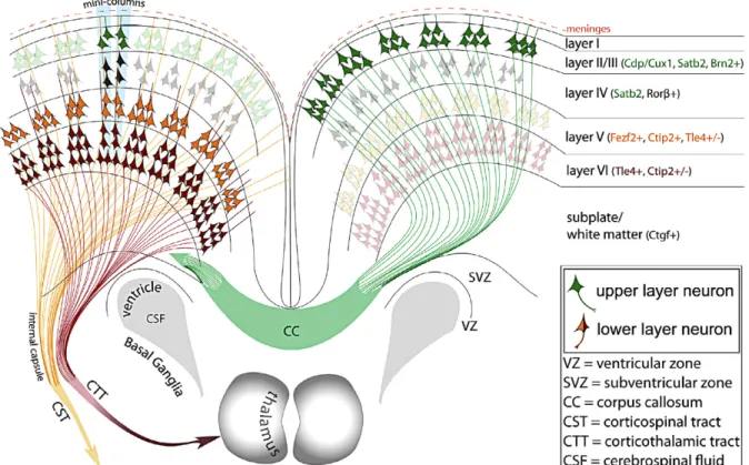
Each layer of the cortex contains pyramidal neurons with a specific morphology and projection pattern. Briefly, deeper layers of the cortex project subcortically while upper layers project intracortically (Gaspard and Vanderhaeghen 2011; Deboer, Kraushar et al.
2013). More specifically, layer VI neurons extend corticothalamic projections while layer V neurons develop mostly corticobulbar, coticospinal and corticotectal projection patterns with a few callosally projecting neurons mixed in (Hallman, Schofield et al. 1988; Molnar and Cheung 2006; Deboer, Kraushar et al. 2013) [Figure 2.4]. Layer IV neurons receive sensory input from thalamocortical axons and project short-range axons to cortical targets (Gaspard and Vanderhaeghen 2011). Neurons in the outermost layer II-III, and the focus of my thesis work, project contralaterally through the corpus callosum as well as within the same cortical hemisphere (Molyneaux, Arlotta et al. 2007; Deboer, Kraushar et al. 2013).
Axonal targeting after initial polarization has been studied in multiple model systems and several diffusible cues have been identified. Receptors in the growth cone respond to
chemoattractant and chemorepulsive cues that enable proper targeting. Netrin is an axon chemoattractant and required for the formation of commisures in the spinal cord and the corpus callosum (Lai Wing Sun, Correia et al. 2011; Fothergill, Donahoo et al. 2013).
Interestingly, Netrin signaling can act as both a chemoattractant and a chemorepellant
(Colamarino and Tessier-Lavigne 1995). In the cortex, semaphorin-neuropilin1 (Luo, Raible et al. 1993; Pasterkamp 2012) ligand receptor signaling acts as a repulsive cue for developing axons. Dual purpose signaling is also evident in semaphorin signaling where semaphorin 3A can act as a chemoattractant for apical dendrites of cortical pyramidal neurons and a repellant for their axons (see below) (Polleux, Morrow et al. 2000).
Morphological development of pyramidal neurons and radial migration are closely associated processes. At the most basic level, during radial-guided migration the leading process will become the apical dendrite and project towards the pial surface (Miller 1981).
Branching of the apical dendrites occurs mostly far away from the cell soma in the apical tuft located in the marginal zone. Thinner basal dendritic arbors will develop later and branch robustly in the area near the cell soma (Jan and Jan 2001). Several signaling cascades have been identified as regulators of dendrite formation and arborization. Among these signals, diffusible semaphorins have been identified as key signals required for apical dendrite orientation (Polleux, Morrow et al. 2000; Jan and Jan 2001; Pasterkamp 2012). Semaphorin 3A signaling through its receptor, neuropilin-1 and cofactor soluble guanylate cyclase, attracts the leading edge of a bipolar neuron causing extension towards the pial surface and maturation into an apical process. Remarkably semaphorin signaling acts on the axon to
repel its growth away from the pial surface (Song, Ming et al. 1998; Polleux, Morrow et al.
2000) (see chapter 3).
Importantly for my thesis, GSK-3 has been implicated downstream of semaphorin signaling (see chapter 3).
2.6 Disruptions in Early Cortical Development
In human syndromes, mispositioned neurons in the cerebral cortex can result from changes in progenitor cell cycle or migration failure. In humans the disruption of neuronal migration can result in developmental delays, seizures and death. For example, classic lissencephaly is the result of improper neuronal migration and is characterized by widely separated or absent gyri, decreased cortical lamination and multipolar or inverted neuronal morphology (Kerjan and Gleeson 2007). Groundbreaking genetic studies in humans have identified several genes mutated in migration disorders: LIS1 (Miller-Dieker syndrome, Type I lissencephaly) (Reiner, Carrozzo et al. 1993), doublecortin (X-linked lissencephaly, type I lissencephaly) (des Portes, Pinard et al. 1998; Gleeson, Allen et al. 1998) or tubulin α 1A (TUBA1) (Keays, Tian et al. 2007). These genes have been deleted in mouse models for studies on the mechanisms of migration (Dobyns, Reiner et al. 1993; Forman, Squier et al.
2005; Jaglin and Chelly 2009).
Human Reelin-linked lissencephaly (Norman-Roberts Syndrome) is another form of lissencephaly that is characterized my abnormal lamination (Hong, Shugart et al. 2000).
Reelin, a secreted glycoprotein from the Cajal Retzius cells, signals through Dab1 for proper lamination of the cerebral cortex, hippocampus and cerebellum (Rice and Curran 2001;
Lakatosova and Ostatnikova 2012). Mutations in this pathway lead to altered neuronal morphology and cortical lamination and can be studied using the classic mouse model, the Reeler mouse (Tissir and Goffinet 2003; Lakatosova and Ostatnikova 2012).
Importantly for my thesis work, many of the proteins implicated in lissencephaly converge onto the GSK-3 regulatory hub. Reelin has been shown to increase
phosphorylation of an inhibitory GSK-3 site, serine 9/21 (Beffert, Morfini et al. 2002) decreasing the phosphorylation of tau. However, reelin also induces the phosphorylation of GSK-3 on an activating tyrosine residue (Tyr216) which correlates with increased
phosphorylation of MAP1B (Gonzalez-Billault, Del Rio et al. 2005). These data suggest GSK-3 regulation of cytoskeletal dynamics may be influenced by Reelin signaling in complex ways. Additionally, GSK-3 mediated phosphorylation of DCX is thought to be required for DCX’s actions in restricting axon branching (Bilimoria, de la Torre-Ubieta et al.
2010) and migration (Bai, Ramos et al. 2003). Finally, inactivation of GSK-3 by Disrupted In Schizophrenia-1 (DISC1) is essential for progenitor proliferation and cortical neuronal migration. Abnormal regulation of GSK-3 by mutated DISC1 may underly the development of schizophrenia and bipolar disorder (Mao, Ge et al. 2009).
2.7 FIGURES AND FIGURE LEGENDS
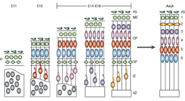
Figure 2.1 Cortical Development.
During the neurogenic phase of cortical development radial glia cells asymmetrically give rise to the first neurons that form the preplate (PP) (green circles and diamonds). The next generation of neurons (blue circles) splits the PP into the marginal zone (MZ) (green circles) and subplate (SP) (green diamonds). The neurons that split the preplate populate the cortical plate and become the deepest layer, layer VI, of the cortex (blue circles). Newly generated neurons migrate past the SP and layer VI to populate the outermost layer of the CP) and become layer V (red). Subsequent neuronal generation by radial glia and
intermediate progenitor cells (IPCs) form the remaining layers of the cortex in an inside-out fashion (remaining colored circles). At late embryonic stages, the radial glia cells generate glial cells and SP degenerates to form the final 6-layered cortex observed in the adult.
Image adapted from Gupta et al. 2002.
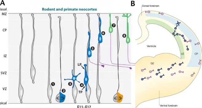
Figure 2.2 Neuronal Migration in the developing cortex
(A) Pyramidal Neuron Migration. In the cortex, radial glia cells retract their apical process to somally translocate towards the pial surface and form the preplate (grey radial glia cells). During the neurogenic phase, radial glia (1) asymmetrically divide (2) to produce a radial glia cell and multipolar daughter neuron (3) that migrates to the subventricular zone (SVZ). The newly born neuron will polarize into a bipolar morphology (4) with a leading process (LP) and trailing process (TP). Upon polarization the neuron will begin glial-guided migration (4-6) until reaching the outermost layer of the cortical plate. During migration the axon extends from the trailing process (purple process). Upon migration termination the neuron leaves the radial glia scaffold, somally translocates to its final position (7) and the leading process becomes the apical dendrite (8) and the axon continues to extent and target.
(B) Interneuron Migration. Interneurons born in the ganglionic emanance (GE) migrate tangentially into the cortex without the glial scaffold. Interneurons migrate in two streams (blue and purple) before diving into the cortical plate to adopt their final positions.
Image adapted from Barnes and Polleux 2009 and Nadarajah and Parnavelas 2002.
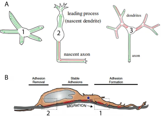
Figure 2.3 Cytoskeletal Organization and Adhesion for Migration
(A) Microtubule rearrangement during migration and maturation. (1) multipolar neurons sense the environment with plus-end microtubules oriented distally (green arrows). (2) Migrating neurons orient plus-end microtubules distally in the leading process (green arrows) and both minus-end distal (red arrows) and plus-end distal (green arrows) microtubules in the trailing process. (3) Developing dendrites increase in minus-end distal (red) microtubules in the dendrites while the axon converts to a plus-end distal microtubule network.
(B) Adhesion requirement for migration. (1) In order for migration to occur adhesion molecules must be inserted into the leading edge for adherence to the radial glia scaffold. (2) Physical movement along the radial scaffold requires removal of adhesions at the rear of the cell soma. Stable adhesions provide stable adherence to the scaffold and are subsequently removed at the rear of the cell after saltatory migration.
Figure Adapted from Sakakibara et al. 2013 and Solecki DJ 2012.
Figure 2.4 Cortical Projections and Laminar Identity
Neurons in each area of the cortex express specific markers and project to specific targets. The deepest layer, layer VI (pink/brown) project to the thalamus while layer V neurons (yellow/orange) project to the spinal cord. Upper layer neurons from layers II and III (green) project intracortically and through the corpus callosum to contralateral cortical targets. Image adapted from DeBoer et al. 2013.
CHAPTER 3: Glycogen Synthase Kinase-3 3.1 Overview
The literature on cortical neuronal migration and morphogenesis suggests that important kinases, like CDK5 and GSK-3, that regulate the functions of microtubule
associated proteins might have key roles. Functions of CDK5 related to neuronal migration and layer formation in the cortex have been well studied (Ohshima, Gilmore et al. 1999; Xie, Sanada et al. 2003; Tanaka, Serneo et al. 2004; Xie, Samuels et al. 2006; Ohshima, Hirasawa et al. 2007). I therefore have focused my thesis on functions of GSK-3. This chapter
provides an introduction to GSK-3 signaling.
3.2 Glycogen Synthase Kinase-3 Structure and Function
Glycogen Synthase Kinase 3 (GSK-3) is a serine/threonine kinase that was originally identified as a key regulator of glycogen synthase in glycogen metabolism and insulin responses (Woodgett and Cohen 1984). GSK-3 is highly conserved from the Drosophila homologue zeste-white3/shaggy (Bourouis, Moore et al. 1990; Siegfried, Perkins et al. 1990) to humans. Despite it’s name, GSK-3 is now known to be a multifunctional kinase with a broad range of targets and a key role in several signaling cascades.
GSK-3 α and β are ubiquitously expressed and both are highly expressed in the brain during development (Woodgett 1990) in both neurons and in glia (Ferrer, Barrachina et al.
2002; Kaidanovich-Beilin and Woodgett 2011). In mammals, GSK-3 has two isoforms,
GSK-3α and GSK-3β (Woodgett 1990) that are 85% identical and have a catalytic domain similarity of over 90% (Ali, Hoeflich et al. 2001; Kaidanovich-Beilin and Woodgett 2011).
The main differences between isoforms lie outside the catalytic domain in the N-terminus, where the α isoform has an N-terminal extension that may regulate nuclear transport (Azoulay-Alfaguter, Yaffe et al. 2011). Mice and humans have an alternatively spliced variant of GSK-3β of unknown function that contains a 13 amino acid inserted sequence in the kinase domain and is highly expressed in the nervous system (Mukai, Ishiguro et al.
2002).
Differences in isoform function are evident in mouse models where GSK-3α knockout mice are viable (MacAulay, Doble et al. 2007) while GSK-3β knockouts are embryonic lethal (Hoeflich, Luo et al. 2000). These results suggest that in many cell types GSK-3β is the more important family member. However, we have found that one isoform can clearly compensate for the other and that deletion of both isoforms is required to get a full picture of GSK-3 function in vivo (Kim, Zhou et al. 2006; Kim, Wang et al. 2009). In the work presented in this thesis both isoforms of GSK-3 have been deleted in neurons of the developing dorsal telencephalon.
3.2.1 GSK-3 Signaling Downstream of RTKs
GSK-3 has high basal activity towards many of its substrates in resting cells and inhibits their function at baseline (Hughes, Nikolakaki et al. 1993; Kaidanovich-Beilin and Woodgett 2011). Unlike most kinases, GSK-3 is inhibited by several upstream signaling pathways, transiently relieving phosphorylation of its substrates and enhancing their function (Hur and
Zhou 2010). GSK-3 is inactivated when phosphorylated on N-terminal Serine 9 (ser9) of GSK-3β (Sutherland, Leighton et al. 1993; Stambolic and Woodgett 1994) and Serine 21 (ser21) of GSK-3α (Sutherland and Cohen 1994). These phosphorylation events are typically mediated by Akt, downstream of receptor tyrosine kinase(RTK)/Phosphoinositide 3-kinase (PI3K) signaling. EGF, PDGF, neurotrophins, IGF and Insulin activate RTKs that, in turn, activate PI3K and Akt culminating in ser9/21 GSK-3 phosphorylation (Cross, Alessi et al.
1995; Kaidanovich-Beilin and Woodgett 2011) [Figure 3.1]. Additionally, signals transduced from the RTKs to PAR-6/PAR-3/aPKC can also inactivate GSK-3 kinase activity (Etienne-Manneville and Hall 2003). N-terminal Ser9/21 phosphorylation has been used in vitro studies to determine the level of GSK-3 kinase activity in multiple pathways and cell
types. It should be noted that these residues may not be a correct readout of kinase activity in all signaling paradigms (see below). Making the situation more complicated, GSK-3 can be activated by the phosphorylation of tyrosine residues, Tyr216 of GSK-3β and Tyr279 of GSK-3α (Hughes, Nikolakaki et al. 1993).
In general, GSK-3 phosphorylates downstream targets with a Serine/Threonine-X-X-X- Serine/Threonine motif. Most of GSK-3 substrates require another phosphorylation event near the GSK-3 site (‘priming’), mediated by a different kinase, for maximal GSK-3 activity (Sutherland 2011). Many GSK-3 priming kinases have been identified including, CK1, CK2, JNK, ERK, and PKA (for review see Sutherland C 2011). Though priming is not absolutely required for GSK-3 substrate phosphorylation, priming strongly enhances GSK-3 effect (Thomas, Frame et al. 1999). Of specific interest to this thesis, CDK5 is able to prime and enhance GSK-3 phosphorylation of MAPS and will be a point of discussion in Chapter 7.
3.2.2 GSK-3 Signaling in the Wnt pathway
Importantly, the Drosophila GSK-3 homologue, Shaggy/zeste white 3, was identified in the context of Wnt rather than RTK signaling. In contrast to RTK signaling where GSK-3 is regulated primarily via phosphorylation, GSK-3 regulation via Wnt is based on protein- protein interactions mediated by Disheveled (van Noort, Meeldijk et al. 2002; Doble and Woodgett 2003). Again, GSK-3 is active at baseline, phosphorylating and inactivating β- catenin, and GSK-3 activity towards this substrate is transiently released when Wnt signaling is activated (see below).
The Wnt/β-catenin Signaling Pathway is crucial for embryonic patterning, cell fate determination, polarity and proliferation (Logan and Nusse 2004). Wnts (wingless in Drosophila) are secreted glycoproteins that bind to the Frizzled-LRP5/6 receptors and act via
Dishevelled (Dvl) and GSK-3 signaling (For review see Logan and Nusse 2004). At basal levels GSK-3 is sequestered by the scaffolding protein Axin (Ikeda, Kishida et al. 1998;
Yasuda, Whitmarsh et al. 1999) into a destruction complex that includes the tumor suppressor Adenomatous polyposis coli (APC), the GSK-3 priming kinase, casein kinase 1 (CK1)(Amit, Hatzubai et al. 2002), and β-catenin (Rubinfeld, Souza et al. 1993). In this complex GSK-3 phosphorylates β-catenin on N-terminal residues Thr41, Ser37, and Ser33 (Liu, Li et al.
2002). Phosphorylation of cytoplasmic β-catenin on these residues signal for its degradation via the proteosomal pathway (Liu, Kato et al. 1999) [Figure 2.1].
The exact mechanism for Wnt-induced inactivation of GSK-3 is still under intensive investigation. Upon Wnt stimulation of Frizzled-LRP5/6, Dvl removes GSK-3 from the
destruction complex, probably by protein-protein interaction, and mediates GSK-3 phosphorylation of the accessory Wnt receptor LRP5/6 (Zeng, Tamai et al. 2005; Dale 2006).
The disruption of the destruction complex results in the accumulation of non-phosphorylated β-catenin and its subsequent translocation to the nucleus (van Noort, Meeldijk et al. 2002).
Nuclear β-catenin binds to T-cell Factor (TCF)/lymphoid enhancer factor (LEF) transcription factors (Behrens, von Kries et al. 1996; van de Wetering, Cavallo et al. 1997) [Figure 3.1].
In general, GSK-3 deletion strongly enhances β-catenin accumulation and signaling (Kaidanovich-Beilin and Woodgett 2011), however this predication has not been carefully assessed in all cell types. GSK-3’s role in Wnt/β-catenin signaling in developing cortical neurons is addressed in Chapter 5.
GSK-3 activity usually inhibits substrate functions. For example, GSK-3 phosphorylation of β-catenin, cyclin D1 and c-myc targets these proteins for degradation, while inhibition of GSK-3 promotes their stabilization and function (Diehl, Cheng et al.
1998; Alt, Cleveland et al. 2000; Sears, Nuckolls et al. 2000). Additionally, phosphorylation of some microtubule associated proteins (MAPs) inhibit tubulin binding [Figure 3.1]. For example, GSK-3 phosphorylation of tau decreases its affinity for microtubules (Lindwall and Cole 1984; Drechsel, Hyman et al. 1992; Hanger, Hughes et al. 1992; Brownlees, Irving et al.
1997; Utton, Vandecandelaere et al. 1997). Similarly, GSK3 phosphorylation of APC (Rubinfeld, Albert et al. 1996; Etienne-Manneville and Hall 2003) and CRMP-2 (Inagaki, Chihara et al. 2001; Fukata, Itoh et al. 2002; Yoshimura, Kawano et al. 2005) decrease their association with microtubules. Obviously, changes in microtubule binding properties will alter cytoskeletal dynamics.
However, the situation is complex in that GSK-3 phosphorylation of MAP-1B increases its association with microtubules (Lucas, Goold et al. 1998). Further, GSK-3 activity is required for the activity of some of its substrates, including doublecortin and CRMP-2 downstream of semaphorin signaling. I will address several of these GSK-3 substrates in Chapter 5.
3.2.3 GSK-3 signaling in the semaphorin pathway
Though originally identified as axonal repellants (Luo, Raible et al. 1993), semaphorins are diffusible guidance cues that can be both chemoattractive and repulsive (Pasterkamp 2012). In the cortex, axons are chemorepulsed by the sema signal and, as a result, project their axons away from the sema gradient (Chedotal, Del Rio et al. 1998; Montolio, Messeguer et al. 2009) and limit branching (Dent, Barnes et al. 2004). In addition, sema signaling is also used to mediate axon-axon interaction and by balancing repulsion with adhesion/fasiculation is involved with axon defasiculation and targeting (Vactor, Sink et al.
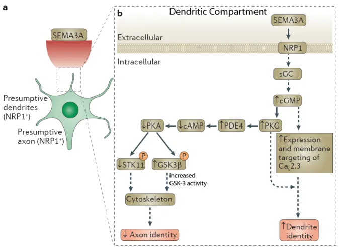
1993; Pasterkamp 2012). In the opposite direction, semaphorin 3A-neuropilin-1 signaling is a chemoattractive cue that orients the apical dendrite to the pial surface (Polleux, Morrow et al. 2000) [Figure 3.2]. Elevating cGMP levels relieves the inhibitory phosphorylation of GSK-3 resulting in increased signaling to substrates and subsequent dendrite orientation.
Increasing evidence suggests that an increase GSK-3 kinase activity is an essential mediator of sema signaling (Eickholt, Walsh et al. 2002; Uchida, Ohshima et al. 2005;
Pasterkamp 2012). Indeed a key GSK-3 target, collapsing response mediator protein-2 (CRMP-2) was discovered as downstream of of semaphorin signaling (Uchida, Ohshima et
al. 2005). CRMP-2 is a microtubule associated protein whose phosphorylation by GSK-3 at site Thr514 required for cytoskeletal rearrangements that are an essential to semaphorin biological actions (Yoshimura, Kawano et al. 2005) [Figure 3.2].
Semaphorin signaling via GSK-3 as been implicated in axon and dendrite specification of hippocampal and cortical neurons (Tran, Kolodkin et al. 2007; Shelly, Cancedda et al. 2011) In non-polarized neurons a strong dendrite-specifying semaphorin gradient asymmetrically increases cGMP levels, increasing GSK-3 signaling, and decreasing the phosphorylation of LKB1 on ser431, the residue responsible for specifying the axon (Polleux, Morrow et al.
2000; Shelly, Cancedda et al. 2011; Shelly and Poo 2011; Pasterkamp 2012) [Figure 3.2].
This signaling cascade specifies dendrite identity, suppresses axon formation and promotes dendritic growth. In the opposite direction, pser431 LKB1 (Barnes, Lilley et al. 2007;
Shelly, Cancedda et al. 2007) and an inactive pool of GSK-3 becomes asymmetrically localized in the neurite being specified as the axon (Jiang, Guo et al. 2005) (see below).
After the axon is initiated, semaphorin 3A treatment rapidly activates the GSK-3 pool in the growth cone to facilitate axon repulsion from the semaphorin gradient (Eickholt, Walsh et al.
2002; Montolio, Messeguer et al. 2009).
These actions of GSK-3 in the semaphorin signaling cascade may have strong
relevance to GSK-3 regulation of excitatory neuron morphogenesis as outlined in my soon to be submitted manuscript (Chapter 5).
3.3 GSK-3 regulation of polarity; Problems with in Vitro Models
As noted above, after a neuron is born it polarizes to form an axon and a somatodendritic domain and subsequently dendritic processes are elaborated. This process is highly regulated and has been mostly delineated through in vitro hippocampal culture models (Dotti, Sullivan et al. 1988; Craig and Banker 1994). Multiple proteins have been implicated as essential components of the polarity cascade (Polleux and Snider 2010). These include PI3K (Menager, Arimura et al. 2004), Akt (Yan, Guo et al. 2006), PTEN (Jiang, Guo et al. 2005), CRMP2 (Inagaki, Chihara et al. 2001; Arimura, Menager et al. 2004), LKB1 (Barnes, Lilley et al. 2007; Shelly and Poo 2011) and GSK-3 (Jiang, Guo et al. 2005; Yoshimura, Kawano et al. 2005; Yoshimura, Arimura et al. 2006).
In vitro experiments suggest a strong role for GSK-3 in regulating neuronal polarity
(Kim, Hur et al. 2011). Inactivation of GSK-3 by Ser9 phosphorylation is essential in transitioning the immature neurite into an axon, while pharmacological inhibition of GSK-3 in vitro leads to multiple axons per individual neuron (Jiang, Guo et al. 2005). In contrast,
expression of a S9A mutant GSK-3 prevents axon formation (Jiang, Guo et al. 2005;
Yoshimura, Kawano et al. 2005). Inhibition of GSK-3 and relief of phosphorylation of microtubule associated proteins is thought to enhance microtubule stability, which is important in specifying an axon (Witte, Neukirchen et al. 2008) (for review see Stiess and Bradke 2010).
In an effort to address the role of ser9/21 in an in vivo environment, GSK-3 point mutation knock-in mice have been created. Mutant mice expressing GSK-3 with Ser9/21 converted to alanine create, in theory, a ‘constitutively active’ GSK-3. Thus, these mutations
should result in steady basal activation of GSK-3 and excessive and uncontrolled phosphorylation of downstream targets. However, these animals exhibit normal neuronal polarity and develop with no overt abnormalities of neuronal morphology (McManus, Sakamoto et al. 2005; Gartner, Huang et al. 2006). These results raise the possibility that ser9/21 phosphorylation may not be important to the morphological regulation of neurons in vivo. The dramatic difference between in vitro and in vivo GSK-3 phenotypes strongly
supports the need to study the role of GSK-3 in a natural in vivo environment. My thesis directly addresses the role of GSK-3 in neuronal morphogenesis in vivo (Chapter 5).
3.4 GSK-3 Functions in vivo in Progenitors
GSK-3 signaling is known to be essential for the regulation of progenitor proliferation in the developing dorsal telencephalon in vivo. Kim et al. convincingly showed that deletion of both GSK-3 isoforms in the cortical radial progenitors resulted in an expansion of the progenitor pool and suppressed neuronal differentiation (Kim, Wang et al. 2009) [Figure 3.3]. Wnt, Sonic hedgehog (Shh) and Notch signaling were all implicated as enhanced by GSK-3 deletion. Kim and colleagues observed a strong increase in β-catenin levels in the GSK-3 deleted cells demonstrating GSK-3 regulation of β-catenin is essential for progenitor homeostasis. Consistent with these results, Walsh and colleagues have shown that increasing β-catenin levels in progenitors increases the production of neural precursors (Chenn and Walsh 2002). Importantly, the schizophrenia-associated protein, DISC1, is thought to act in part by regulating neural progenitor proliferation via GSK-3 (Mao, Ge et al. 2009). Recently, work from the Ip lab has demonstrated a novel role for GSK-3 IPC amplification via the
to neurons and was shown to bind GSK-3 as a part of its actions. Taken together, these studies establish critical roles for GSK-3 in the regulation of progenitors in the developing dorsal telencephalon in vivo and suggest that GSK-3 signaling importantly controls neuronal number in the developing brain. However, functions of GSK-3 in developing neurons in vivo remain elusive and form the subject of my thesis work (Chapter 5).
3.5 GSK-3 Function in Mature Neurons and Human Diseases
Not surprisingly, GSK-3 exhibits important functions in mature neurons. For the most part this regulation has been studied in in vitro models. For example, pharmacological inhibition of GSK-3 decreases vesicular transport in axons by reducing tau phosphorylation and binding to the motor protein kinesin-1 (Cuchillo-Ibanez, Seereeram et al. 2008).
Additionally, GSK-3 phosphorylates Dynamin 1 (Clayton, Sue et al. 2010) to regulate bulk endocytosis during periods of high activity when clatherin-dependent endocytosis is unable to endocytose large amounts of membrane. Finally, it has been demonstrated that GSK-3 is essential for LTD and over expression blocks LTP (Hooper, Markevich et al. 2007; Peineau, Taghibiglou et al. 2007; Zhu, Wang et al. 2007), possibly through PSD-95 regulation (Nelson, Kim et al. 2013). Though this thesis will not address the role of GSK-3 in mature neurons, this topic will be discussed in Chapter 6.
GSK-3 has important functions in psychiatric and neurologic diseases making further understanding of its role in neural development an urgent priority. For example, abnormal GSK-3 activity may underlie mood disorders. Thus Lithium, a known GSK-3β inhibitor, is the mainstay of treatment for bipolar disorder (Klein and Melton 1996; O'Brien, Harper et al.
2004)(Stambolic, Ruel et al. 1996; De Sarno, Li et al. 2002; Freland and Beaulieu 2012).
Additionally, the drug valproate, also sometimes used to treat mood disorders, is also an inhibitor of GSK-3 kinase activity (Chen, Huang et al. 1999; De Sarno, Li et al. 2002).
GSK-3 may also be important in the pathogenesis of schizophrenia (Emamian 2012).
Administration of antipsychotics, haloperidol or clozapine inhibits GSK-3 activity via ser9 phosphorylation (Emamian, Hall et al. 2004; Emamian 2012). Additionally, a regulator of GSK-3, Disrupted-in-Schizophrenia 1 (DISC1) has been implicated as a major susceptibility factor for Schizophrenia (De Rienzo, Bishop et al. 2011). DISC1 functions in party by affecting the GSK-3 functions related to progenitor proliferation in both neonates and adults (Mao, Ge et al. 2009).
Finally in Alzheimer’s disease, two of the hallmarks of the disease are aggregation of amyloid-β peptides from amyloid precursor protein and the development of neurofibrially tangles composed of hyper-phosphorylated tau (Glenner and Wong 1984). Interestingly GSK-3 inhibition blocks the production of amyloid-β peptides suggesting that GSK-3 is required for normal amyloid precursor protein function (Phiel, Wilson et al. 2003).
Additionally, GSK-3 strongly regulates the phosphorylation status of the MAP tau (Ishiguro, Shiratsuchi et al. 1993) (see above). Hyper-phosphorylated tau is found in the neurofibrially tangles in the brains of Alzheimer’s patients (Hanger, Hughes et al. 1992; Pei, Braak et al.
1999; Lucas, Hernandez et al. 2001). Hyper-phosphorylated tau also underlies a class of neurodegenerative diseases called tauopathies.
Although my dissertation does not address the functions of GSK-3 in adult neurons, better understanding of GSK-3 regulation during neuronal development, including the regulation of microtubule associated proteins, may inform approaches to inhibiting GSK-3 for therapeutic intervention.
3.6 FIGURES AND FIGURE LEGENDS
Figure 3.1 GSK-3 Signaling Cascades
(A-B) Canonical Wnt Signaling. (A) At basal levels GSK-3 is associated with casein kinase, APC, Axin and β-catenin in a ‘destruction‘ complex. GSK-3 phosphorylates β-catenin to signal its degradation. (B) Upon Wnt stimulation GSK-3 is sequestered away from the destruction complex leading to the accumulation of β-catenin and altered gene expression.
(C) Receptor Tyrosine Kinase Signaling. GSK-3 is downstream of multiple cascades.
Signaling through PI3K/AKT leads to ser9 phosphorylation or GSK-3 and GSK-3 inhibition.
GSK-3 signals to multiple downstream Microtubule Associated Proteins (MAPS).
Figure 3.2 Semaphorin signaling in dendrite development
(A) Schematic of semaphorin gradient specifying dendrite formation. (B) A strong semaphorin signal asymmetrically increases cGMP levels to specify dendrite identity.
Increased cGMP in the dendrites decreases the phosphorylation of STK11 (LKB1) and GSK-3, increasing GSK-3 signaling in the dendrite. Both Ser431 phosphorylation of LKB1 and GSK-3 inactivation in a single neurite are required initiating an axon. Figure adapted from Pasterkamp 2012.
Figure 3.3 GSK-3 regulation of Progenitor Homeostasis
(A-A’) Coronal sections of E13.5 control and GSK-3 deleted mice. GSK-3 mutants have an increased progenitor pool (sox2). Nestin labels Radial Glia. Scale 900uM. (B-B’) Coronal sections of E13.5 control and GSK- 3 deleted mice. GSK-3 mutants have fewer Tuj1-labeled neurons and increased progenitor pool (Sox2). Scale bar = 500uM. Figure adapted from Kim et al. Nat. Neuroscience 2009
CHAPTER 4: TGF-β Activated Kinase 1 4.1 Overview
Neurons are some of the most highly polarized cells in the body and the polarization of axons and dendrites underlies the proper flow of information in the brain. Currently, the molecular mechanisms responsible for neuronal polarization during brain development in vivo are unclear. However, recent work from several labs have started to defined a signaling
cascade required for cortical neuron polarization in vivo. At the core of this cascade is the phosphorylation of a serine/threonine kinase, LKB1, on its serine residue 431. Here I demonstrate in vitro that TGF-β Activated Kinase-1 (TAK1) is capable of associating with LKB1 and phosphorylating LKB1 on serine 431. Additionally, in vitro knock-down of TAK1 in dissociated cortical cultures results in delayed polarization, consistent with a role in axon formation. Finally, genetic deletion of TAK1 in progenitors resulted in no overt change in lamination or polarization in vivo. Despite promising in vitro data, our results indicate that TAK1 is not the major upstream kinase responsible for transducing the polarization cascade in vivo.
The work in this chapter was conducted under the direction of Franck Polleux, PhD.
Following the completion of the TGF-β Activated Kinase-1 project I generated LKB1:Nex conditional mutants and conducted initial characterization on this mutant line (data not shown). This work was continued by Dr. Polleux at Scripps and was recently published
4.2 Introduction
Following cell cycle exit, neurons polarize by undergoing drastic morphological changes to form two distinct domains: a single axon and multiple dendrites. While many proteins have been implicated in the establishment of neuronal polarity and axon specification in vitro, such as APC (Shi, Cheng et al. 2004), PI3K (Yoshimura, Arimura et al. 2006), and
GSK-3 (Shi, Cheng et al. 2004; Jiang, Guo et al. 2005; Yoshimura, Kawano et al. 2005), few of these genes have been shown to be required for neuronal polarization in vivo. However, recent work demonstrated that LKB1, the ortholog of Caenorhabditis elegans Partitioning- defective 4 (Par4 or STK11), is critical for the establishment of neuronal polarity in vivo
(Kishi, Pan et al. 2005; Barnes, Lilley et al. 2007; Shelly, Cancedda et al. 2007; Shelly and Poo 2011).
The vast majority of LKB1 protein is inactive and located in the nucleus with STRADβ (Dorfman and Macara 2008). Upon a polarization cue, LKB1 is shuttled into the cytosol by STRADα and MO25 and this translocation increases the catalytic activity of LKB1 by approximately 10-fold (Baas, Boudeau et al. 2003). In epithelial cells, activation of LKB1 via an inducible STRADα was sufficient to induce polarity (Baas, Kuipers et al. 2004) indicating a critical role for LKB1 in this process. As in epithelial cells, the translocation of LKB1 to the cytoplasm in cortical neurons allows for LKB1 to be activated by phosphorylation of an axon-specifying residue, serine 431 (Barnes, Lilley et al. 2007). This phosphorylation event leads to pLKB1 being sequestered to the a single neurite, phosphorylation of downstream SAD kinases and finally, axon initiation (Barnes, Lilley et al.
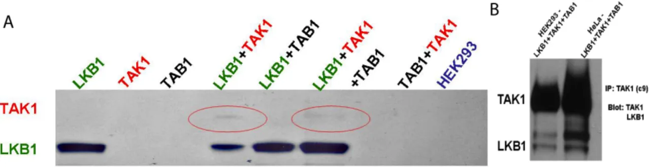
2007; Shelly, Cancedda et al. 2007). The kinase upstream of the LKB1 phosphorylation of ser431 is unknown. Recently, the Schneider lab demonstrated that the kinase TAK1 is capable of phosphorylating and inducing LKB1 activity in vivo (Xie, Zhang et al. 2006) suggesting that TAK1 could be acting upstream of LKB1 in the polarization cascade.
TGF-β activated kinase 1 (TAK1) is a serine/threonine kinase initially identified as a mitogen-activated protein kinase (Yamaguchi, Shirakabe et al. 1995). While it can be activated by TGF-β, TAK1 has been implicated in both the JNK (Shirakabe, Yamaguchi et al.
1997; Wang, Zhou et al. 1997) and P38 MAPK pathways (Moriguchi, Kuroyanagi et al.
1996) and has critical roles in cell survival, proliferation, stress response and immune regulation (Delaney and Mlodzik 2006). Interestingly, the role of TAK1 has not been assessed in cortical neurons.
TAK1 requires a combination of adaptor proteins, TAK1 Binding proteins 1, 2 and 3 (TAB1, TAB2 and TAB3), for its activation (Shibuya, Yamaguchi et al. 1996; Kishimoto, Matsumoto et al. 2000; Takaesu, Kishida et al. 2000). While TAK1 association with either TAB2 or TAB3 associates the complex to specific receptors, K63-linked ubiquitination along with TAB1 physical association enable TAK1 kinase activity (Shibuya, Yamaguchi et al.
1996; Kishimoto, Matsumoto et al. 2000; Sakurai, Miyoshi et al. 2000; Takaesu, Kishida et al. 2000; Landstrom 2010). Of particular note, TAK1 knockout mice are embryonic lethal (Sato, Sanjo et al. 2005) and therefore the majority of the studies related to TAK1 have been conducted in vitro.
Here I have addressed the function of TAK1 in neuronal polarity both in vitro and in vivo using Emx1-Cre (Gorski, Talley et al. 2002) and a conditional TAK1loxp/loxp mouse (Sato,
Sanjo et al. 2005). I demonstrate in vitro that TAK-1 is capable of associating with LKB1 and phosphorylating LKB1 on ser431. The phosphorylation of LKB1 is enhanced by TAB1 co-expression. Additionally, overexpression of TAK1 in newly born neurons results in a delay in axon initiation in vitro, consistent with a role for TAK1 in polarization upstream of LKB1. Finally, deletion of TAK1 in vivo does not inhibit axon initiation, as seen with LKB1 deletion with Emx1-cre (Barnes, Lilley et al. 2007). We conclude that in vivo TAK1 is not a critical regulator of polarity in newly born neurons of the developing dorsal telencephalon.
4.3 Results
TAK1 expression in the developing murine cortex
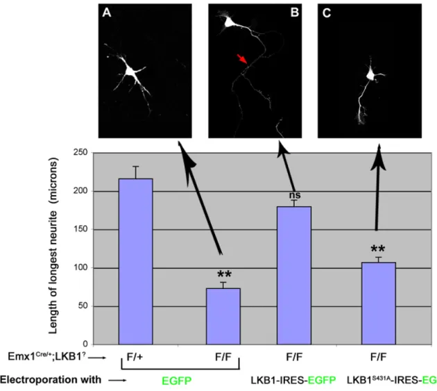
In vivo deletion of LKB1 in cortical pyramidal neurons results in a loss of axon formation
(Barnes, Lilley et al. 2007) [Figure 4.1]. Furthermore, re-expression of LKB1 via cortical electroporation of the conditional LKB1 knockout neurons was able to rescue axon formation [Figure 4.1]. Interestingly, Barnes et al. identified a residue in LKB1, serine 431, that specifies axon formation. A mutation in LKB1, serine 431 to alanine mutation (LKB1S431A), was not able to rescue axon formation [Figure 4.1]. These data indicate the in vivo importance of LKB1, specifically the phosphorylation of LKB1 on serine 431, in neuronal polarization (Barnes, Lilley et al. 2007).
Recently, TAK1 expression has been shown to regulate LKB1 kinase activity (Xie, Zhang et al. 2006) making it an upstream candidate for LKB1 phosphorylation. However, the role of TAK1 in the developing cortex has never been explored. LKB1 is expressed throughout the cortex but is enriched in the progenitor zone (VZ) of the developing cortex [Figure