R E S E A R C H
Open Access
Early sedation and clinical outcomes of
mechanically ventilated patients: a prospective
multicenter cohort study
Lilian Maria Sobreira Tanaka
1, Luciano Cesar Pontes Azevedo
2,3, Marcelo Park
2,3, Guilherme Schettino
2,
Antonio Paulo Nassar Jr
4, Alvaro Réa-Neto
5, Luana Tannous
5, Vicente Ces de Souza-Dantas
6,7, André Torelly
8,
Thiago Lisboa
8, Claudio Piras
9, Frederico Bruzzi Carvalho
10, Marcelo de Oliveira Maia
11, Fabio Poianas Giannini
12,
Flavia Ribeiro Machado
13, Felipe Dal-Pizzol
14, Alexandre Guilherme Ribeiro de Carvalho
15, Ronaldo Batista dos Santos
16,
Paulo Fernando Guimarães Morando Marzocchi Tierno
17, Marcio Soares
18,19, Jorge Ibrain Figueira Salluh
18,19*and for the ERICC study investigators
Abstract
Introduction:Sedation overuse is frequent and possibly associated with poor outcomes in the intensive care unit (ICU) patients. However, the association of early oversedation with clinical outcomes has not been thoroughly evaluated. The aim of this study was to assess the association of early sedation strategies with outcomes of critically ill adult patients under mechanical ventilation (MV).
Methods:A secondary analysis of a multicenter prospective cohort conducted in 45 Brazilian ICUs, including adult patients requiring ventilatory support and sedation in the first 48 hours of ICU admissions, was performed. Sedation depth was evaluated after 48 hours of MV. Multivariate analysis was used to identify variables associated with hospital mortality.
Results:A total of 322 patients were evaluated. Overall, ICU and hospital mortality rates were 30.4% and 38.8%, respectively. Deep sedation was observed in 113 patients (35.1%). Longer duration of ventilatory support was observed (7 (4 to 10) versus 5 (3 to 9) days,P= 0.041) and more tracheostomies were performed in the deep sedation group (38.9% versus 22%,P= 0.001) despite similar PaO2/FiO2ratios and acute respiratory distress
syndrome (ARDS) severity. In a multivariate analysis, age (Odds Ratio (OR) 1.02; 95% confidence interval (CI) 1.00 to 1.03), Charlson Comorbidity Index >2 (OR 2.06; 95% CI, 1.44 to 2.94), Simplified Acute Physiology Score 3 (SAPS 3) score (OR 1.02; CI 95%, 1.00 to 1.04), severe ARDS (OR 1.44; CI 95%, 1.09 to 1.91) and deep sedation (OR 2.36; CI 95%, 1.31 to 4.25) were independently associated with increased hospital mortality.
Conclusions:Early deep sedation is associated with adverse outcomes and constitutes an independent predictor of hospital mortality in mechanically ventilated patients.
* Correspondence:jorgesalluh@gmail.com 18
IDOR–D’Or Institute for Research and Education, Rua Diniz Cordeiro 30, Rio de Janeiro 22281-100, Brazil
19
Postgraduate Program, Instituto Nacional de Câncer, 10° Andar, Praça Cruz Vermelha 23, Rio de Janeiro 20230-130, Brazil
Full list of author information is available at the end of the article
Introduction
Sedation is an important component of care for patients under mechanical ventilation (MV) in the ICU. Significant distress is related to MV itself or to routine procedural in-terventions [1] and minimizing pain, anxiety and distress is a major recommendation in recent guidelines [2]. Pain and anxiety control is usually obtained with analgesics and sedatives that ensure comfort, improve synchrony with the ventilator and decrease work of breathing [3]. Some studies, however, have shown that oversedation is associ-ated with poor outcomes, including delirium, prolonged MV, ventilator-associated pneumonia, long ICU and hos-pital length of stay [3-6], posttraumatic stress disorder [7] and cognitive impairment [8,9] as well as increased costs [10-12]. Nevertheless, the issue of early sedation has seldom been evaluated, especially in randomized controlled studies. Despite the current recommendations [2], there is still a significant gap between evidence from recent trials and implementation in clinical practice [2,6,13,14]. Moreover, to date no large randomized controlled trials of sedation strategies used mortality as the primary outcome. In addition, clinical trials of sedation have until now enrolled patients mostly after 24 to 48 hours following initiation of MV, resulting in inadequate assessment of early sedation practice and its association with clinically relevant out-comes [15-17].
The aim of this study is thus to describe the association of early sedation strategies (sedation depth and sedative choice) with clinical outcomes of mechanically ventilated adult ICU patients, with hospital mortality as the primary outcome.
Materials and methods
Study design, patient selection and definitions
This study was a secondary analysis of a multicenter prospective cohort conducted in 45 Brazilian ICUs (from the Brazilian Research in Intensive Care Network) from 12 states between 1 June 2011 and 31 July 2011 [18]. Additional file 1 presents the participating ICUs in more detail. In the present study, adult patients (≥18 years old) requiring invasive ventilatory support in the first 48 hours of ICU admission using sedatives on the second day of MV were included.
We excluded patients submitted exclusively to noninva-sive MV and those presenting with primary neurological disorders. We also excluded those with missing data re-garding sedation depth on the second day of MV. Demo-graphic, clinical and laboratory data were collected during the ICU stay and included cause of respiratory failure, chronic health status, Charlson Comorbidity Index [19], Simplified Acute Physiology Score 3 (SAPS3) [20] and the Sequential Organ Failure Assessment (SOFA) score [21]. Data regarding the need for vasopressors, tracheostomy and renal replacement therapy were also reported. Sepsis
diagnosis was made based on current definitions [22]. A patient was considered to have an infection when present-ing with suggestive clinical, laboratory, radiographic and microbiological findings that justified the administration of antibiotics (excluding prophylaxis) [23]. Acute respira-tory distress syndrome (ARDS) was defined and classified according to the Berlin definition [24].
As the primary study was not designed to evaluate sed-ation strategies [18], the Glasgow Coma Scale (GCS) – originally reported for SOFA score calculation–was used as a surrogate for sedation depth. Light sedation was de-fined as GCS≥9 whereas those patients with GCS <9 were considered deeply sedated. Reliability of GCS as a surro-gate for the Richmond Agitation–Sedation Scale (RASS) as well as selection of GCS = 9 as the cutoff value were based on the study by Ely and colleagues that demon-strated an excellent correlation (r= 0.91, P <0.001) be-tween the GCS and RASS, where GCS = 9 would be equivalent to RASS =−2 [25].
Our primary hypothesis was that early sedation strat-egies, namely sedation depth, could be associated with clinical outcomes including hospital mortality, so we chose to evaluate the sedation level on the second day of MV.
The study was approved by the institutional review board of Hospital Sírio-Libanês at the coordinating center (Comitê de Ética em Pesquisa; approval number HSL 2010/ 51) and subsequently by local review boards. Additional file 1 describes the local review boards by participating hospital. Due to the observational nature of the study, the following institutions waived the need for informed con-sent: Comitê de Ética em Pesquisa–Hospital São Camilo; Comitê de Ética em Pesquisa – Hospital Sírio-Libanês; Comissão de Ética para Análise de Projetos de Pesquisa– HCFMUSP; Comitê de Ética em Pesquisa–Hospital das Clinicas de Niteroi; Comitê de Ética em Pesquisa–Hcor; Comitê de Ética em Pesquisa – Hospital Israelita Albert
Einstein; Comitê de Ética em Pesquisa – HCFMRP –
USP; Comitê de ética do Hospital Sao Jose; Comitê de Ética em Pesquisa do Vitória Apart Hospital; Comitê de Ética em Pesquisa do Hospital Madre Teresa; Comitê de Ética em Pesquisa do Hospital da Mulher Heloneida Studart; Comitê de Ética em Pesquisa–Hospital Moin-hos de Vento; Comitê de Ética em Pesquisa do Hospital Mater Dei; Comitê de Ética em pesquisa – Hospital Mario Lioni; Comitê de Ética e Pesquisa do Hospital Espanhol; Comitê de Ética em Pesquisa – Instituto de Pesquisa Clínica Evandro Chagas, Fundação Oswaldo
Cruz; Comitê de Ética em Pesquisa – UNIFESP/EPM;
Statistical analysis
Continuous variables are reported as the median (25 to 75% interquartile range). Univariate and multivariate analyses using a binary logistic regression were used to identify factors associated with the dependent variable (hospital mortality). Variables yieldingP<0.2 by univariate analysis, or those considered clinically relevant despite P values, were entered into the multivariate analysis to esti-mate the independent association of each covariate with the dependent variable. Results are presented as the odds ratio with 95% confidence interval. A Kaplan–Meier curve with log-rank test was used to compare patients with light versus deep sedation for hospital mortality (censored at day 30). Two-tailed P <0.05 was considered statistically significant. All statistical tests were carried out using the commercial SPSS19.0 package for Windows (SPSS Inc., Chicago, IL, USA).
Results
Characteristics of the study population
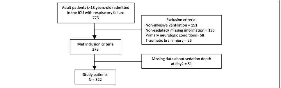
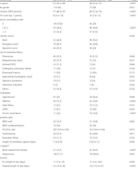
From 773 patients derived from the original cohort, a total of 322 patients fulfilled the eligibility criteria (Figure 1). Patients excluded for missing data about sedation depth on day 2 (n= 51) were comparable with the included population regarding demographic data, comorbidities, physiologic and disease severity variables and outcomes (data not shown). The main patient characteristics are pre-sented in Table 1. Median (25 to 75% interquartile range) age was 59 (41 to 74) years. The median SAPS3 was 61 (50 to 72) and SOFA score was 8 (6 to 11) on the first day in the ICU. Overall ICU and hospital mortality rates were 30.4% and 38.8% respectively. When comparing vor and survivor subgroups, we observed that nonsurvi-vors showed higher median age (68 (52 to 77) years vs. 53 (34 to 69) years,P<0.001), higher SAPS3 and SOFA score (68 (57 to 79) vs. 57 (48 to 67),P< 0.001 and 10 (7 to 12) vs. 8 (5 to 10),P<0.001) and higher percentage of patients with a Charlson Comorbidity Index >2 (37.6% vs. 16.2%,
P<0.001). Those who died during hospital admission also
had a higher frequency of moderate/severe ARDS (24% vs. 9.6%,P= 0.004), need for vasopressors (85.6% vs.71.1%,P= 0.003) and renal replacement therapy (33.6% vs.13.7%,
P<0.001).
Sedative choice and sedation depth
The most frequently used sedatives were the association of midazolam and fentanyl (39.4%) or of propofol and fen-tanyl (14%) and the use of fenfen-tanyl (12.4%) or midazolam (8.1%) as single drugs. Sedative drug type was also associ-ated with sedation levels: the sole administration of fen-tanyl and dexmedetomidine was more frequent in lightly sedated patients (15.3% vs. 7.1% for fentanyl, P= 0.033; 8.6% vs. 0.9% for dexmedetomidine,P= 0.005).
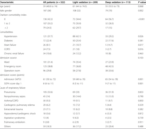
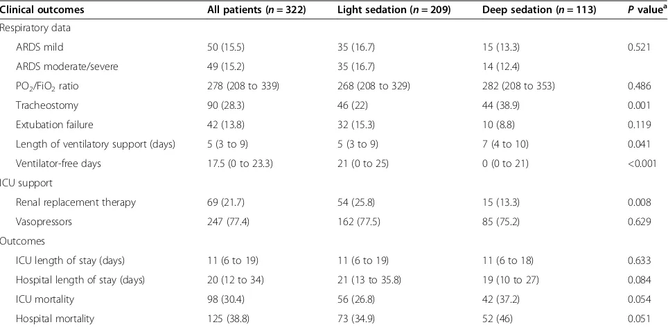
Deep sedation was observed in 113 patients (35.1%) and was associated with a higher overall disease severity, as demonstrated by higher median SAPS 3 (65 (54 to 78) for deep sedation vs. 59 (48 to 70) for light sedation, P= 0.001) (Table 2). Moreover, longer duration of ventilatory support was observed (7 (4 to 10) vs. 5 (3 to 9) days,P= 0.041) and more tracheostomies were performed in the deep sedation group (38.9% vs. 22%,P= 0.001) despite simi-lar arterial partial pressure of oxygen/fraction of inspired oxygen ratios and ARDS severity. Finally, trends to in-creased ICU and hospital mortality (37.2% vs. 26.8%,P= 0.054 and 46% vs. 34.9%,P= 0.051 respectively) were also associated with deep sedation (Table 3). A survival curve comparing patients with deep and light sedation is shown in Figure 2.
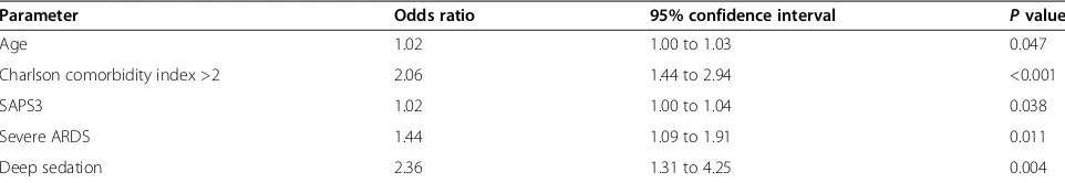
In a multivariate analysis, age, Charlson Comorbidity Index >2, SAPS3, severe ARDS and deep sedation were in-dependently associated with increased hospital mortality (Table 4).
Discussion
[image:3.595.58.541.568.720.2]In the present multicenter cohort study we evaluated the association of early sedation strategies with outcomes of adult mechanically ventilated patients in the ICU. Deep sedation in the first 48 hours of MV is a common
Table 1 General characteristics of study patients according to survival status
Characteristic Survivors (n=197) Nonsurvivors (n=125) Pvaluea
Age (years) 53 (34 to 69) 68 (52 to 77) <0.001
Male gender 114 (58) 73 (58) 0.925
Admission SAPS3 (points) 57 (48 to 67) 68 (57 to 79) <0.001
SOFA score day 1 (points) 8 (5 to 10) 10 (7 to 12) <0.001
Charlson comorbidity index
0 106 (53.8) 30 (24)
1 to 2 59 (29.9) 48 (38.4) <0.001
> 2 32 (16.2) 47 (37.6)
Admission source 0.038
Ward 52 (26.4) 49 (39.2)
Emergency room 79 (40.1) 46 (36.8)
Operation room 66 (33.5) 30 (24)
Cause of respiratory failure
Pneumonia 66 (33.5) 39 (31.2) 0.668
Nonpulmonary sepsis 30 (15.2) 15 (12) 0.415
Asthma/COPD 23 (11.7) 7 (5.6) 0.068
Cardiogenic pulmonary edema 11 (5.6) 9 (7.2) 0.558
Extracranial trauma 11 (5.6) 12 (9.6) 0.173
Hypovolemic/cardiogenic shock 10 (5.1) 8 (6.4) 0.614
Aspirative syndromes 10 (5.1) 3 (2.4) 0.234
Pulmonary embolism 4 (2) 5 (4) 0.296
Others 32 (16.2) 27 (21.6) 0.226
Comorbidities
Hypertension 63 (32) 58 (46.4) 0.008
Diabetes 30 (15.2) 42 (33.6) <0.001
Heart failure 12 (6.1) 14 (11.2) 0.101
COPD 12 (6.1) 12 (9.6) 0.243
Chronic renal failure 11 (5.6) 23 (18.4) <0.001
Respiratory data
ARDS mild 29 (14.7) 21 (16.8) 0.004
ARDS moderate/severe 19 (9.6) 30 (24)
PO2/FiO2ratio 287 (220 to 350) 252 (184 to 328) 0.019
Tracheostomy 54 (27.4) 36 (28.8) 0.817
Extubation failure 23 (11.7) 19 (15.2) 0.182
Length of ventilatory support (days) 5 (3 to 8) 6 (3 to 11) 0.260
ICU support
Renal replacement therapy 27 (13.7) 42 (33.6) <0.001
Vasopressors 140 (71.1) 107 (85.6) 0.003
Outcomes
ICU length of stay (days) 11 (7 to 19) 11 (5 to 18.5) 0.389
characteristic, observed in 35.1% of patients. Deep sedation was associated with increased disease severity, longer dur-ation of ventilatory support and higher need for tracheos-tomy. In a multivariate analysis, deep sedation was an independent factor associated with increased hospital mor-tality (odds ratio = 2.36).
[image:5.595.57.540.103.145.2]The more frequently used sedative regimens in our study were the association of midazolam and fentanyl or of propofol and fentanyl. When compared by sedation depth, the administration of fentanyl or dexmedetomidine as single drugs was more frequent in lightly sedated pa-tients (15.3% vs. 7.1% for fentanyl,P= 0.033; 8.6% vs. 0.9%
Table 1 General characteristics of study patients according to survival status(Continued)
Sedation depth
Light sedation 136 (69.0) 73 (58.4) 0.051
Deep sedation 61 (31.0) 52 (41.6)
Data presented as median (25 to 75% interquartile range) orn(%). ARDS, acute respiratory distress syndrome; COPD, chronic obstructive pulmonary disease; PO2/FiO2, arterial partial pressure of oxygen/fraction of inspired oxygen; SAPS3, Simplified Acute Physiology Score 3; SOFA, Sequential Organ Failure Assessment. a
Pvalue of the comparison between survivors and nonsurvivors.
Table 2 General characteristics of study patients according to sedation depth
Characteristic All patients (n= 322) Light sedation (n= 209) Deep sedation (n= 113) Pvaluea
Age (years) 59 (40.8 to 74) 61 (44 to 74.5) 55 (33.5 to 73) 0.068
Male gender 187 (58) 108 (52) 79 (70) 0.002
Charlson comorbidity index
0 136 (42.2) 72 (34.4) 64 (56.7) <0.001
1 to 2 107 (33.2) 75 (35.9) 32 (28.3)
> 2 79 (24.5) 62 (29.7) 17 (15)
Comorbidities
Hypertension 121 (37.7) 88 (42.1) 33 (29.2) 0.026
Diabetes 72 (22.4) 50 (25.4) 22 (17.6) 0.381
Heart failure 26 (8.1) 21 (10.7) 5 (14.7) 0.077
COPD 24 (7.5) 21 (10) 3 (2.7) 0.016
Chronic renal failure 34 (10.6) 24 (12.2) 10 (8) 0.463
Admission source 0.103
Ward 101 (31.4) 74 (35.4) 27 (23.9)
Emergency room 125 (38.8) 77 (36.8) 48 (42.5)
Operation room 96 (29.8) 58 (27.8) 38 (33.6)
Admission scores (points)
Admission SAPS3 61 (50 to 72) 59 (48 to 70) 65 (54 to 78) 0.001
SOFA score day 1 8 (6 to 11) 8 (5 to 11) 9 (7 to 11) 0.001
Cause of respiratory failure
Pneumonia 105 (32.6) 69 (33) 36 (31.9) 0.833
Nonpulmonary sepsis 45 (14) 30 (14.4) 15 (13.3) 0.790
Asthma/COPD 30 (9.3) 19 (9.1) 11 (9.7) 0.850
Cardiogenic pulmonary edema 20 (6.2) 15 (7.2) 5 (4.4) 0.329
Extracranial trauma 23 (7.1) 13 (6.2) 10 (8.8) 0.382
Hypovolemic/cardiogenic shock 18 (5.6) 12 (5.7) 6 (5.3) 0.872
Aspirative syndromes 13 (4) 9 (4.3) 4 (3.5) 0.739
Pulmonary embolism 9 (2.8) 6 (2.9) 3 (2.7) 0.911
Others 59 (18.3) 36 (17.2) 23 (20.4) 0.488
[image:5.595.59.536.296.717.2]for dexmedetomidine,P= 0.005). These findings may be attributed to a rapid onset and offset of action of these drugs and their easiness to titrate, while benzodiaze-pines are more likely to accumulate, especially during con-tinuous infusion and when associated with other drugs [26,27]. Recent studies demonstrate that sedation with benzodiazepines aiming at the same target is usually asso-ciated with a higher rate of sedation depth beyond the target as compared with dexmedetomidine [28].
Although important studies were published recently, a systematic review published in 2010 showed that clinical trials on sedation are commonly reported as low quality,
mainly due to their design (for example, before and after studies), heterogeneous protocols and the relatively small number of patients in a single center or few cen-ters [29]. These factors generate limited evidence to guide sedation drug choice and administration strategies in mech-anically ventilated patients. Moreover, most studies – in-cluding recent randomized clinical trials – have enrolled patients 24 to 48 hours after the initiation of MV, making the clinical relevance of sedation depth in the early period more difficult to understand. A recent multicenter pro-spective cohort study by Shehabi and colleagues enrolled 251 patients, aiming to characterize the patterns of early sedation practice in 25 Australia and New Zealand (ANZ) ICUs and to assess its relationship with relevant clinical outcomes. After adjusting for potential confounding, early deep sedation was a predictor of time to extubation and mortality [30]. These results were corroborated by a co-hort study in 11 Malaysian hospitals including medical/ surgical patients (n= 259) who were sedated and venti-lated≥24 hours [31]. Likewise, in our study we could as-sess sedation strategies in the first 48 hours of MV and its relevant clinical outcomes in a large number of patients (n= 322), and demonstrated that early deep sedation was an independent predictor of hospital mortality.
[image:6.595.57.539.100.336.2]Unlike the other variables that were also found to be in-dependently associated with mortality in our study, such as ARDS severity, comorbidities and disease severity, sedation depth is a potentially modifiable risk factor for mortality. Implementing sedation protocols to achieve light sedation has been proven feasible and reproducible [32,33]. This Table 3 Clinical outcomes according to sedation depth
Clinical outcomes All patients (n= 322) Light sedation (n= 209) Deep sedation (n= 113) Pvaluea
Respiratory data
ARDS mild 50 (15.5) 35 (16.7) 15 (13.3) 0.521
ARDS moderate/severe 49 (15.2) 35 (16.7) 14 (12.4)
PO2/FiO2ratio 278 (208 to 339) 268 (208 to 329) 282 (208 to 353) 0.486
Tracheostomy 90 (28.3) 46 (22) 44 (38.9) 0.001
Extubation failure 42 (13.8) 32 (15.3) 10 (8.8) 0.119
Length of ventilatory support (days) 5 (3 to 9) 5 (3 to 9) 7 (4 to 10) 0.041
Ventilator-free days 17.5 (0 to 23.3) 21 (0 to 25) 0 (0 to 21) <0.001
ICU support
Renal replacement therapy 69 (21.7) 54 (25.8) 15 (13.3) 0.008
Vasopressors 247 (77.4) 162 (77.5) 85 (75.2) 0.629
Outcomes
ICU length of stay (days) 11 (6 to 19) 11 (6 to 19) 11 (6 to 18) 0.633
Hospital length of stay (days) 20 (12 to 34) 21 (13 to 35.8) 19 (10 to 27) 0.084
ICU mortality 98 (30.4) 56 (26.8) 42 (37.2) 0.054
Hospital mortality 125 (38.8) 73 (34.9) 52 (46) 0.051
Data presented as median (25 to 75% interquartile range) orn(%). ARDS, acute respiratory distress syndrome; PO2/FiO2, arterial partial pressure of oxygen/fraction
of inspired oxygen;.a
Pvalue of the comparison between light and deep sedation.
[image:6.595.57.291.537.695.2]evidence is important because deep sedation still seems to be current practice in many ICUs worldwide [5,13] and is present in 35% of the patients enrolled in the present study. Accordingly, Payen and colleagues observed in their survey a large proportion of patients in a deep state of sedation, and in addition no major changes in sedation depth or sedative dosages occurs during the first week of the ICU stay [34].
Finally, our findings provide validation of the concept that early sedation has a major impact on outcomes. Demonstrating this in a middle-income country in South America is an important step to provide generalizability of previous findings because the main previously pub-lished results on this issue are from ANZ ICUs. Several ANZ trials are well known for frequently presenting dif-ferent results compared with similar interventions in other parts of the world, as demonstrated by studies in the last decade in the critical care field [35,36] and even in the sedation area [37]. This discrepancy is attributed to substantial differences due to the high standards in process of care and staffing patterns, and is consequently translated into lower mortality in ANZ ICUs [38,39].
The present study has some limitations that must be considered. First, the sedation level was not assessed using a specific sedation scale. Nonetheless, although originally created to assess the level of consciousness after head injury, it is known that GCS (the chosen scale) <9 denotes a comatose state even in nontraumatic settings, such as cerebral depression by pharmacological cause [40,41]. Moreover, the GCS shows excellent cor-relation and discrimination with the RASS (r= 0.91,P< 0.001), as described by Ely and colleagues from 1,360 paired observations among 275 adult patients in med-ical and coronary ICUs [25]. This strong correlation be-tween the RASS and the GCS was also shown in other sedation scale validation studies [42,43], which allows its applicability as a surrogate for sedation depth in the present study.
Another limitation is the potential influence of previous neurological status on GCS values. Data regarding GCS on day 1 were collected in patients who were already se-dated and under MV. A reported low GCS could therefore be influenced by the baseline neurological status and not
only by sedation strategies, because it may also reflect acute neurological conditions associated with the current illness presented by the critically ill, such as encephalop-athy. However, this is not an exclusive limitation of the GCS because this may also occur with the more specific sedation assessment scales such as the RASS or Sedation-Agitation Scale (SAS). Moreover, it is possible that certain patients may have required larger sedative doses on day 2 of MV for other reasons (ICU proce-dures, for instance) and we did not collect these data. In order to minimize the impact of other conditions in GCS values, we excluded patients with primary or known acute neurological disorders and included only patients under sedation in the present study. Consider-ing the potential confounders mentioned above, the strong association between deep sedation and mortality must be analyzed regarding our interpretation of GCS values mainly as a surrogate of sedation depth.
Finally, we have not evaluated the presence of delirium. In mechanically ventilated patients, delirium is an inde-pendent predictor of hospital and 6-month mortality [6,44]. These outcomes are shown even in light sedated patients, where a dose–effect response was described [45]. Despite this clinical relevance, the incidence of delirium could not be evaluated in our study, since it was not the aim of the primary analysis. Finally, participant ICUs re-ported the existence of a sedation protocol, but the actual adherence for local recommendations was not assessed in the present study as it was beyond its initial scope.
Conclusions
Early deep sedation is associated with worse outcomes and constitutes an independent predictor of hospital mor-tality in a prospective cohort of mechanically ventilated patients. Randomized clinical trials should be designed to address the impact of light versus deep sedation in the early phase (<48 hours) of MV.
Key messages
Early deep sedation is associated with longer
[image:7.595.57.538.100.183.2]duration of ventilator support and higher need for tracheostomy in mechanically ventilated patients.
Table 4 Factors associated with hospital mortality in a multivariate analysis
Parameter Odds ratio 95% confidence interval Pvalue
Age 1.02 1.00 to 1.03 0.047
Charlson comorbidity index >2 2.06 1.44 to 2.94 <0.001
SAPS3 1.02 1.00 to 1.04 0.038
Severe ARDS 1.44 1.09 to 1.91 0.011
Deep sedation 2.36 1.31 to 4.25 0.004
Area under receiver operating curve for predicted mortality (95% confidence interval) = 0.768 (0.718 to 0.813),P<0.001. Hosmer–Lemeshowχ2
Early sedation depth is independently associated with increased hospital mortality.
Sedation depth is a potentially modifiable risk factor for adverse outcomes; future clinical trials of light sedation with early (<24 hours) randomization of patients should therefore be encouraged.
Additional file
Additional file 1:Is a table listing the sites, investigators and institutional review boards that participated in the Epidemiology of Respiratory Insufficiency in Critical Care study.
Abbreviations
ANZ:Australia and New Zealand; ARDS: acute respiratory distress syndrome; GCS: Glasgow Coma Scale; MV: mechanical ventilation; RASS: Richmond Agitation–Sedation Scale; SAPS3: Simplified Acute Physiology Score 3; SOFA: Sequential Organ Failure Assessment.
Competing interests
This study was performed with institutional funds. JIFS received honoraria and unrestricted research grants from Hospira, Inc. São Paulo, Brazil, but this did not interfere with the design, analysis or the elaboration of the manuscript and therefore with adherence to the ethical requirements of the journal. The remaining authors declare that they have no competing interests.
Authors’contributions
LMST participated in study conception, data acquisition, data analysis and interpretation, and drafting of the manuscript. LCPA participated in study conception, data acquisition, data analysis and interpretation, and drafting of the manuscript. MP participated in study conception, data analysis and revising the manuscript for important intellectual content. GS participated in study conception, data acquisition, data analysis and drafting of the manuscript. APNJr participated in data acquisition, drafting of the manuscript and revising the manuscript for important intellectual content. AR-N participated in data acquisition and revising the manuscript for important intellectual content. LT participated in data acquisition and revising the manuscript for important intellectual content. VCdS-D participated in data acquisition and revising the manuscript for important intellectual content. AT participated in data acquisition and revising the manuscript for important intellectual content. TL participated in data acquisition and revising the manuscript for important intellectual content. CP participated in data acquisition and revising the manuscript for important intellectual content. FBC participated in data acquisition and revising the manuscript for important intellectual content. MdOM participated in data acquisition and revising the manuscript for important intellectual content. FPG participated in data acquisition and revising the manuscript for important intellectual content. FRM participated in data acquisition and revising the manuscript for important intellectual content. FD-P participated in data acquisition and revising the manuscript for important intellectual content. AGRdC participated in data acquisition and revising the manuscript for important intellectual content. RBdS participated in data acquisition and revising the manuscript for important intellectual content. PFGMMT participated in data acquisition and revising the manuscript for important intellectual content. MS participated in study conception, data analysis and interpretation, and drafting of the manuscript. JIFS participated in study conception, data analysis and interpretation, and drafting of the manuscript. All authors approved the final copy of the manuscript.
Acknowledgements
This study was funded by the Research and Education Institute from Hospital Sírio-Libanês, São Paulo and the D’Or Institute for Research and Education, Rio de Janeiro, Brazil. The funding institutions had no active role in the design, analysis and interpretation of the results or in the publication process. The study was supported by the Brazilian Research in Intensive Care Network.
Author details
1
Hospital Copa D’Or, Rua Figueiredo de Magalhães 875, Rio de Janeiro 22031-010, Brazil.2Research and Education Institute, Hospital Sírio-Libanês, Rua Cel. Nicolau dos Santos 69, São Paulo 01308-060, Brazil.3ICU, Emergency Medicine Department, Hospital das Clinicas da Faculdade de Medicina da Universidade de São Paulo, Av. Eneas Carvalho Aguiar 255, São Paulo 05403-000, Brazil.4ICU, Hospital São Camilo Pompeia, Av. Pompeia 1178, São Paulo 05022-000, Brazil.5CEPETI–Centro de Estudos e Pesquisas em Terapia Intensiva, Rua Monte Castelo 366, Curitiba 82530-200, Brazil.6ICU, Instituto Nacional de Câncer–Hospital do Câncer I, Praça Cruz Vermelha 23, Rio de Janeiro 20230-130, Brazil.7ICU, Hospital Pasteur, Av. Amaro Cavalcanti 495, Rio de Janeiro 20735-040, Brazil.8Rede Institucional de Pesquisa e Inovação em Medicina Intensiva (RIPIMI), Irmandade da Santa Casa de Misericórdia de Porto Alegre, Rua Professor Annes Dias 285–Centro Histórico, Porto Alegre 90020-090, Brazil.9ICU, Vitoria Apart Hospital, Rodovia BR-101 Norte, Km 2, 38 Boa Vista II, Serra, ES 29161-900, Brazil.10ICU, Hospital Mater Dei, Rua Gonçalves Dias 2700 (Bloco I), Belo Horizonte 30140-093, Brazil.11ICU, Hospital Santa Luzia, SHLS 716–Conjunto E, Brasília 70390-902, Brazil.12ICU, Hospital São Luiz, Unidade Itaim, Rua Doutor Alceu de Campos Rodrigues 95, São Paulo 04544-000, Brazil.13ICU, Anesthesiology, Pain and Intensive Care Department, Universidade Federal de São Paulo, Rua Napoleão de Barros, 715 6° andar, São Paulo 04024-900, Brazil.14ICU, Hospital São José Criciúma, Rua Coronel Pedro Benedet 630, Criciúma 88801-250, Brazil.15ICU, UDI Hospital, Av Professor Carlos Cunha 2000, São Luis 65076-820, Brazil.16ICU, Hospital Universitário da Universidade de São Paulo, Av Prof. Lineu Prestes 2565– Cidade Universitária, São Paulo 05508-000, Brazil.17ICU, Surgical Emergency Department, Hospital das Clinicas da Faculdade de Medicina da Universidade de São Paulo, Av. Eneas Carvalho Aguiar 255, São Paulo 05403-000, Brazil.18IDOR– D’Or Institute for Research and Education, Rua Diniz Cordeiro 30, Rio de Janeiro 22281-100, Brazil.19Postgraduate Program, Instituto Nacional de Câncer, 10° Andar, Praça Cruz Vermelha 23, Rio de Janeiro 20230-130, Brazil.
Received: 1 March 2014 Accepted: 23 June 2014 Published: 21 July 2014
References
1. Puntillo KA, Max A, Timsit JF, Vignoud L, Chanques G, Robleda G, Roche-Campo F, Mancebo J, Divatia JV, Soares M, Ionescu DC, Grintescu IM, Vasiliu IL, Maggiore SM, Rusinova K, Owczuk R, Egerod I, Papathanassoglou ED, Kyranou M, Joynt GM, Burghi G, Freebairn RC, Ho KM, Kaarlola A, Gerritsen RT, Kesecioglu J, Sulaj MM, Norrenberg M, Benoit DD, Seha MS,et al:Determinants of procedural pain intensity in the intensive care unit: the Europain Study.
Am J Respir Crit Care Med2014,189:39–47.
2. Barr J, Fraser GL, Puntillo K, Ely EW, Gélinas C, Dasta JF, Davidson JE, Devlin JW, Kress JP, Joffe AM, Coursin DB, Herr DL, Tung A, Robinson BR, Fontaine DK, Ramsay MA, Riker RR, Sessler CN, Pun B, Skrobik Y, Jaeschke R:Clinical practice guidelines for the management of pain, agitation, and delirium in adult patients in the intensive care unit.Crit Care Med2013,41:263–306. 3. Arroliga AC, Thompson BT, Ancukiewicz M, Gonzales JP, Guntupalli KK, Park PK,
Wiedemann HP, Anzueto A, Acute Respiratory Distress Syndrome Network:Use of sedatives, opioids, and neuromuscular blocking agents in patients with acute lung injury and acute respiratory distress syndrome.Crit Care Med 2008,36:1083–1088.
4. Arabi Y, Haddad S, Hawes R, Moore T, Pillay M, Naidu B, Issa A, Yeni B, Grant C, Alshimemeri A:Changing sedation practices in the intensive care unit– protocol implementation, multifaceted multidisciplinary approach and teamwork.Middle East J Anesthesiol2007,19:429–447.
5. Patel RP, Gambrell M, Speroff T, Scott TA, Pun BT, Okahashi J, Strength C, Pandharipande P, Girard TD, Burgess H, Dittus RS, Bernard GR, Ely EW: Delirium and sedation in the intensive care unit: survey of behaviors and attitudes of 1384 healthcare professionals.Critical Care Med2009, 37:825–832.
6. Salluh JI, Soares M, Teles JM, Ceraso D, Raimondi N, Nava VS, Blasquez P, Ugarte S, Ibanez-Guzman C, Centeno JV, Laca M, Grecco G, Jimenez E, Árias-Rivera S, Duenas C, Rocha MG, DECCA (Delirium Epidemiology in Critical Care) Study Group:Delirium epidemiology in critical care (DECCA): an international study.Crit Care2010,14:R210.
7. Granja C, Gomes E, Amaro A, Ribeiro O, Jones C, Carneiro A, Costa-Pereira A, JMIP Study Group:Understanding posttraumatic stress disorder-related symptoms after critical care: the early illness amnesia hypothesis.
8. Jackson JC, Girard TD, Gordon SM, Thompson JL, Shintani AK, Thomason JW, Pun BT, Canonico AE, Dunn JG, Bernard GR, Dittus RS, Ely EW:Long-term cognitive and psychological outcomes in the awakening and breathing controlled trial.Am J Respir Crit Care Med2010,182:183–191.
9. Nelson BJ, Weinert CR, Bury CL, Marinelli WA, Gross CR:Intensive care unit drug use and subsequent quality of life in acute lung injury patients.
Crit Care Med2000,28:3626–3630.
10. Dasta JF, Kane-Gill SL, Pencina M, Shehabi Y, Bokesch PM, Wisemandle W, Riker RR:A cost-minimization analysis of dexmedetomidine compared with midazolam for long-term sedation in the intensive care unit.
Crit Care Med2010,38:497–503.
11. Awissi DK, Bégin C, Moisan J, Lachaine J, Skrobik Y:I-SAVE study: impact of sedation, analgesia, and delirium protocols evaluated in the intensive care unit: an economic evaluation.Ann Pharmacother2012,46:21–28. 12. Mascia MF, Koch M, Medicis JJ:Pharmacoeconomic impact of rational use
guidelines on the provision of analgesia, sedation, and neuromuscular blockade in critical care.Crit Care Med2000,28:2300–2306.
13. Salluh JI, Dal-Pizzol F, Mello PV, Friedman G, Silva E, Teles JM, Lobo SM, Bozza FA, Soares M, Brazilian Research in Intensive Care Network:Delirium recognition and sedation practices in critically ill patients: a survey on the attitudes of 1015 Brazilian critical care physicians.J Crit Care2009, 24:556–562.
14. Shinotsuka CR, Salluh JI:Perceptions and practices regarding delirium, sedation and analgesia in critically ill patients: a narrative review.Rev Bras Ter Intensiva2013,25:155–161.
15. Mehta S, Burry L, Cook D, Fergusson D, Steinberg M, Granton J, Herridge M, Ferguson N, Devlin J, Tanios M:Daily sedation interruption in
mechanically ventilated critically ill patients cared for with a sedation protocol: a randomized controlled trial.JAMA2012,308:1985–1992. 16. Jakob SM, Ruokonen E, Grounds RM, Sarapohja T, Garratt C, Pocock SJ,
Bratty JR, Takala J, Dexmedetomidine for Long-Term Sedation Investigators: Dexmedetomidine vs midazolam or propofol for sedation during prolonged mechanical ventilation: two randomized controlled trials.
JAMA2012,307:1151–1160.
17. Riker RR, Shehabi Y, Bokesch PM, Ceraso D, Wisemandle W, Koura F, Whitten P, Margolis BD, Byrne DW, Ely EW, Rocha MG, SEDCOM (Safety and Efficacy of Dexmedetomidine Compared With Midazolam) Study Group:
Dexmedetomidine vs. midazolam for sedation of critically ill patients: a randomized trial.JAMA2009,301:489–499.
18. Azevedo LC, Park M, Salluh JI, Rea-Neto A, Souza-Dantas VC, Varaschin P, Oliveira MC, Tierno PF, Dal-Pizzol F, Silva UV, Knibel M, Nassar AP Jr, Alves RA, Ferreira JC, Teixeira C, Rezende V, Martinez A, Luciano PM, Schettino G, Soares M, ERICC (Epidemiology of Respiratory Insufficiency in Critical Care) investigators:Clinical outcomes of patients requiring ventilator support in Brazilian intensive care units: a multicenter, prospective, cohort study.
Crit Care2013,17:R63.
19. Charlson ME, Pompei P, Alex KL, MacKenzie CR:A new method of classifying prognostic comorbidity in longitudinal studies: development and validation.J Chronic Dis1987,40:373–383.
20. Moreno RP, Metnitz PG, Almeida E, Jordan B, Bauer P, Campos RA, Iapichino G, Edbrooke D, Capuzzo M, Le Gall JR, SAPS 3 Investigators:SAPS 3–from evaluation of the patient to evaluation of the intensive care unit.
Intensive Care Med2005,31:1345–1355.
21. Vincent JL, Moreno R, Takala J, Willatts S, De Mendonça A, Bruining H, Reinhart CK, Suter PM, Thijs LG:The SOFA (Sepsis-Related Organ Failure Assessment) score to describe organ dysfunction/failure. On behalf of the Working Group on Sepsis-Related Problems of the European Society on Intensive Care Medicine.Intensive Care Med1996, 22:707–710.
22. Levy MM, Fink MP, Marshall JC, Abraham E, Angus D, Cook D, Cohen J, Opal SM, Vincent JL, Ramsay G, SCCM/ESICM/ACCP/ATS/SIS:International Sepsis Definitions Conference.Crit Care Med2003,31:1250–1256.
23. Rosolem MM, Rabello LS, Lisboa T, Caruso P, Costa RT, Leal JV, Salluh JI, Soares M:Critically ill patients with cancer and sepsis: clinical course and prognostic factors.J Crit Care2012,27:301–307.
24. Definition Task Force ARDS, Ranieri VM, Rubenfeld GD, Thompson BT, Ferguson ND, Caldwell E, Fan E, Camporota L, Slutsky AS:Acute respiratory distress syndrome: the Berlin Definition.JAMA2012, 307:2526–2533.
25. Ely EW, Truman B, Shintani A, Thomason JW, Wheeler AP, Gordon S, Francis J, Speroff T, Gautam S, Margolin R, Sessler CN, Dittus RS, Bernard GR:Monitoring
sedation status over time in ICU patients. Reliability and validity of the Richmond Agitation–Sedation Scale (RASS).JAMA2003,289:2983–2991. 26. Devlin JW, Roberts R:Pharmacology of commonly used analgesics and
sedatives in the ICU: benzodiazepines, propofol, and opioids.Crit Care Clin2009,25:431–449. vii.
27. Hughes CG, McGrane S, Pandharipande PP:Sedation in the intensive care setting.Clin Pharmacol2012,4:53–63.
28. Pandharipande PP, Pun BT, Herr DL, Maze M, Girard TD, Miller RR, Shintani AK, Thompson JL, Jackson JC, Deppen SA, Stiles RA, Dittus RS, Bernard GR, Ely EW:Effect of sedation with dexmedetomidine vs lorazepam on acute brain dysfunction in mechanically ventilated patients: the MENDS randomized controlled trial.JAMA2007, 298:2644–2653.
29. Jackson DL, Proudfoot CW, Cann KF, Walsh T:A systematic review of the impact of sedation practice in the ICU on resource use, costs and patient safety.Crit Care2010,14:R59.
30. Shehabi Y, Bellomo R, Reade MC, Bailey M, Bass F, Howe B, McArthur C, Seppelt IM, Webb S, Weisbrodt L, Sedation Practice in Intensive Care Evaluation (SPICE) Study Investigators; ANZICS Clinical Trials Group:Early intensive care sedation predicts long-term mortality in ventilated critically ill patients.Am J Respir Crit Care Med2012,186:724–731. 31. Shehabi Y, Chan L, Kadiman S, Alias A, Ismail WN, Tan MA, Khoo TM, Ali SB,
Saman MA, Shaltut A, Tan CC, Yong CY, Bailey M, Sedation Practice in Intensive Care Evaluation (SPICE) Study Group Investigators:Sedation depth and long-term mortality in mechanically ventilated critically ill adults: a prospective longitudinal multicentre cohort study.Intensive Care Med 2013,39:910–918.
32. Pun BT, Balas MC, Davidson J:Implementing the, PAD guidelines: top ten points to consider.Semin Respir Crit Care Med2013,2013:223–235. 33. Mansouri P, Javadpour S, Zand F, Ghodsbin F, Sabetian G, Masjedi M,
Tabatabaee HR:Implementation of a protocol for integrated management of pain, agitation, and delirium can improve clinical outcomes in the intensive care unit: a randomized clinical trial.J Crit Care2013,28:918–922.
34. Payen JF, Chanques G, Mantz J, Hercule C, Auriant I, Leguillou JL, Binhas M, Genty C, Rolland C, Bosson JL:Current practices in sedation and analgesia for mechanically ventilated critically ill patients: a prospective multicenter patient-based study.Anesthesiology2007,106:687–695. quiz 891–892.
35. NICE-SUGAR Study Investigators, Finfer S, Chittock DR, Su SY, Blair D, Foster D, Dhingra V, Bellomo R, Cook D, Dodek P, Henderson WR, Hébert PC, Heritier S, Heyland DK, McArthur C, McDonald E, Mitchell I, Myburgh JA, Norton R, Potter J, Robinson BG, Ronco JJ:Intensive versus conventional glucose control in critically ill patients.N Engl J Med 2009,360:1283–1297.
36. Ho BCH, Bellomo R, McGain F, Jones D, Naka T, Wan L, Braitberg G:The incidence and outcome of septic shock patients in the absence of early goal directed therapy.Crit Care2006,10:R80.
37. Bucknall TK, Manias E, Presneill JJ:A randomized trial of protocol-directed sedation management for mechanical ventilation in an Australian intensive care unit.Crit Care Med2008,36:1444–1450.
38. Judson JA, Fisher MM:Intensive care in Australia and New Zealand.
Crit Care Clin2006,22:407–423. vii–viii.
39. Bellomo R, Stow PJ, Hart GK:Why is there such a difference in outcome between Australian intensive care units and others?Curr Opin Anaesthesiol2007,20:100–105.
40. Moulton C, Pennycook A, Makower R:Relation between Glasgow coma scale and the gag reflex.BMJ1991,303:1240–1241.
41. Teasdale G, Knill-Jones R, van der Sande J:Observer variability in assessing impaired consciousness and coma.J Neurol Neurosurg Psychiatry1978, 41:603–610.
42. Sessler CN, Gosnell MS, Grap MJ, Brophy GM, O’Neal PV, Keane KA, Tesoro EP, Elswick RK:The Richmond Agitation–Sedation Scale. Validity and reliability in adult intensive care unit patients.Am J Respir Care Med2012, 166:1338–1344.
44. Ely EW, Shintani A, Truman B, Speroff T, Gordon SM, Harrell FE Jr, Inouye SK, Bernard GR, Dittus RS:Delirium as a predictor of mortality in mechanically ventilated patients in the intensive care unit.JAMA2004,291:1753–1762. 45. Shehabi Y, Riker RR, Bokesch PM, Wisemandle W, Shintani A, Ely EW,
SEDCOM (Safety and Efficacy of Dexmedetomidine Compared With Midazolam) Study Group:Delirium duration and mortality in lightly sedated, mechanically ventilated intensive care patients.Crit Care Med 2010,38:2311–2318.
doi:10.1186/cc13995
Cite this article as:Tanakaet al.:Early sedation and clinical outcomes of mechanically ventilated patients: a prospective multicenter cohort study.Critical Care201418:R156.
Submit your next manuscript to BioMed Central and take full advantage of:
• Convenient online submission
• Thorough peer review
• No space constraints or color figure charges
• Immediate publication on acceptance
• Inclusion in PubMed, CAS, Scopus and Google Scholar
• Research which is freely available for redistribution