ePrints Soton
Copyright © and Moral Rights for this thesis are retained by the author and/or other
copyright owners. A copy can be downloaded for personal non-commercial
research or study, without prior permission or charge. This thesis cannot be
reproduced or quoted extensively from without first obtaining permission in writing
from the copyright holder/s. The content must not be changed in any way or sold
commercially in any format or medium without the formal permission of the
copyright holders.
When referring to this work, full bibliographic details including the author, title,
awarding institution and date of the thesis must be given e.g.
AUTHOR (year of submission) "Full thesis title", University of Southampton, name
of the University School or Department, PhD Thesis, pagination
lj]JIV)33ITY I? SOUT}'.'/ PTCK
ACTIVATION AiTD SECHZTICII OF Ir:AGTIV£ RS'IIK
A thesis presented for the degree of Doctor of philosophy
Laura l.Lary Ginesi
I would like to extend ray gratitude to my supervisor, Dr. Alan L'oble, for introducing ne to the subject of inactive renin, and for his constant encouragement and constructive criticisn during the preparation of the manuscript. I would also like to thank prof.K.A. Hunday for the provision of excellent research facilities in the department of Physiology and Pharmacology, and the Science Research Council for their financial support during my studies.
The staff of the Animal House have provided me with a continual supply of fine, healthy rabbits during the last three years. I would like especially to thank Lir. Colin junce, the superintendant and Ruth Slake.
I must also mention my colleagues in the laboratory for their friendship and for their help in ny work. My parents,
members of my family and friends have provided a constant source of encouragement , and have never ceased to amaze me with their interest in what I have been doing in the last few years. I am especially grateful to my brother, peter Hurray, for typing this thesis for me.
COHTE'ITS
G M B R A L DiTRODUGTI® 1
CIL4PTER 1 LITERATURE EEVIE^.V
EENIK 4
BEI'im SUBSTRATE 5
AI7GI0TSKSIN I CmVERTHIG EI-JZYIffi > 6
INACTIVE BEHU; 8
Multiple molecular weight forms of renin 9 The physiological role of Inactive renin 11 The Control of Renin Secretion I5 Internal "baroreceptor 15
Fiacula Densa receptor I7
Renal sympathetic nerves 18
Circulating catecholamines 20
Humoral factors 22
Plasma- electrolytes 25
E;(TR.tRI2JAL SOURCES OF RENIN $1 E/lLP-ilPE Al^lD nraiBITORS AIJD
ACTI-VATORS OF K E M m 55
ANGIOTENSIN
Pressor action of angiotensin II Angiotensin II in the heart Angiotensin II and fluid and
electrolyte "balance
Angiotensin II and thirst
Intrarenal effects of angiotensin II Actions of angiotensin I and
angiotensin III
55 55
58
59 41 42
45
THE KALLIKREIN-iailHI SYSTEM Plasma kallikrein
Glandular kallikreins
Renal kallikrein-kinin system
REI^'DJ ASSAY METHODS Introduction
Radio -immunoassay of angiotensin I Generation of angiotensin I
Activation of Inactive Renin Dis^cussion 55 59
66
74 80ASSAY OP KAULIKREIKS Introduction
Assay of kallikreins in Urine Assay of plasma kallikrein Discussion
83 87 97 104
PI VITRO BICUBATI® OF KIDNEY CORIEX SLICES
Introduction
Materials and Methods Discussion
106
107
111
CHAPTER 3 DOES KALLIKREIN ACTIVATE BIACTIVE R M I H ?
niTRODUCTiai
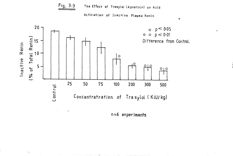
I The in vitro Effect of Trasylol on Acid Activation
Discussion
II The Effect of Trasylol (in vivo) on Plasma- Renin in the Rabbit Discussion
III Tlie in vitro Effect of Trasylol on Renin Release
IV The Effect of Trasylol on Acid Activation of Inactive Renin from
CHAPISa 4 THE ROLE OF CALCrOL IK REGULATION OF REKIK SECRETIOII
INTRODUCTION 156
I The effect of calcium ion
concentra-tion on renin release in vitro 158
Discussion 'l64
II The effect of potassium ion
concen-tration on renin release in vitro l64
Discussion I75
III The effect of sodium in
concen-tration on renin release in .vitro 174
Discussion . 184
IV The effect of magnesium ion
concen-tration on renin release in vitro <186
Discussion I96
V The effect of lithium ions on
renin release in vitro I9Y
Discussion 202
GENERAL DISCUSSION 20)
CHAPTER 5 TEE EFFECTS OF TWO CALCIUU ANTAGONISTS AND THE CALCIUM lOHOPHORE ON RENIN RELEASE D'T VITRO
n^TRGDUCTION 208
Materials and Methods 209
I The effects of verapamil and flunarizine on renin secretion in the presence and absence of
calcium 210
Discussion 220
II The effects of the calcium iono-phore A23187 on renin secretion in the presence and absence of
calcium 222
Discussion 232
CONCLUSIONS 236,
Fig.No. Title
1:1 The renin-angiotensin system 2
1:2 The juxtaglomerular apparatus 14
1:5 The enzymatic conversion of angiotensinogen 36 1:4 The relationship between plasma Isllikrein
and clotting mechanisms 46
2:1 Estimation of active and inactive renin
concentrations 60
2:2 Antibody dilution curves 63
2:5 The standard curve 65
2:4 Angiotensin I production vs time 68 2:5 The effect of renin concentration on
angiotensin I production 7O
2:6 The effect of substrate concentration
angiotensin I production 75
2:7 The effect of pH on plasma renin 75
2:8 The standard curve for methanol 89
2:9 The effect of kallikrein concentration
on methanol production $0
2:10 Methanol.production vs time 92
2:11 The effect of TAJ/Ie concentration on
methanol production 95
2:12 Assay of kallikreins in plasma 98 2:15 Production of p-nitroaniline ivith time 100 2:14 Effect of Chromoz^m PK dilution on
absorbance at 405nm 102
2:15 Experimental protocol for in vitro
investigations 108
5:1 Proposed mechanism whereby acidification of plasma leads to activation of inactive
renin 113
5:2 The effect of Trasylol during dialysis
of plasma 121
5:5 The effect of Trasylol on acid activation
of inactive plasma- renin 125
Fig.No. _ Title parre
5:5 The effect of Trasylol on active plasna
renin I30
3:6 The effect of Trasylol on inactive renin
in vivo 131
3:7 The effect of Trasylol on plasna
kalli-krein > I35
3:8 The effect of Trasylol on urine flow I35 3:9 The effect of Trasylol on urinary
kalli-krein I36
3:10 The effect of Trasylol on electrolyte
excretion 139
3:11 The effect of Trasylol on renin release
in vitro 145
3:12 The effect of Trasylol on inactive renin
release in vitro 147
3:13 The effect of Trasylol during dialysis of
slice supernatant 150
3:14 The effect of Trasylol on inactive renin in
slice supernatant 151
4:1 The effect of calcium concentration on
renin release in vitro 'I6I
4:2 The effect of calcium concentration on
inactive renin release in vitro l62 4:3 The effect of potassium concentration on
renin release iia vitro I66
4:4 The effect of K^on inactive renin release 167 4:5 The effect of extracellular [ K"*" ] on renin
2
release in the presence and absence of Ca 169
4:6 The effect of extracellular [K*] on inactive 2 +
renin in the presence and absence of Ga *.172 4:7 The effect of changing 3 on renin f
release 176
4:8 The effect of changing ] on inactive
renin release ' 178
4:9 The effect of changing on active 2 +
renin in the presence and absence of Ca 182 4:10 The effect of changing on inactive
4:11 The effect of on renin release in
vitro (2'3ml,I Ca^"*") I9I 4:12 The effect of|i\ig^]on inactive renin
release in vitro (2-3inIvI C a ^ I92
r 2 + 1
4:13 The effect of [teg Jon active renin in 2 +
y the presence and absence of Ca I93
4:14 The effect of[Mg^^]on inactive renin in 2 +
the presence and absence of Ca I94 4:15 The effect of lithium ions on renin
release in vitro I99
4:16 The effect of lithium ions on inactive
renin release in vitro 201
5:1 The effects of calcium antagonists on
basal renin release in vitro 212
5:2 The effects of calcium antagonists on
renin release in response to low [Ca^*] 215 5:3 The effect of calcium antagonists on
2 + renin release in response to Ca
218
deprivation
5:4 The structure of A23187 222
5:5 The effect of the calcium ionophore on
basal renin release in vitro 226
5:6 The effect of the calcium ionophore on
r 2 1
renin release in response to low I Ca J 229 5:7 The effect of the calcium ionophore on
2 + renin release in response to Ca
LIST OF T.ABLES
Table No. Title Page
1:1 Comparison between plasma and glandular
kallikreins 48
2:1 Antibody dilution curves 62
2:2 The standard curve for angiotensin I 64 2:3 Angiotensin I production during
incuba-tion of rabbit renin with sheep renin
substrate 69
2:4 The effect of renin concentration on
angio-tensin I production during the incubation 7I
2:5 The effect of substrate concentration on
angiotensin I production 72
2:6 The effect of pH on plasma renin
concen-tration 76
2:7 The effect of dialysis on recovery of renin 77
2:8 The standard curve for methanol 88
2:9 The effect of kallikrein dilution on methanol
production $1
2:10 Methanol production during incubation of
of kallikrein with TAMe 95
2:11 The effect of substrate concentration on
methanol production 94
2:12 Hydrolysis of TAMe by protease enzymes 96
2:13 Change in absorbance at 405nm 99
2:14 The effect of substrate dilution on
absorbance at 4 0 5 ™ 101
2:15 Hydrolysis of Chromozym PK by protease
enzymes IO3
2:16 Composition of I[rebs'-Ringer bicarbonate 110 3:1 The effect of Trasylol on active and total
plasma renin concentration 120
3:2 The effect of Trasylol on inactive renin measured after acidification of rabbit
plasma 122
3:3 Plasma active and total renin in 8 control
3:4 Plasma active and total renin in 10
experimental animals given i.v. Trasylol 128 3:5 Inactive renin in control and
experi-mental animals 132
3:6 Plasma kallikrein activity in Group 1
and Group 2 rabbits 132>
3:7 Urine flow in Group 1 and Group 2 rabbits 134 3:8 The effect of intravenous Trasylol on the
excretion of kallikrein in urine I34 3:9 The effect of Trasylol administration on
sodium and potassium excretion I38 3:10 ^^^I-Trasylol in various tissues in mice
30 minutes after intravenous injection I40 3:11 The effect of Trasylol on renin release
from rabbit kidney cortex slices 144 3:12 The effect of Trasylol on inactive renin
released by rabbit kidney cortex slice I46 3:13 The effect of Trasylol on renin
concentra-tion in pooled kidney slice supernatants 149 3:14 Inactive renin in pooled kidney cortex
slice supernatants: effect of Trasylol 149 4:1 The effect of calcium concentration on
renin release in vitro •I60
4:2 Changes in renin secretion in response to
calcium I60
4:3 The effect of potassium concentration on 2 +
renin release in vitro (Ca present) I68 4:4 Changes in renin secretion in response to
potassium (Ca^^ present) 168
4:5 The effect of potassium concentration on
release in calcium-depleted media I69 4:6 Changes in renin release in vitro in
response to potassium (Ca^*-deprived) I7I 4:7 The effect of sodium concentration on
renin release in vitro (Ca^"*" present) 177 4:8 Changes in renin secretion in response to
Table Ko. Title Pa_£e
4:9 The effect of changing jjla renin 2 +
release in Ca -deprived media ]_qo 4:10 Changes in renin secretion in response
to changing Qla J (Ca^ -deprived) igi 4:11 The effect of magnesium concentration on
renin release in vi-jro (Ca^* present) iss 4:12 The response of renin release in vitro
to changes in [ ] (Ca^* present) 135 4:15 The effect of [l.ig^"^ 1 on renin release
2 +
in Ca .-deprived media . I90
4:14 The effect of magnesium ions on the response
to calcium deprivation I95
4:15 The effect of lithium ions on renin
release in vitro I98
4:16 The response of renin secretion in vitro
to lithium., ions 200
2 +
5:1 1 The effect of Ca -antagonists on basal
5:2 J renin release 211
5:5 The effect of calcium antagonists on renin 2 +
release in Ca -free media 214
5:4 The effect of Ca-antagonists on the response . of renin release to low extracellular
concentrations of calcium 216
5:5 -The effect of calcium antagonists on renin
release - during Ca-depletion 217
5:6 The effect of calcium antagonists on the
response of renin release to Ca depletion 219 5:7 The effect of the calcium ionophore on
basal renin release <225
5:8 Basal renin release in the presence of A25187 225 5:9 The effect of A23187 on renin release
(Ca^^-free media) 227
5:10 The effect of A 25187 on the response to
low extracellular [ Ca^"*" ] 1228 5:11 The effect of A23187 on renin release in
Ca-depleted media ^31
5:12 The effect of the calcium ionophore on
PHYSIOLOGY AND PHARMACOLOGY Doctor of Philosophy
ACTIVATiai AlID SECRETION OF INACTIVE EEL^TO by Laura Mary Ginesi
The renin-angiotensin system is important in the regulation of extra-cellular fluid volume and the maintenance of arterial blood pressure. The control of this system is complex, but a major determinant of its activity is the secretion of renin by granular cells in the kidney cortex. In recent years, an inactive, but activatable form of this enzyme has been identified in the kidneys of most species; and it is thought to be secreted into plasma. A potential site for control of the renin-angiotensin system could involve differential secretion of these two forms of renin, and this may involve a sodium-sensitive mechanism.
A recent hypothesis suggests that a kallikrein could be the physio-logical activator of inactive renin. In accordance with this pro-posal, the serine protease inhibitor, Trasylol, vras found to inhibit acid-activation of inactive renin in rabbit plasma. However, in ure-tha-ne-anaesthetised rabbits, the inhibition caused no immediate change in circulating levels of either forrPi of renin. In contrast, both forms of renin were reduced after a delay which appeared to be related to the accumulation of Trasylol within renal tissue. Fur-thermore ^ high concentrations of the inhibitor reduced the release of both forms of renin by rabbit kidney cortex slices in vitro.
It is concluded that plasma kallilcrein is not the primary factor which determines the relative amounts of circulating active and in-active renin.
Calcium is important for stimulus-secretion coupling in gland _ and muscle cells, where it it thought to play a stimulatory role. In contrast, recent work has shown that active renin secretion by the kidney is inhibited by calcium. Studies using the rabbit kidney cor-tex slice preparation confirmed these findings, and demonstrated that the secretion of inactive renin was also reduced by calcium. The effects of Ka.. K and llg ions on active and inactive reriin secretion in vitro appeared to depend on the presence of external calcium. However, studies vdth the two calcium antagonist drugs, verapamil and
flunarizine, and the calcium ionophore A 23187, indicated that renin secretion may depend not only on a change in the intracellular con-centration of calcium, but also on consequent changes in intracell-ular sodium.
GZTEHAL niTRODUGTIOH
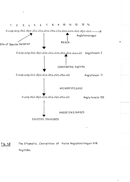
The proteolytic enzyme renin plays an important part in the maintenance of arterial blood pressure and extracellular fluid volume. It initiates the sequence of reactions which leads to the production of the physiologically active peptides, angiotensins II and III (See Fig. l-l).
Renin is mainly synthesised and stored in modified smooth muscle cells (granular cells) in the wall of the afferent arteriole as it enters the gloraerulus of the kidney. After it is released into the circulation, renin acts on its endogenous substrate, angiotensin_ ogen, to liberate the decapeptide angiotensin I. Converting enzyme (Kininase II), which has been identified in the vascular endothelium of many tissues, removes a His^^ - Leu^^ fragment from the carboxj' terminal portion of angiotensin I to relea.se angiotensin II.
By means of its potent pressor activity, angiotensin II parti-cipates in the regulation of systemic arterial blood pressure. The peptide also promotes the conservation of salt and water, both
directly and indirectly through its action in the adrenal cortex. It is also a potent dipsogen. Aminopeptidases in plasma produce angio-tensin III, the des-l-aspartyl heptapeptide, which has recently been shown to have biological activity. Further proteolysis of angiotensin is thought to produce only inactive fragments.
The control of renin secretion from the kidney is complex, and involves changes in arterial blood pressure, sodium transport,
sympa-thetic nerves and a variety of humoral factors. A new site for control of the renin-angiotensin system may involve an,inactive form of renin which has been shown to exist in kidney and plasma.. Acid, cold storage and protease enzymes have all been shown to activate these forms of renin in vitro. A recent hypothesis suggests that the serine protease enzyme kallikrein may take part in the physiological activation of inactive renin. This possibility lia-s been investigated in the rabbit in the experiments w M c h will be described in Chapter 5 of this thesis, using a commercially-ava.ila.ble protease inhibitor
knoT/n as Trasylol (aprotinin, Bayer).
Acfi v a f o r
Prostaglandins
Inhibffor
/ KPhospholipid
Pr einhibifor
Binding Protein?
/
RENIN
Prorenin?
ANGIOTENSIN
Converting
' Enzyme
ANGIOTENSIN
f
-(Ammopeptidase)
•
ANGIOTENSIN I I I
(Angiotensinase A)
contrast to*their stimulatory role in other secretory tissues, calcium ions inhibit the release of active renin from the kidney. Since differential release of inactive renin from Juxtaglomerular cells could be a new site for control of the renin-angiotensin system, the effect of changing the concentration of calcium and other ions in the incubation medium on the release of both active and inactive renin has been investigated using rabbit kidney cortex slices.
Stiin->
RENIN (EG 1:4.99.19)
Renin is a highly specific proteolytic enzyme which is synthes-ised, stored and secreted by the granular cells of the juxtagloner-ular apparatus of the 1-d.dney. Although it has no physiological ^ activity of its own, renin cleaves the Leu^- Leu^^ bond of both natural and synthetic forns of its substrate to liberate angiotensin I (Ang l). Its catalytic activity is not affected by heavy metal ions dimercaprol, EDTA or by diisopropyl fluorophosphate (SP?) which shows that disulphide bridges are not an essential part of its active site, and it is not a metalloenzyme.(Lee,1969; Skeggs et al,1977) Benin binds to concanavalin A (Printz & Dworachack,1977) and therefore is a glycoprotein.
Although it was first discovered in 1898 (Tigerstedt & Bergman, 1898), renin has only recently been completely purified. Eaas et al. (1955a) achieved a 56,000-fold purification of renin by salt and solvent precipitation. This preparation had a specific activity of 780 GU/ mg protein. Before 1977, many groups had tried to improve the purification of renin, but were unable to prepare renin with signifi-cantly greater specific activity. This can be attributed to the very low concentration of renin in the kidney, to confusion with other enzymes which have similar properties, and to the instability of renin which appeared to increase as purification progressed. How-ever, the development of affinity chromatography and the use of protease inhibitors have now allowed complete purification of hog
(inagami & Murakami, 1977^; Gorvol et a-1.1977). rat (iviatoba, Mura-kami & Inagami,1977), dog (Dzau et al,1979) and human (Yokosawa et al, I98O) renins.
Human renin appears to have a number of properties which dis-tinguish it from'kidney renins of other species (Yokosawa et al, 1980) The molecular weights of pure pig, rat and dog renins are in the range 35-37,000 daltons while human kidney renin has molecular weight of 40,000. It also has a higher isoelectric point (pH 5*7) than renin from other species (pH 4'5-5'4). In addition, antibodies raised against human renin had negligible cross-reactivity with other prepa-rations of renin, while other renins appear to have similar antigenic properties.
of renin which are present in the kidneys and plasma of most species (see page 8 ) .
EM.JIN SIBSTRATE.
The presence of a substrate for renin in plasma was first re-ported by Plentl, Page & Davis (1943) who found that "renin activator" was a component of the^^C^- globulin fraction of plasma. In recent years, purification and partial characterisation of angiotensinogen has been achieved. Tewksbury et al (1977) prepared homogeneous
human renin substrate and later (Tewksbury et al. 1978) reported that it wa.s a glycoprotein with molecular weight 56,800 dsltons and carbo-hydrate content of 14 %. Eokubu et al (1980) found that electropho-retically pure human angiotensinogen had a carbohydrate content of 15 per cent and molecular weight of 60,900. Skeggs et al (1957) isolated and sequenced a 14 amino acid polypeptide chain which functioned as a substrate for renin (see Pig.l.j) to release angiotensin I and a 4 amino acid fragment. The tetradecapeptide substrate is linked to the remainder of the angiotensinogen molecule by an ester linkage
(lee &Mil8on, 1971).
The existence of several forms of renin substrate in plasma was first suggested by Skeggs et al in I963 and has been confirmed by other groups. Printz et al (1977) found 4 major and 2 minor peaks of substrate activity in human plasma by isoelectric focussing and Lentz et al (1978) resolved 2 major peaks which accounted for
The functional significance of the various foriis of angioten-sinoeen in plasiiia. remains to be clarified. Eggena et al (1976) suggested that heterogeneity may account for the altered kinetics of the renin-substrate reaction which is observed in sone hyper-tensive states.
It is now known that angiotensinogen is synthesised in the
liver and disappears from the circulation after hepatectomy (Tateishi & Ifesson, 1972). Furthermore, renin substrate is^released by perfused livers (llasjletti & Lfesson .I971), liver slices (?reenan & Rostorfer, 1972) and by isolated liver cells (Weigard et al. I977). The rate of renin substrate release can be influenced by a variety of factors. Both glucocorticoids and oestrogens increase angiotensinogen levels in rats (Menard et al. 1973) dogs (Seid et al,1973) and humans (Eggena et al, 197$). The effect of oestrogens may be mediated by mechanisms that are controlled by hormone receptors since immature rats did not respond to ethinyl oestradiol, but gave an adult response to dexametlia-sone (Krakoff & Eisenfeld, 1977).
Haemodilution (Reid et al. 1974) and angiotensin II (Blair-Rest et al, 1974) increase renin substrate levels in dogs. Nephrectomy and ureteric ligation (Eiwada et al, 1976) and thyroidectomy (Bouhick et al, I98I) also increase its production. Kato et al (1979) found that furosemide increased angiotensinogen levels in hypertensive patients.
ANGIOTENSIN I CONVEKPING EKZYi^IB (EC 5.4.15.1)
In 1954> Skeggs et al found that two forms of the -hypertensive peptide hypertensin (angiotensin) were present after incubation of partially purified pig renin and horse angiotensinogen. They later
1971), kidney (Hall et al.l976). testes (Cushnan & Cheung, 1971) and parts of the brain and pituitary gland (Yang & Ileff, 1972; Poth et
al, 1975).
Converting enzyme is a membrane-bound glycoprotein with molec-ular weight of 129,000-480,000 daltons (Oshima, Gesce & Erdos, 1974; Das & Soffer, 197S). It is a metalloenzyme v/hich requires one molar equivalent of zinc for its catalytic activity. In I968, Bakhle
reported that conversion of angiotensin I by canine pulmonary particles Ti-as inhibited by a bradykinin-potentiating factor (EPP) which had
been extracted from the venom of a pit viper, BothroDs .jara-cara (Perreira, I96S). It is now clear that converting enzyme, which is also kno?ra as Kininase II, catalyses the sequential removal of
Phe-^.rg and Ser-Pro from the carboxy terminus of bra dykinin in addition to removing His-Leu from Ang I (Soffer, Reza & Caldwell,1974; Dorer et al, 1974). Thois the net physiological effect of increasing
converting enzyme activity is vasopressor since it inactivates brady-kinin and increases circulating levels of angiotensin II. By means of antibodies raised against pure preparations of pulmonary
con-verting enzyme, it has been established that the enzyme is located on the luminal surface of the pulmonary endothelium ( Ryan et al, 1975), vascular endothelium and proximal tubular cells (Caldwell, Seegal & Hsu, 1976; Soffer & Case, 1978).
8
INACTIVE RENIN
The earliest evidence that more than one f o m of renin existed
vias obtained by Haas et al (l953b) who found tliree intercon-vertible forms of the enzyme in a partially purified preparation of hog kidney renin. Skeggs et al (1967) reported that the specific activity of hog renin which had been prepared at pH 7*0 tos increased by exposure to acid. A similar increase in renin activity of human amniotic fluid after acidification was also observed by Lumbers (1971) who suggested that this effect could be due to activation of an
inactive form of renin. Since then, many groups of workers have confirmed that acid-activatable inactive renin is present in human amniotic fluid and plasma (Skinner et al, 1972; Day & Leutscher, 1974; Leckie & McConnell, 1975a; Derkx et al, 1976). Inactive renin is also present in plasma of the pig (Bailie et al, 1979; Boyd, 1974)» ' the rabbit (Leckie et al. 1973; Richards et el, 1979)> the dog
(Funakav/a, Funae & Yamamoto, 1978), the mouse (lleilsen, Llalling & Poulsen, 1979) and the sheep (Lush et al, I98O). Although Ilakane
et al (1979) found no evidence for inactive renin in the rat, other workers (Vandongen et al, 1977) reported that 15 % of rat renin was in an inactive form.
Renin activity in human plasma also increases when plasma is stored at -5°C for 4 days (Osmond, Ross & Scaiff, 1973; Sealey et al, 1976) but cryo-activation is not observed at 4°C (riseuh et al, 1978; Millar et al. I98O),
In recent years it has become apparent that both acid- and cryo-activation of inactive plasma renin depends on the presence of endo-genous serine protease enzymes. Indeed, a wide variety of proteolytic enzymes are powerful activators of inactive renin in vitro. Urinary kallikrein has been reported to be more potent than trypsin in activ-ating inactive renin in plasma (Sealey, Atlas & Laragh, 1978).
The role of serine proteases in activation of inactive renin is an interesting area for study and there is currently o great deal of interest in this subject. It will be discussed in greater detail in Chapter J of this thesis.
MULTIPLE MOLECULAR WEIGHT FORMS OF RH'TIN
The biochemical nature of inactive renin rer,tains to be clar-ified. Multiple forms of the enzyme have been identified and many of these appear to have low enzymatic activity. From a purified
preparation of porcine kidney renin, Inagami & Murakami (1977) isolated three forms of renin when acidification had been avoided during the purification. "Big renin" (molecular weight (MW) 61,000 daltons) had specific activity which was 21 % of that of fully active renin (MW 42,000). Two forms of "big big renin" (MV/ 140,000) were also found and had specific activities of 0«19 % and 0«05 % respectively when compared with the fully active form. Overturf et al (1979) have, also purified renin with molecular weight 62,000 from pig kidney, and Slater & Haber (1979) found a renin vd.th molecular weight of 58,000 in a purified preparation of human kidney renin. . In a very recent abstract, (Takii, Kurakami & Inagami,1981) complete purification of inactive renin from hog kidney was reported. A 1*2 million-fold
purification yielded a protein vdth a. molecular weight of 51,000 w M c h could be activated by treatment with trypsin or pepsin. The effect of acidification or cold exposure on the activity of this protein was not mentioned.
Other groups have found high molecular weight forms of renin by gel filtration of impure kidney extracts. Boyd (1972) found two forms of pig renal renin with molecular weights of $8,000 and 60,000. He later (Boyd, 1975? Boyd, 1974) isolated a binding protein which was associated with the high molecular weight form and which was capable of converting the small, fast-acting form to the larger,
slow-acting renin. A similar binding protein (ifff 13,000) was isolated from rabbit kidney extracts (Leckie & McConnell,1975b). The inhibitor was
10
renin (60,000 daltons) which occurred when it was incubated with a 40,000 dalton form (Kawamura et al. 1979).
In contrast to these findings, Day & Leutscher (1974) reported that acid-activation of "big renin" (OT 60,000) from kidneys of
patients with renal carcinoma ivas not associated with a change in its molecular weight. Levine et al (1976) found a. high molecular weight form of renin (Lfiff 57,000-59,000X in purified>pig kidney extracts despite an acidification step during the preparation. Further acidi-fication did not result in increased renin activity. Similarly, a 65,000 dalton form of renin was present in an acid extract of dog kidney (potter et al.l978).
Thus there appears to be general agreement that a renin with mol-ecular weight of approximately 60,000 daltons is present in the kidney of many species. An even larger form {WI greater that 140,000) has also been reported in the pig ( Inagami & Kural-cami, 1977) and human (Barrett et al, 1977) kidney extracts. By gel filtration, the mol-ecular weight of pure, active renin is about 42,500 (inagami &
riurak&mi, 1977)* The relationship between changes in molecular weight and activation of the enzyme is far from clear.
High molecular weight forms of renin are also present ih plasma. However, there appears to be greater controversy over the molecular weight. Day & Leutscher (1975a) found varying proportions of "big renin" (MW 65,000) and normal renin (MW 45,000) in the plasma of patients with diabetic neuropathy and renal carcinoma, while normal plasma contained only the low molecular weight form. "Big renin" could be activated by acid and protease enzymes but this did not involve any change in molecular weight (Day & Leutscher, 1975b). Hseuh et al (1975) found both "big renin" (iJW 60,000) and normal renin (l.!W 40,000) in plasma of normal men taking a high salt diet. Both active and inactive renin were present in both peaks. Only the low molecular weight form was present in subjects taking a low salt diet. Recently, Nubuo et al 1981) reported that two high molecular weight forms
(ivIW 52,000 and 60,000) which could be activated by acidification were present in normal plasma in addition to a 40,000 dalton form of renin.
reported tha-t the nolecular rei^ht is approxiriately 55,000 daltons (Atlas et al. 1979, Leckie et el, 1977a; leckie et al, 1977b;
Yokosara et al, 1979). Thus, there is no general concensus as to the molecular weight of inactive plasns renin. Host of the reports
suggest that activation of plasma inactive renin, at least in humans, does not involve any change in its molecular weight (Day & Leutscher, 1975; Sggena et al, 1979; Hseuh et al, 1978; Schulkes et al, 1978). In the mouse, there are two high molecular weight forms of plasma renin {lITfl 70,000 and 800,000). Both forms are activated by acidi-fication, but this is only accompanied by a reduction in molecular weight in the larger form (iJeilsen, dialling & Poulsen, 1978).
The possibility that inactive renin is either a true zymogen of renin or an inhibitor-bound form of the enzyme, and the different physiological implications of these two hypotheses have recently been reviewed by Leckie (1981).
THE PHYSIOLOGICAL ROLE OF I M C T I V E RENIN
Until recently, relatively few studies concerning inactive renin had been published, and so its physiological role is rather unclear at present. Inactive renin in plasma increases during pregnancy (Skinner et al, 1975) and in patients with renal carcinoma (ley et al, '1975). Inactive renin forms a large proportion of total renin in the plasma of hypertensive patients (Sealey et al, 1977), of patients with diabetic neuropathy (Hseuh et al, I98O; Leckie et al, 1978) and in anephric patients ( Sealey et al, 1977)' This suggest that inactive renin could be a potentially important site for control- of the renin system.
12
and in rabbits (Grace et al,1979). Durinj furosenide treatment, inactive renin disappeared from the circulation of rabbits (Richards et al, I98I) and dogs (James & Hall, 1974)• These findings have led to the suggestion that a sodium-sensitive mechanism could control activation or release of inactive renin from the kidney (Richards _ejt al, I98I). This concept is supported by studies usinj kidney cortex slices. Reducing the concentration of sodium bathing the slices was found to increase active, but reduce inactive inactive renin secretion (Kunday, Koble & Richards, I98O).
Inactive renin has also been reported to increase while active renin is suppressed by propranolol in both hypertensive (Atlas ejb al, 1977) and normotensive (Derkx et al. 1976) subjects, and after clonidine treatment in hypertensive patients ( Atlas et al, 1977)• In contrast to the above studies, where there was a. degree of independence between the changes in active and inactive renin, the two forms increased in parallel during haemorrhage in dogs (James & Hall, 1974) a'nd rabbits (Richards et al , 1979)- In the pig, both forms of renin were increased by isoprenaline, furosemide, saline and propranolol (Bailie et al, 1979). Isoprenaline infusion into the rabbit caused an increase in both forms of renin (Richards et al - in press). However, the same authors found that in vitro, isoprenaline increased active renin secretion in a dose-related fashion, whilst inactive renin remained unchanged. An increase in both active and inactive renin vra,s observed in hypertensive patients during dietary sodium restriction (Atlas et al, 1977).
Despite the many reports where changes have been observed, other groups have failed to record any alteration in inactive renin during stimulation or suppression of the renin-angiotensin system. IIo change in circulating inactive renin vas observed after furosemide (Rumpf _et al, 1978)» saralasin infusion (Kapelgaard et al, 1978; Leckie et al. 1977b) or captopril administration (Millar et al, I98O),although active renin increased. Similarly, saline loading (Weinberger et al, 1977)» indomethacin (Rumpf et al. 1978) and propranol (Birkenhager et al, 1978) suppressed the renin-angiotensin system without a change in inactive renin.
related to differences in species and methodologies. Further
investigation of activation and release of inactive renin are clearly needed to clarify the role that it plays within the renin system.
THE GONTHOL OP R M I K SBGRETIQIJ.
Before the control mechanisms which are thought to regulate the release of renin can be discussed, it is necessary to describe briefly the structure of the juxtaglomerular apparatus (see Pig. 1.2")
The close anatomical relationship between the cells of the afferent
and efferent arterioles, the macula densa cells of the distal tubule and the juxtaglomerular granular cells is an important factor in renin secretion.
Granular cells are usually found in the media of the afferent arteriole and synthesise, store and secrete renin. These cells are differentiated vascular smooth muscle cells. They contain myofibrils, but are distinguished from typical smooth muscle cells in the afferent arteriole by the presence of numerous cytoplasmic processes and
infoldings of the plasma membrane (Gorgas, 1378). The endoplasmic reticulum and Golgi membranes are well-developed in granular cells, a characteristic of secretory function. The dense, membrane-bound granules of juxtaglomerular cells contain renin (Morris & Johnstone, 1976J Gross & Barajas, 1978).
In 1952, De Muylder identified sympathetic nerves which travelled to the afferent arterioles, and the presence of non—myelinated, norad«. renergic nerve fibres which terminate close to the granular cells has been confirmed in most species (Barajas, 1964; Biava & West, 1966; Nilson, 1965; Wagermark et al, 1968). These sympathetic fibres
terminate in varicosities which are separated from the cells by 1200-2000 A, with a basement membrane intervening between the two struct-ures (Bara.jas et al, 1977; 3ara jas & Huller, 1973)- Thus there is morphological evidence for a functional relationship between the
sympathetic nervous system and renin release. Cholinergic innerv-ation of granular cells is less well-defined. McKenna & Angelakos
(1968) found that the afferent arterioles of the dog receive cholinergic innervation from ganglion cells in the hilar region, riowever, the distribution of renal, nerves which contain acetylchol-inesterase appears to be similar to that of adrenergic fibres
14
( T a k e n f r o m D a v i s , 1 9 7 1 )
RENAL
INTERSTITIUM
GLOMERULUS
%
ARTERIO
RENAL
NERVES
Na Load x C
m
ifKNa ConcenfrafionA
MACULA DENSA
JUXTAGLOMERULAR
CELLS
AFFERENT
ARTERIOLE
The macula densa marks the transition between the ascending limb of the loop of "lenle and the distal convoluted tubule. The cells on the glomerular side of the tubule in this segment are heavily nucleated and lie in close contact with granular cells in
the vascular pole of the tubule of origin. Ilacula densa cells may be columnar or cuboidal depending on the species v;hich is studied. The Polkissen, or polar mass, consists of mostly agranular cells which lie in >-the angle which is formed between the afferent and efferent arteriole. These cells are in close contact with the macula densa and are continuous with cells from the arteriolar walls, where they may replace vascular smooth muscle cells.
During the past two decades, five basic mechanisms for the control of renin secretion have been generally
agreed;-1 ) An intrarenal baroreceptor.
2) A macula densa receptor which responds to changes in sodium (or chloride) load in tubular fluid.
3) Renal sympathetic nerves and circulating catecholamines.
4) Humoral factors including angiotensin II and prostaglandins.
5) Plasma electrolytes.
These control mechanisms and the pharmacological alteration,- of renin release have recently been thoroughly reviewed by Fray (198O) and by Eeeton & Campbell (198O), Earlier major reviews include those by Vander (I967)» Davis & Freeman (1976) and Peart (1978). These reviews, and many of the studies which will be discussed in the following section of this thesis, have been concerned only with the release of active renin. Where possible, studies which include inactive renin will also be included,
IHTRAHMAL BfiJaORDGEPTOR
16
in renal "bipod flow, but Kolff (1958) reported that renin was released under these circumstances whether flow v.-as pulsatile or non-pulsatile. The presence of an intrarenal "stretch" receptor ivas proposed by Tobian (1962) who suggested tliat renin release was
inversely proportional to the degree of stretch of renal afferent arteriolar walls. This, in turn, is determined by renal perfusion pressure.
Some of the most convincing evidence that an intrarenal vascular receptor exists and determines renin release from granular cells has been obtained by Blaine and his co-workers. This group developed the non-filtering dog kidney preparation (Blaine, Davis and Witty, 1970) to investigate the baroreceptor hypothesis and found that haemorrhage and constriction of the aorta above the renal arteries caused a two-fold increase in plasma renin activity (PHA) in conscious dogs even after the influence of the macula densa (see page 17 ), renal
sympathetic nerves and circulating catecholamines (see page 18 ) had been removed (Blaine, Davis & Prewitt, I97I). The intrarenal baroreceptor appeared to be located at the level of the afferent arterioles since papaverine, which dilates these vessels and there-fore prevents renal autoregulation, abolished the increase in renin secretion (Witty et al, 1971). However, it must be noted that papaverine, which is a phosphodiesterase inhibitor, itself reduces basal renin secretion from rat kidney slices (Churchill, lIcDonald & Churchill,1980).
The nature of the stimulus detected by the baroreceptor is not clear. According to Tobian's hypothesis, the degree of stretch of the afferent arterioles regulates renin secretion. However, hypo-tension-induced renin release is usually associated with renal vasodilatation and a reduction in renal vascular resistance (Cowley & Guyton, 1972; Eide et al, 1973; Gotshall et al, 1974; Schmidt et al, 1972; Skinner et al. I964). In a review, Davis & Freeman(197^) proposed that the intrarenal baroreceptor could respond to a change in wall tension of the afferent arteriole. According to Laplace's Law, wall tension is the product of the transmural pressure gradient
in elastic components of the vessel wall.
MACULA DEHSA. RECEPTOR
The " macula densa hypothesis " can be summarised as follows;-a decrefollows;-ase in tubulfollows;-ar concentrfollows;-ation of sodium in the region of the macula densa stimulates renin release from granular cells. It was first proposed by Vender in 196? who also reviewed the anatomical
>
basis for such a relationship. At first sight this theory appears logical in view of the fact that dietary sodium depletion is
associated with increased plasma renin activity in man (Brovm et al.
1963),
dog (Erubacher & Vender, 1968% Ilogil et al.I969)
rats(Keeton., Pettinger & Campbell, 1976) and rabbits (Grace et al. 1979). Early studies in w M c h " loop diuretics " were used to inves-tigate the macula densa hypothesis provided conflicting results. Ethacrynic acid increased both renin release and sodium excretion in anaesthetised dogs (Cooke et al, 1970; Birbari et al, 1972) even
when volume depletion v/as prevented. . lleyer et 31(1972) found that
furosemide produced a rapid release and sodium excretion in rabbits. Thus, it appeared that a direct relationship between tubular sodium excretion and renin secretion existed. However, in the light of more recent findings, these results can be reconciled with Yander's hypothesis since furosemide and ethacrynic acid themselves stimulate renin release (Bailie, Crosslan & Hooke, I97&; Bide et al. 1975; O s b o m , Hoolce-i!; Bailie, 1977)* These diuretics also appear to. inhibit sodium transport at the macula densa and therefore prevent the cells from sensing the increase in sodium load (Schnermann, Ploth
& Hermle, 1976).
18
Purosemide diuresis in pentobarMtone-anaesthetised, volume-deplete sheep maintained in an upright posture was accompanied by an increase in plasma active renin and concurrent decrease in the inactive form. These observations throw some doubt on the existence of a macula densa receptor at all.
In anaesthetised dogs, volume expansion with isotonic saline was found to increase the fractional excretion of sodium and suppress
renin release (Hash et al, I968) %hile infusion of hypertonic saline
reduced fractional sodium excretion and increased renin secretion. Further evidence in support of the macula densa hypothesis has been provided by Churchill and his co-workers. They studied early distal tubular fluid which had been obtained by micropuncture of cortical distal tubules of anaesthetised rats. They (Churchill, Churchill & LIcDonald, 1978) found that early distal tubular sodium load and not sodium concentration was directly related to dietary sodium intake. The rate of renin secretion was inversely related to sodium intake. More recently Churchill et al (1979) reported that both saline- and mannitol- induced diuresis increased sodium load in the distal tubule and reduced renin release. However, the concentration of sodium in tubular fluid was increased by saline and reduced by mannitol. They concluded therefore, that a change in sodium load, rather than its concentration regulates renin release .
Although much evidence suggests that renin release is inversely related to sodium at the macula densa, some workers maintain that the converse hypothesis is true (Thurau et al, 19^7; Thurau et al. 1972)» From studies using a microgmde injection technique, they conclude that an increase in sodium load as a result of increased glomerular filtration (GFR) stimulates renin release and that this system is involved in autoregulation of the blood flow to individual nephrons. This controversy remains to be clarified.
RENAL SYMPATHETIC NERVES
(1965) was the first to report the effect of electrical stimulation of the afferent arteriole and its surrounding sympathetic fibres. Renin release increased and GFR, RBF and sodium excretion were all reduced. Since the increase in renin secretion was prevented by mannitol diuresis, he proposed that the fall in sodium excretion after nerve stimulation had caused the increase in renin release. This interpretation was in accordance with the macula densa hypo-thesis.
Hoeffer et al (1972) also reported that stimulation of the renal artery and nerves of anaesthetized dogs increased plasma renin activity. However, this response was completely abolished by the adrenergic antagonist, propranolol. Pretreatment with phenoxy-benzamine,an • flT-adrenoreceptor antagonist, had no effect on the in-crease in renin release in response to renal nerve stimulation. The authors therefore proposed that renal nerve stimulation altered renin release by way of a fl -adrenergic receptor. Kerve stimulation-induced renin release was not antagonised by d propranolol, which has only 1^6 of the B-antagonistic effect of the 1 isomer, in anaesthetized dogs, (Taher et al, 1976) and cats, (Johns & Singer, 1974) while a racemic mixture of the two forms prevented the increased renin release, but not the vasoconstriction of renal vessels.
In most of the above studies, changes in renal sodium handling were not measured, and so renin release via the macula densa receptor could not be excluded. When renal nerves of a non-filtering kidney were stimulated in anaesthetized dogs pretreated with papaverine,
(Johnson, Ifevis & Witty, 1971) plasma renin activity still increased. Similarly, Taher et al (1976) stimulated the renal nerves of anaes-thetized dogs at a frequency which did not change EEF, G?R or sodium excretion, but produced a consistent rise in PRA. Thus, neurally-induced changes in renin release were confirmed as being mediated by afi-adrenoreceptor and were considered independent of the intra-renal baroreceptor and macula densa receptor.
20
and the dorsolateral pons (Richardson et al. 1974). ?hen it was reported, renal denervation and propranolol, but not phenoxyben-zamine abolished the increase in renin release (l5atcheff et al, 1977; Passo et al. 1971a, 1971b; Richardson et al. 1974; Zehr & Peigl, 1973).
Thus, plasma renin activity can be altered by neural activity in higher centres of the central nervous system via changes in the activity of renal sympathetic %-adrenergic nerve tracts.
CIRCULaTII^TG GATECHOL&JvilNES
The first evidence that suggested that catecholamines could be involved in regulating the secretion of renin was obtained by Vander (1965)> who reported that both adrenaline and noradrenaline increased renin activity in anaesthetized dogs when perfusion press-ure was held constant. Since GFR and sodium excretion were reduced, he concluaed that the renin response vjas mediated by activation of the macula densa. However, Nash et al
(I968)
found that noradren-aline still induced renin release after snoradren-aline infusion had restored sodium excretion to normal levels.In the non-filtering dog kidney preparation, pretreatment with papaverine abolished the increase in renin release after adrenaline, but not after noradrenaline (Johnson, Davis & Witty, 1971)• Since the response to both catecholamines vias accompanied by reduced renal blood flow, these workers concluded that adrenaline stimulated renin through activation of the intrarenal baroreceptor, while noradren-aline exerted a direct effect on juxtaglomerular cells.
excretion in anaesthetized dogs (Ueda et si. 1970). The (^-adren-ergic antagonist, dibenarnine, reduced tlie effect of noradrenaline on renal function, but did not affect the renin response. In contrast, propranolol antagonised noradrenaline-induced renin secretion. Thus, these results suggest tliat both adrenaline and noradrenaline increase renin release via a^-adrenoreceptor on juxtaglomerular cells.
Although some early results suggested that isoprenaline did not change renin secretion, (Ayers, Harris & Hefer, I969; Brjag, Page a McCubbin,
I966),
subsequent investigators have reported that iso-prenaline stimulates renin release when infused intravenously in anaesthetized dogs, (Assaykeen, Tanigara. & Allison, 1974; Chokslii, Yeh & Samet, 1972; Tanigawa., Allison & Assaykeen, 1972), isolated perfused rat kidneys, (VanDongen, Peart & Boyd, 1973; Viskoper et al. 1977)J aad when added to rat (Weinberger, Aoi & Henry, 1975)j and rabbit (Richards et al. In press) kidney cortex slices in vitro. Recently, Himori, Eayagawa & Ishimori (1979) tried to classify the ^-adrenoreceptor involved in isoprenaline-induced renin release. Since the -antagonist, atenolol, suppressed both the hypotension and increased renin release, which were induced by isoprenaline, while the -antagonist, IPS-399 reduced only the hypotensive res-ponse, these workers concluded that isoprenaline acted at a/fg" sdrenoreceptor on granular cells to stimulate renin secretion.22
Ein>!0R.1L FACTORS
A variety of blood-bone factors are known to influence renin secretion. Angiotensin II appears to suppress the release of renin and thus form part of a negative feedback loop. Intravenous infusion of ang II reduced plasma renin activity in anaesthetized dogs when renal perfusion pressure maintained by a suprarenal aortic clamp
(McDonald et al, 1975; Vander & Geelhoed, I965), and when the renin-angiotensin system had been stimulated by sodium depletion in dogs, (Bunag, Page & llcCubbin, I967) sheep, (Blair-West et al, 1971) aod human subjects ( De Champlain et al,
I966).
Furthermore, plasma renin activity increased in rabbits which had been immunized against ang II (stokes, Dates & Weber, 1975)Angiotensin II suppressed renin release in the isolated, per-fused rat kidney, (Eofbauer et al, 1974; Van Dongen, Peart & Boyd, 1974) and reduced renin release in vitro from kidney cortex slices of the rat (Capponi et al, 1977; I'lichelakis, 1971). In contrast,
Morris, Nixon & Johnston (I976) have reported that renin release by isolated glomeruli was unchanged by ang II. The inhibitory effect of ang II on renin release from rat renal cortex slices in vitro vjas abolished by the specific antagonist l-Sar-6-4.1a-ang II (Capponi £t al, 1977; Naftilan & Oparil, 1978). It now appears that the inhi-bitory response to ang II depends on the presence of calcium in the incubation medium, (Van Dongen & Peart, 1974) and is abolished by the Ca^"*-antagonist, verapamil (Park, Han & Fray,
I98I),
but not by its methoxy derivative, D6OO
(Churchill,I98O).
In recent years, the role of prostaglandins in the regulation of renin secretion has been the subject of numerous investigations. In 1971, Werning et al reported that plasma renin activity increased after PGB^ was injected into the aorta of anaesthetized dogs.
Arterial blood pressure fell, while heart rate, urinary volume and urinary excretion of sodium and potassium were increased. This group therefore concluded that PGE^ stimulated renin release through changes in renal sodium handling. However, when urinary losses were replaced, intrarenal infusion of PG£^ still increased renin activity
perfusion maintained by a clamp on the aorta, infusion of PGEg
increased ERA as well as increasing urine flov.', sodium excretion and renal blood flow (Yun et al, 1977)•
In order to eliminate the effects of changes in sodium handl-ing at the macula densa, the renal sympathetic nerves and endoren-ous prostejlandins, Gerber et al (1978) studied the response to intravenous infusion of PGE^ in anaesthetized dojs with a single, de-nervated non-filtering kidney. The animals were pretreated with
indomethacin and given an infusion of propranolol. Since both renal blood flow and renin release increased, this group suggested tliat the
effect of PGBg vras mediated by activation of the renal-baroreceptor or by a direct effect of on granular cells. In vitro PGE^ and PGE^ did not change renin secretion from rat (Corsini, Crosslan &
Bailie, 1974) or rabbit, (Weber et al, 197^ a & 1976 b) renal cortex slices. These results support the concept that the release of renin by PGE is due to the activation of the intrarenal baroreceptor.
In contrast to PGE^, the prostaglandin precursor arachidonic acid appears to stimulate renin release by a direct action on gran-ular cells, since it increases renin secretion from rabbit renal cortex slices in vitro, (Weber et al, 1976; Whorton et si, 1978) and from isolated rat glomeruli (Beierr/altes et al. 1980). These
effects are abolished by inhibitors of prostaglandin synthesis. When infused into the aorta of saline-loaded anaesthetized rats, ara-chidonic acid caused an increase in plasma, renin activity, (Weber et al. 1975) even though urine volume, sodium excretion and mean arter-ial blood pressure were unchanged. Similar findings have been repor-ted after intrarenal infusion of arachidonic acid in intact dogs,
(Bolger et al, 1976) and in dogs with a single, denervated, non-filtering kidney (Data et al, 1978).
It is not yet clear which prostaglandin is the renin-releasing metabolite of arachidonic acid, but prostacyclin (PGI^) is emerging as a possible candidate. Bolger et al (1978) reported that renal blood flow, urinary volume and sodium excretion increased together with renal venous PEA. after intrarenal infusion of PGI^ in anaes-thetized dogs. Gerber et al (1978) found that prostacyclin was more potent in stimulating renin release in anaesthetized dogs with a denervated, non-filtering kidney than were PGE^ or D^. PGI
24
workers have found that both basal and arachidonic acid-stimulated renin release were abolished by 9, ll-azoprosta-5, IJ-denoic acid. This compound inhibits the synthesis of prostaclyclin without
affect-ing PGEg or . In a recent paper, Jackson et al
(I98I)
foundthat an active metabolite of PGI^, 6-Jceto-PGE^ was about five times more potent than prostaclyclin in causing- renin secretion from
non-filtering, ^-blocked kidneys of anaesthetized dogs.
In contrast to other prostaglandins, PGE^^ inhibits renin se-cretion from rabbit renal cortex slices in vitro (ffeber et al. 1976).
Furthermore, PEA. is suppressed in women receiving PGFg^ intra
-amnio-tecally to induce abortion ( Pyhrquist, Soveri & Widholm, 1976). The enzyme PGE^-9-ketoreductase converts PGE^ to PGFg^. weber,
Larson & Sherer (1977) found that its activity was increased in rats taking a high salt diet, and suggested that activation of the enzyme could be involved in adjusting PEA during periods of altered sodium intake. However3 Lusman et al (1973) found no changes in the plasma concentrations of PGE^ and PGF^^ in normal subjects during changes in sodium intake. Similarly, the relative excretion of PGE^ and PGFg^ in urine was unchanged during alteration of dietary sodium intake
(Campbell et al.
I98I).
There have been no reports to date of theeffect of prostaglandins on the secretion of inactive forms of renin. In vitro separations would appear to be particularly appropriate for such an investigation.
Variations in the plasma concentration of antidiuretic hormone, (ABE) witliin the physiological range, also appear to be involved in the regulation of renin release. Vasopressin reduced PEA in
anaes-thetized dogs, (Shade et al, 1973) - even in the absence of a
functional macula densa, and in anaesthetized rats (Churchill,
Churchill & McDonald, 1973) • Exogenous ADK also suppressed PEA after it had been increased in dogs by dietary sodium depletion, (Johnson, ICinter & Beeuwkes, 1979> Tagawa et al, 1971) aortic constriction, (Bunag, Page & McCubbin,
I967)
or ureteral occlusion (Vander,I968).
Furthermore, plasma renin is elevated in rats with hereditary dia-betes insipidus, (Henderson et al. 1978) and is very low in human subjects with the syndrome of inappropriate secretion of AIH(Fichman, Iviichelakis & Horton, 1974). These patients have high •
levels of vasopressin.
renin release by isoprenaline (Konrads et al, 1976; Van Dongen, 1975) but did not itself change renin release. Similarly, vasopressin did not alter renin secretion from rat renal cortex slices in vitro (De Vito et al. I970). In human subjects, the reduction in PRA which isas observed after the administration of vasopressin could be due to expansion of the extracellular fluid volume, (Annat et al,
I976;
Hewsome & Bartter, I968) or to a direct intrarenal effect of AT)TT(Hesse & Nielsen, 1977). However, Goodwin, LedingiiamLaragh (1970) observed only slight changes in PHA. of human subjects after administration of vasopressin both in the presence and absence of fluid expansion. The physiological relevance of interactions between ADH and renin release mechanisms remains to be clearly demonstrated.
In summary, there are complex interactions between renin re-lease mechanisms and plasma or local tissue levels of other hormones, particularly ang II, prostaglandins and antidiuretic horraone. The evidence elicited from in vivo experiments, while clearly important to the overall picture, is often difficult to interpret. Secondary changes in salt and water homeostasis will themselves affect renin release mechanisms. Studies carried out in vivo often provide more direct evidence of the site of action of the hormones. They have, however, sometimes failed to provide consistent results from diff-erent laborotories. A possible source of such confusion could be the wa,y in Vc'hich samples have been processed for as say. . In retrospect, we can see that some studies were made of active renin only and others of total renin. Secretion control for inactive renin in this context" is almost entirely unexplored territory.
PLASM ELECTROLYTES
The effects of various ions on the intake of renin secretion have been studied extensively using in vitro techniques in order to eliminate the influence of the intrarenal baroreceptor, macula densa, sympathetic nervous system and circulating hormones.
26
concentration of sodium in the medium increased renin release from rat kidney cortex slices (Braverman et si, 1971;; Eamerson e t ^ , I97I; Lyons & Churchill, 1975a; Oelkers et si. 1970). Similarly, reducing the concentration of sodium suppressed renin release
(Capponi & Talloton, 1976). One ^roup of workers reported that when the osmolality of the medium was unchanged, increasing the concen-tration of sodium stimulated the release of renin (Blendstrup et al. 1975). However, $hey also found that this effect of sodium was reversed (prederiksen, Leyssac L Skinner, 1975) when the osmolal-ity increased. They concluded therefore that sodium elicited renin release by virtue of its osmotic effect on water movement rather than by a direct effect on granular cells. In contrast to these findings, Weinberger & Rosner ^1971) found no change in renin re-lease from rat kidney cortex slices when the concentration of sodium was increased, provided osmolality was constant. In contrast to the investigators described above, other studies have shown that increas-ing the concentration of sodium suppressed renin release from dog,
(I'lichelakis, 1971) and rabbit, (Richards et al,
I98O)
renal cortex slices, and from suspensions of rat renal cortical cells (Lyons & Churchill, 1975%)Unlike its stimulatory role in secre tory processes in many other tissues, (Rubin,I970) there is a growing body of evidence that calcium ions inhibit the secretion of renin. Studies with isolated, perfused kidneys show that hypercalcaemia inhibits renin release (iCotchen et al, 1974; Wat kins et al, 1976). Removal of calcium
from the medium, and complete depletion of calcium by the addition of chelating agents such as EDTA. and EGTA. are powerful stimulators of renin release (Fray, 1977; Fray & Park, 1979; Harada- & Rubin, 1978; Tan Dongen & Peart, 1974).
Calcium may play a central role in the regulation of renin secretion. The inhibitory effect of ang II requires the presence of calcium in the medium (Van Dongen 6 Peart, 1974). Similarly, low perfusion pressure stimulates renin release only when calcium is pre-sent in the medium, and becomes less effective as the concentration of calcium is raised (Fray & Park, 1979). It has also been reported
(Park & MaIvin, 1977) that renin release induced by adrenaline, and the suppression of its release by ouabain were abolished after cal-cium removal.
24
influx of Ca into cells have been described. Angiotensin II, ADH, ouabain and high concentrations of potassium ions all inhibited the release of renin by dog renal cortex slices (Park, Kan & Fray, 1981). All of these effects were abolished by the calcium antagonist, vera-pamil. The methoxy-derivative of verapamil, D oOO, abolished the inhibitory effect of potassium ions, (Churchill, 1980) but did not antagonise the effect of ang II, even at doses 2 - 6-fold greater than had been required to antagonise the effect of potassium.
Thus, according to these studies, an increase in the intra-cellular concentration of calcium, which is achieved by ang II, oua-bain, or depolarization by high extracellular concentrations of po-tassium, inhibits the secretion of renin. On the other hand, calcium chelators, isoprenaline or low extracellular concentrations of Ca reduce intracellular levels of calcium and appear to stimulate renin secretion.
The juxtaglomerular granular cells are modified smooth muscle cells. Normal smooth muscle contraction is associated with influx of Ca^* ions, and Peart (1978) drew analogies between situations which cause smooth muscle contraction, and control of renin release. A vasoconstrictive agent such as angiotensin is associated with in-creased influx of Ca into smooth muscle and inhibition of renin re-lease. Isoprenaline, a vasodilator , stimulates renin release. When our understanding of the intracellular control of secretion of renin eventually approaches the degree of sophistication currently
tissues
available for other secretory^such as the gastric mucosa and the pancreatic islet tissues, intracellular interactions between Ca ions and renin secretory mechanisms for the two forms of renin will probably prove crucially important.
1970)-28
During- this ^period, the plasma concentration of potassium fell, but plasma sodium was unchanged. Potassium repletion restored renin to control levels. Similar inverse relationships between potassium and plasma renin have been reported in conscious dogs, (McCaa, McCaa & Gayton, 1975; Young et al. 1976) conscious rats, (Sealey et al. 1970) and normal human subjects (Brunner et al. 1970; Hollenberg et al,
1975).
Miller and his co-workers (1975) studied the effects of po-tassium loading in adrenalectomized patients and in patients with primary adrenal insufficiency, all of whom were taking a fixed daily dose of mirieralooorticoids. Potassium loading was associated with negative sodium balance and increased plasma renin in these subjects. This was in contrast to the suppression of PRA which had been observed in normal subjects. T/hen sodium losses were replaced in Addisonian patients, potassium loading did not change plasma renin activity. It was concluded that the suppression of P M by potassium required high circulating levels of mineralocorticoids, which were absent in Addisonian patients.
Kotchen, Galla & Luke (l97o) studied the effects of oral in-gestion of iCCl and KECCy in conscious sodium-depleted rats. They found that KCl reduced plasma reninj JECO^ did not. They later
(Kotchen, Galla & Luke, 1978) reported tliat the chloride ion may itself inhibit renin release, and concluded that the anion which accompanies potassium may modify the signal that is delivered to the macula, densa.
linked action on granular cells and by altering the delivery of so-dium at the macula densa.
Intrarenal infusion of magnesium chloride in dors, at a level which caused a four-fold increment in the concentration of magnes-ium in plasma, resulted in an increase in plasma renin activity (Churchill & Lyons, I976). ilean arterial blood pressure was reduced and renal blood flow and glomerular filtration rate were unchanged. Since urine volume and sodium excretion increased, which would be expected to suppress renin release, it was concluded that magnesium influenced plasma- renin activity, either by means of renal sympa-thetic nerves or by acting directly on granular cells. Wilcox
(1978) investigated these possibilities by auto-transplanting a kidney to the neck of dogs. Intravenous infusions of MgClg in-creased the release of renin into plasma and inin-creased renal blood flow. GER, sodium excretion and systemic arterial pressure were unchanged. In addition, the stimulation of renin release by mag-nesium was antagonised by concurrent hypercalcaemia and saline in-fusion.
In isolated rat kidney, increasing the concentration of mag-nesium in the perfusate, stimulated renin release (Fray, 1977). This effect was suppressed by increasing the concentration of potass-ium , or by an increase in renal perfusion pressure (Ettienne & Fray, 1979)• Since magnesium did not cause a further increase in the rate of reain release after removal of calcium from the medium, (Fray, 1978) it vss concluded that magnesium acted directly on the granular cell, to hyperpolarize the cell membrane. It was suggested that this resulted in an increase in the rate of renin release by lowering the intracellular concentration of calcium.
Morimoto et al (1970) reported that reducing the concentration of magnesium caused a small fall in renin released by dog renal
cortex slices. A further reduction in renin release was observed when both calcium and magnesium were removed. In contrast to this, no significant change in the release of renin from isolated, super-fused glomeruli was observed when magnesium concentration was reduced
(Baumbach & Heyssac, 1977)• Furthermore, plasma renin activity in normal human subjects taking a low-sodium diet did not change
30
From these findings, it therefore appears that magnesium stim-ulates renin release, while low concentrations of magnesium inhibit its secretion. However, the mechanism by which it alters the rate of secretion from granular cells is unclear.
Lithium ions may also stimulate the rplease of renin. Lith-ium therapy is often used in the treatment of manic states, and such patients have been reported to have a concentrating defect and prim-ary polydipsia (^Forrest et al, 1974; Singer et al. 1972). Acute in-fusion of lithium causes natriuresis and diuresis (Cox & Singer, 1978; Vander, I968). Using toad bladder in vitro, lithium ions have been found to antagonise the hydro-osmotic effect of ADH and the increase in sodium transport that is induced by aldosterone (Cox & Singer, 1978; Singer & Pranko, 1973).