ISSN Online: 2158-2750 ISSN Print: 2158-2742
DOI: 10.4236/ajps.2017.89150 Aug. 18, 2017 2238 American Journal of Plant Sciences
In Vitro
Propagation and Recovery of Eight
Apple and Two Pear Cultivars Held in a
Germplasm Bank
Analí Lizárraga
1, Marga Fraga
2, Javier Ascasíbar
3, María Luz González
11Department of Plant Physiology, University of Santiago de Compostela, Santiago de Compostela, Spain 2Cultigar, Brión, Spain
3Centro de Investigaciones Agrarias de Mabegondo INGACAL-CIAM, Abegondo, Spain
Abstract
Eight apple (Malus x domestica) and two pear (Pyrus communis) traditional cultivars of commercial interest were selected from the germplasm bank CIAM (Centro de Investigaciones Agrarias de Mabegondo, NW of Spain) one of the most important collections of The Iberian Peninsula. In order to define the establishment protocol, apical buds 1.0 - 1.5 millimetre length of the se-lected cultivars were established in solid mineral medium Murashige and Skoog (MS) with modified vitamins (thiamine 10X) and supplemented with 1 mg∙L−1 6-benzyladenine (BA), 0.2 mg∙L−1 gibberellic acid (GA
3) and 0.3 mg∙L−1
of indole-3-butyric acid (IBA). The highest shoot initiation rates were ach- ieved with apple cultivar “Principe Grande” (55%) and “Barburiña” pear cul-tivar (50%); the lowest rate was obtained with apple culcul-tivar “Camoesa” (11%). Initiation rates for shoots culture varied between 22% and 37% for the other cultivars. The cytokinins BA, zeatin, 2-isopentyladenine (2iP) and thi-diazuron (TDZ) were tested at concentrations of 0, 0.25, 0.5 and 1 mg∙L−1 in
the absence or presence of IBA (0.1 mg∙L−1) for the multiplication step. For all
cultivars, the highest multiplication rates were obtained with medium sup-plemented with TDZ and also in media containing BA. The in vitro rooting success of shoots in medium with 0.1 mg∙L−1 IBA without cytokinins, and the
survival rate of plantlets acclimatized under greenhouse conditions depended on the genotype of the different cultivars. In vitro propagation of these ten traditional apple and pear cultivars in NW Spain has not previously been re-ported.
Keywords
Apple Shoots, Pear Shoots, Axillary Bud, Multiplication, Cytokinin, Gene Bank
How to cite this paper: Lizárraga, A., Fra- ga, M., Ascasíbar, J. and González, M.L. (2017) In Vitro Propagation and Recovery of Eight Apple and Two Pear Cultivars Held in a Germplasm Bank. American Jour- nal of Plant Sciences, 8, 2238-2254. https://doi.org/10.4236/ajps.2017.89150
Received: July 24, 2017 Accepted: August 15, 2017 Published: August 18, 2017
Copyright © 2017 by authors and Scientific Research Publishing Inc. This work is licensed under the Creative Commons Attribution International License (CC BY 4.0).
DOI: 10.4236/ajps.2017.89150 2239 American Journal of Plant Sciences
1. Introduction
Apple (Malus x domestica) and pear (Pyrus communis) trees, both of which be-long to the Rosaceae family, are very popular fruits in high demand because of their sweet taste and nutritional value. They are included among the best known and most highly consumed and economically important fruit trees in the world, along with citrus, Prunus fruits and table grapes. According to data published by FAO in 2013, an amount of 103,442,237 T of apples and pears were produced worldwide, making them two of the most widely produced fruits in the world [1]. Most of the traditional cultivars of fruit trees, which are held in germplasm banks, are of increasing interest both in Spain and worldwide. The conservation of these cultivars over centuries has remarkable advantages as the trees are high-ly adapted to the environmental conditions of the sites and their fruits have par-ticular organoleptic characteristics; they also represent a potential genetic re-serve for future improvement plans, as well as being an irreplaceable cultural heritage resource. Germplasm banks play an important role in the conservation of fruit trees [2]. In Galicia (NW Spain), the Mabegondo Agricultural Research Center CIAM-INGACAL germplasm bank contains one of the largest numbers of genetically characterized apple [3] and pear trees, and is one of the few germ- plasm banks in Spain dedicated to conserving traditional fruit in field collections
[4]. However, the phytosanitary status of these trees in the field, due to the tradi-tional propagation system, is disturbed and many of them are infected by viruses, endophytic bacterias and mycoplasmas. Following the guidelines of the Euro-pean and Plant Protection Organization [5], which forms part of the World Trade Organization, fruit trees must be free of the most harmful viruses before the fruits can be marketed. The diseases must be eradicated and the material must be cleaned for long-term preservation in germplasm banks [6]. As it is well-known, in vitro culture of apical meristems [7] together with heat therapy are valuable tools for the recovery and eradication of the main viruses present in almost all apple and pear cultivars [8]. To carry this out, first of all it is essential the establishment of in vitro culture for micropropagation [9], and secondly vi-rus removal [10] and recovery of different cultivars.
Specific protocols are required for establishment, multiplication and rooting
[11][12] of the different fruit cultivars in order to be able to sanitize them. In vitro micropropagation has been reported in several cultivars of apple [13] and pear trees propagated in vitro [14] [15] [16] [17]. Therefore, effective in vitro regeneration is a necessary precondition for the implementation of different biotechnological approaches in plant breeding.
cy-DOI: 10.4236/ajps.2017.89150 2240 American Journal of Plant Sciences tokinin applied. Thidiazuron and benzyladenine are the cytokinins most fre-quently used in regeneration systems for apple and pear trees [24][25][26][27] [28], but their efficiency depends on genotype and other factors [22]. Other cy-tokinins such as zeatin, 2iP and kinetin have also been tested in several studies and have generally been found to be less active [28]. The development of axillary buds as a multiplication method has been used in some fruit cultivars [29] as is one of the most effective methods of maintaining the genetic stability of cultivars. The rooting and acclimatization success depends on the genotype of each culti-var, the different growth regulators used and external factors [11].
The aim of this study has been the in vitro establishment of the different se-lected apple and pear trees cultivars for their multiplication and rooting in order to know their hormonal requirements for micropropagation and to acquire enough material for further investigation related with virus eradication.
2. Materials and Methods
2.1. Plant Material
Plant material of eight Galician traditional apple cultivars “Cacharela”, “Camoe-sa”, “Repinaldo”, “Tres en Cunca”, “Gravillán”, “Ollo Mouro”, “José Antonio” and “Príncipe Grande”, and two pear cultivars “Barburiña” and “Manteca Oscu-ra” were obtained from apical buds of genetically characterized dormant cuttings. Plant material belonging to the CIAM Germplasm Bank was collected in January 2012, 2013 and 2014.
2.2. Initiation of Aseptic Cultures, Media and Culture Conditions
Dormant cuttings were washed thoroughly under running tap water for twenty minutes and surface sterilized with 8% (w/v) Capluq-50 fungicide solution, be-fore being dried and wrapped in plastic film. The cuttings were then stored at 4˚C for one month. The cuttings were sprouted in a phytotron, at 25˚C day and 18˚C night under a photoperiod of 16 hours. The new shoots of length more than 2 cm were excised and the basal end was sealed with paraffin wax. Then were sterilized with 10% sodium hypochlorite solution plus one drop of Tween 20® for 15 minutes and were washed three times with sterile distilled water in order to remove traces of chlorine before establishment on the in vitro culture. All shoot tips (1.0 - 1.5 mm) were isolated and cultured for four weeks on basal MS medium with mineral salts [30], modified MS vitamins and 3% (w/v) su-crose, and solidified with 7% microagar (Duchefa) containing 1 mg∙L−1 BA; 0.2mg∙L−1 GA
3 and 0.3 mg∙L−1 IBA [31]. The pH of the medium was adjusted to 5.8
with 1N NaOH before autoclaving at 121˚C for 20 minutes. The medium was dispensed (25 mL) in 90 mm diameter sterile Petri dishes (Sterilin®) and was covered with sterile filter paper to prevent growth of endophytic bacteria. All cultures were transferred to a growth chamber at 25˚C ± 1˚C day and 18˚C ± 1˚C night with a light intensity of 40 µmol∙m−2∙s−1 illuminated by white
DOI: 10.4236/ajps.2017.89150 2241 American Journal of Plant Sciences
2.3. Shoot Multiplication and Rooting
The explants were cultured for four weeks before being transferred to tubes (22 × 150 mm) containing 20 mL of MS basal medium of the same composition as before but without gibberellic acid (GA3). After two subcultures in tubes, the
ex-plants were transferred to 350 mL jars (n = 9) for shoot multiplication, contain-ing each jar six explants (total n = 54) of a minimum length of 10 - 15 mm with 3 - 5 leaves per bud, in 50 mL of MS medium supplemented with different con-centrations (0, 0.25, 0.5 and 1 mg∙L−1) of four cytokinins (BA, Zea, 2iP or TDZ),
with or without auxin IBA (0.1 mg∙L−1). Shoot explants were subcultured onto
fresh medium of the same composition every 4 weeks. After sixth subculture in jars, healthy elongated apple and pear shoots of a minimum length of 15 - 20 mm and with at least two expanded leaves were selected for rooting. These shoots were excised from the explants and transferred to rooting medium con-sisting of MS basal medium supplemented with 0.1 mg∙L−1 of IBA, 30 g∙L−1 of
sucrose, and 7 g∙L−1 agar, or on MS 0 basal medium, without growth regulators,
for 28 days.
2.4. Transplantation and Acclimatization of Plantlets
After 4 weeks culture in rooting medium, the rooted shoots were washed in wa-ter to remove agar debris and placed in 40-well trays containing a mixture of 60% blonde peat substrate, 20% crust, 10% coconut fiber and 10% black peat. Each tray was placed in polystyrene boxes and covered with transparent plastic film to maintain high relative humidity and to allow entry of light. The trays were placed in a rooting chamber at a fixed temperature of 24˚C ± 2˚C with photoperiod of 16 light hours. After one month, the plantlets were transferred to greenhouse conditions, in a tunnel with a water spray fog irrigation system, mesh shading and warm bed to protect the shoots from low temperatures. The plantlets were held for one week under these conditions before the film was re-moved from each tray and the tunnel cover was opened slowly. Finally, the sur-viving rooted plantlets were transferred to individual pots containing substrate of the same composition and placed in a greenhouse, with shading mesh, con-trolled misting and environmental photoperiod. Percentage survival data were determined 4 weeks after transplanting the plantlets.
2.5. Statistical Analysis
The shoot multiplication developed with cytokinins (BA, Zea, 2iP and TDZ) at different concentrations (0.25, 0.5 and 1 mg∙L−1) with and without IBA (0.1
mg∙L−1), plus control in (MS 0) for each apple and pear tree cultivar data resulted
DOI: 10.4236/ajps.2017.89150 2242 American Journal of Plant Sciences
3. Results
3.1. Initiation of Aseptic Cultures and Establishment
The number of apical buds from axillary shoots available in each of the cultivars was variable due to the different size of the cuttings collected in the field. On the other hand, contamination by endophytic organisms, mainly bacteria, greatly affected the development of apex cultures. The use of sterile filter paper covering the plates, slows down the growth of endophytic bacteria, helping the explants development. However, endophytic bacterias proliferate faster on plates without filter paper. The phytosanitary status was therefore a determining factor for the success of the establishment and propagation of each cultivar affecting their de-velopment, and presence of bacterial colonies in the medium just where the bas-al end of shoot cultured was observed. Minerbas-al MS medium supplemented with 1 mg∙L−1 BA, 0.2 mg∙L−1 GA
3 and 0.3 mg∙L−1 IBA was used for establishment in
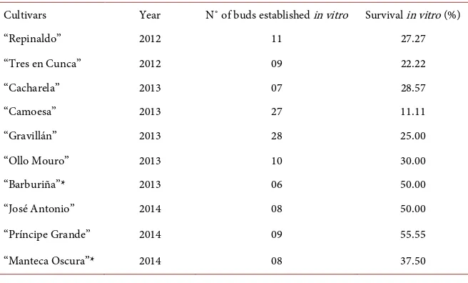
aseptic culture. The highest survival rate at establishment was obtained with the apple cultivar “Príncipe Grande” (55%), followed by the apple cultivar “José Antonio” and the pear cultivar “Barburiña” (both with a survival rate of 50%) (Table 1). The survival rates of the remaining apple and pear cultivars were be-tween 22 and 37%. The lower survival rate was in the apple cultivar “Camoesa” with only 11%.
3.2. Shoot Multiplication
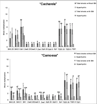
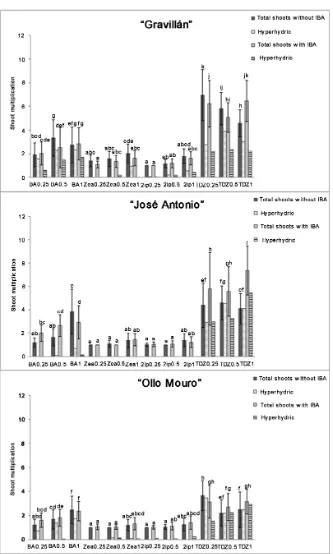
For all cultivars, the effects of different concentrations of cytokinins on in vitro axillary shoot development are presented in the following figures (Figures 1-4).
The best response to micropropagation of the eight apple and two pear culti-vars was always obtained in the presence high concentrations (0.5 and 1.0 mg∙L−1)
[image:5.595.210.538.530.728.2]of a cytokinin (TDZ or BA) in the medium. However, treatments for multiplica-tion of axillary shoots with all different concentramultiplica-tions of zeatin and 2iP, with or
Table 1. Establishment in vitro culture of eight apples and two pears cultivars. The culti-vars marked with *belong to pear trees.
Cultivars Year N˚ of buds established in vitro Survival in vitro (%)
“Repinaldo” 2012 11 27.27
“Tres en Cunca” 2012 09 22.22
“Cacharela” 2013 07 28.57
“Camoesa” 2013 27 11.11
“Gravillán” 2013 28 25.00
“Ollo Mouro” 2013 10 30.00
“Barburiña”* 2013 06 50.00
“José Antonio” 2014 08 50.00
“Príncipe Grande” 2014 09 55.55
DOI: 10.4236/ajps.2017.89150 2243 American Journal of Plant Sciences
Figure 1. Effects of BA, Zea, 2iP and TDZ at three concentrations (0.25, 0.5 and 1 mg∙L−1)
with and without IBA (0.1 mg∙L−1) on shoot multiplication after 4 weeks. Bars represent
the standard deviation (SD). Means denoted by different letters are significantly different (Tukey P = 0.05).
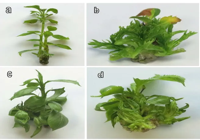
without IBA, yielded very low multiplication rates in all cultivars. The optimum concentration of cytokinin for shoot development varied depending on the cul-tivar. Although TDZ yielded the highest mean rates of shoot formation, at the highest concentrations (1.0 mg∙L−1) many shoots from different cultivars were
hyperhydric showing symptoms like thick stems and leaves, with anomalous leaf morphology and very short stems that made posterior subculture and rooting difficult (Figure 5).
3.3. Rooting and Plantlet Acclimatization
DOI: 10.4236/ajps.2017.89150 2244 American Journal of Plant Sciences
Figure 2. Effects of BA, Zea, 2iP and TDZ at three concentrations (0.25, 0.5 and 1 mg∙L−1)
with and without IBA (0.1 mg∙L−1) on shoot multiplication after 4 weeks. Bars represent
the standard deviation (SD). Means denoted by different letters are significantly different (Tukey P = 0.05).
DOI: 10.4236/ajps.2017.89150 2245 American Journal of Plant Sciences
Figure 3. Effects of BA, Zea, 2iP and TDZ at three concentrations (0.25, 0.5 and 1 mg∙L−1)
with and without IBA (0.1 mg∙L−1) on shoot multiplication after 4 weeks. Bars represent
the standard deviation (SD). Means denoted by different letters are significantly different (Tukey P = 0.05).
DOI: 10.4236/ajps.2017.89150 2246 American Journal of Plant Sciences
Figure 4. Effects of BA, Zea, 2iP and TDZ at three concentrations (0.25, 0.5 and 1 mg∙L−1)
with and without IBA (0.1 mg∙L−1) on shoot multiplication after 4 weeks. Bars represent
the standard deviation (SD). Means denoted by different letters are significantly different (Tukey P = 0.05).
“Manteca Oscura”). The apple cultivar “Tres en Cunca” did not root under any of the conditions studied. The survival rates of the plantlets acclimatized for four weeks were very variable (Table 2) being 100% in “Principe Grande” and values between 50% and 96.8% for the other cultivars of both apple and pear trees. The complete procedure for micropropagation is illustrated in Figure 6.
4. Discussion
A micropropagation protocol was developed from apices of apple and pear shoots cultured in vitro. The effects of several cytokinins and AIB auxin on mi-cropropagation were examined.
The common establishment medium for all apple and pear cultivars consisted of solid MS mineral supplemented with BA (1 mg∙L−1), GA
3 (0.2 mg∙L−1) and IBA
(0.3 mg∙L−1) [31]. The combined effect of BA and GA
3 for establishment of the in
DOI: 10.4236/ajps.2017.89150 2247 American Journal of Plant Sciences
Figure 5. Normal development of apple (a) and pear shoots (c) in media containing BA, and hyperhydric shoots obtained with a high concentration (1 mg∙L−1) of the cytokinin
TDZ in apple and pear shoots (b), (d) respectively.
Table 2. Effect of IBA on in vitro rooting and acclimatization of shoots of apple and pear cultivars, after 4 weeks in ex vitro conditions.
Cultivars Concentration IBA (mg∙L−1) Rooting (%) In vitro roots/shoot Mean n˚ of length (mm) Mean root Acclimatization in greenhouse (%)
“Cacharela” 0.1 0 13 0 1.14 ± 0.38 ab 0.00 ± 0.00 a 33.00 ± 6.63 0.00 ± 0.00 --- 81.25
“Camoesa” 0.1 0 91 59 3.78 ± 2.26 d 5.67 ± 2.46 f 38.31 ± 11.29 33.59 ± 8.06 69.38 96.87
“Gravillán” 0.1 0 65 11 2.97 ± 1.75 cd 2.00 ± 1.26 ab 47.31 ± 11.78 63.50 ± 10.85 57.14 83.33
“José Antonio” 0.1 0 59 39 2.76 ± 2.23 bc 3.53 ± 2.42 d 59.00 ± 25.21 82.43 ± 34.61 81.25 52.38
“Ollo Mouro” 0.1 0 0 4 0.00 ± 0.00 a 1.00 ± 0.00 a 85.66 ± 7.07 0.00 ± 0.00 --- 50.00
“Príncipe
Grande” 0.1 0 4 0 1.00 ± 0.00 a 0.00 ± 0.00 a 61.50 ± 13.43 0.00 ± 0.00 --- 100.00
“Repinaldo” 0.1 0 91 80 3.39 ± 1.56 d 4.12 ± 1.92 e 73.49 ± 33.55 48.86 ± 26.09 87.75 76.74
“Tres en
Cunca” 0.1 0 0 0 0.00 ± 0.00 a 0.00 ± 0.00 a 0.00 ± 0.00 0.00 ± 0.00 --- ---
“Barburiña”* 0.1 0 7 2 2.00 ± 0.82 ab 1.00 ± 0.00 a 90.25± 27.71 1.00 ± 0.00 75.00 0.00
“Manteca
Oscura”* 0.1 0 0 0 0.00 ± 0.00 a 1.00 ± 0.00 a 57.33 ± 6.80 0.00 ± 0.00 --- 66.66
[image:10.595.209.537.385.700.2]DOI: 10.4236/ajps.2017.89150 2248 American Journal of Plant Sciences
Figure 6. Complete procedure for micropropagation (a) apple shoot tips estab-lishment in vitro (b) (c) multiplication stage (d) in vitro shoot rooting (e) Trans-plantation of apple plantlets after for 4 weeks in rooting medium in pots under rooting chamber conditions at a temperature of 24˚C ± 2˚C and (f) acclimatiza-tion under greenhouse condiacclimatiza-tions after one month.
The presence of GA3 together with BA and IBA promotes growth of
estab-lished buds [26]; however, some authors indicate that GA3 inhibits the
develop-ment of apple shoots at the multiplication stage and even at the rooting stage
[34]. In the present study with the eight apple and two pear cultivars, MS me-dium supplemented with BA (1 mg∙L−1) GA
3 (0.2 mg∙L−1) and IBA (0.3 mg∙L−1)
proved suitable for establishing all selected cultivars. However, GA3 was
sup-pressed from multiplication stage media.
BA is one of the most commonly used and effective cytokinins for developing axillary buds in both apple and pear [35][36]. Other cytokinins such as Zea, 2iP and kinetin [37] are less commonly used, and the use of phenylurea derivatives such as thidiazuron and N-2-chloro-4-pyridyl)-N-phenylurea (CPPU) is some-times described on shoot proliferation in pear cultures [38]. However, theeffec-tiveness of any of these cytokinins in the multiplication medium will depend primarily on the genotype of the cultivar [28].
DOI: 10.4236/ajps.2017.89150 2249 American Journal of Plant Sciences the setting medium, avoiding, to a great extent, the rapid proliferation of endo-phytic bacteria in the establishment phase. This allowed the development of shoot tips by improving the survival rate of the in vitro starting material.
At the multiplication stage, the in vitro induction of axillary buds capable of forming shoots is widely used to propagate fruit trees. Furthermore, the risk of mutation in clones derived from axillary buds is much lower than the risk of mutation during organogenesis from callus.
Both BA and TDZ are the most frequently used cytokinins in apple regenera-tion and they have been compared in several studies [28]. TDZ has been found to be more effective than BA for shoot regeneration in numerous trials [25] [41] [42]. The optimal TDZ concentration largely depended on genotype. We found that growth media containing the highest level of TDZ (1 mg∙L−1) yielded the
highest number of shoots in only four of the selected cultivars “José Antonio” and “Tres en Cunca” apple cultivars, as well as the pear cultivars “Barburiña” and “Manteca Oscura”. For the other cultivars, the lowest concentrations of TDZ (0.25 and 0.5 mg∙L−1) were more suitable and favorable. However, at higher
concentration of TDZ, callus formation was observed and smaller shoots devel-oped. Although some authors have reported otherwise [43], we found that the concentration of TDZ affected shoot length in all cultivars, with visibly shorter shoots produced at the highest concentration of 1 mg∙L−1. Even though the
re-generated shoots did not hyperhydric in many apple and pear cultivars, visible differences were observed in the morphology of regenerated shoots: more com-pact shoots with short internodes and small rolled leaves with wavy edges.
Results from studies with Pyrus pyrifolia showed that the presence of hyper-hydricity in the explants was influenced by the type of cytokinin. In our study, more hyperhydric buds appeared when TDZ instead than BA or cytokinins de-rived from adenine were used, and the concentration had little influence [44], TDZ had a stronger effect on hyperhydricity, as also observed in Malus pumila
[42]. The results of the present study showed that high concentrations of TDZ or BA were more important in relation to the formation of hyperhydric shoots than whether the cytokinin was derived from phenylurea or adenine. However, high concentrations (1 mg∙L−1) of other cytokinins tested (2iP and zea) did not
pro-duce hyperhydricity, except in apple trees cultivar “Camoesa”, “Gravillán” and “Ollo Mouro” with an average between 0.02 and 0.96 hyperhydric shoots. This confirms that the type of cytokinin and optimal concentration for micropropa-gation of woody plants may depend on genotype [38].The medium of multipli-cation selected by the rate and quality of the buds that grew in them have been three: MS BA 0.25 mg∙L−1 IBA 0.1 mg∙L−1 for cultivars “Cacharela”, “Camoesa”,
“Gravillán” and “Ollo Mouro”; MS BA 0.5 mg∙L−1 IBA 0.1mg∙L−1 for “José
Anto-nio”, “Príncipe Grande”, “Repinaldo” and “Tres en Cunca” and MS BA 1 mg∙L−1
for two pears “Barburiña” and “Manteca Oscura”.
DOI: 10.4236/ajps.2017.89150 2250 American Journal of Plant Sciences concentration of IBA for maximum rooting differs between species and cultivars
[45][46]. We observed that the presence of IBA in the medium significantly in-creased the in vitro rooting of the shoots in all cultivars, with the exception of “Ollo Mouro” and “Manteca Oscura”, compared to auxin-free medium. Rooting rates were achieved in most of the studied cultivars on medium supplemented with IBA (0.1 mg∙L−1). Regenerated plantlets were successfully transferred to the
greenhouse for acclimatization. However, production of a limited number of plantlets has been achieved in some cultivars due to the low rate of in vitro rooting in MS0 medium. In addition, genotype strongly influenced the process of acclimatization to ex-vitro conditions, which is another factor limiting the success of micropropagation of these fruit trees cultivars.
The most responsive cytokinins for inducing axillary buds from apple and pear shoots were TDZ and BA, while Zea and 2iP were much less effective. TDZ was more effective than BA for producing buds in all apple and pear cultivars tested. In both pear cultivars and the apple cultivars “Jose Antonio” and “Tres en Cunca” better responses were obtained at the highest concentration (1 mg∙L−1) of
TDZ, while the other apple cultivars showed a better response with lower con-centrations (0.25 and 0.5 mg∙L−1) of TDZ. BA was the most effective cytokinin (at a
concentration of 1 mg∙L−1) for most of the apple cultivars tested (“Cacharela”,
“Camoesa”, “osé Antonio”, “Ollo Mouro”, “Principe Grande”, “Repinaldo” and “Tres en Cunca”) and for both pear cultivars, while in the apple cultivar “Gravillán” a better response was obtained with a lower concentration (0.5 mg∙L−1) of BA.
5. Conclusion
To conclude, BA and TDZ proved to be the most effective cytokinins for multip-lication of all apple and pear cultivars tested, varying the optimal concentration according to the genotype. But the use of TDZ was ruled out because of malfor-mations in leaves and stems in all cultivars, although with the three concentra-tions of TDZ higher rates of multiplication were obtained. The presence of IBA 0.1 mg∙L−1 in the rooting medium was effective only on five apple cultivars, but
did not have a positive response (less than 10%) on root formation in the re-maining apple and pear tree cultivars selected.
Acknowledgements
The authors are grateful to the CIAM germplasm bank of the Mabegondo Agri-cultural Research Center (INGACAL) for the agreement signed for the transfer of plant material from Malus and Pyrus to our laboratory at the University of Santiago de Compostela.
References
[1] FAO (2013) Fuente. http://faostat3.fao.org
Kul-DOI: 10.4236/ajps.2017.89150 2251 American Journal of Plant Sciences turpflanzen, 62, 9-16.
[3] Pereira-Lorenzo, S., Ramos-Cabrer, A.M., Ascasíbar-Errasti, J. and Piñeiro-Andión, J. (2003) Analysis of Apple Germplasm in Northwestern Spain. Journal of the American Society for Horticultural Science, 128, 67-84.
[4] Pereira-Lorenzo, S., Ramos-Cabrer, A.M. and Díaz-Hernández, M.B. (2007) Evalua-tion of Genetic Identity and VariaEvalua-tion of Local Apple Cultivars (Malus x domestica Borkh.) from Spain Using Microsatellite Markers. Genetic Resources Crop Evolu-tion, 54, 405-420. https://doi.org/10.1007/s10722-006-0003-7
[5] European Plant Protection Organization (1999) EPPO Standars PM 4/27(1) Certi-fication Schemes Malus, Pyrusand cydonia. European Plant Organization, Paris. [6] Panattoni, A., Luvisi, A. and Triolo, E. (2013) Review. Elimination of Viruses in
Plants: Twenty Years of Progress. Spanish Journal of Agriculture Research, 11, 173- 188. https://doi.org/10.5424/sjar/2013111-3201
[7] Manganaris, G.A., Economou, A.S., Boubourakas, I.N. and Katis, N.I. (2003) Eli-mination of PPV and PNRSV through Thermotherapy and Meristem-Tip Culture in Nectarine. Plant Cell Reports, 22, 195-200.
https://doi.org/10.1007/s00299-003-0681-y
[8] Paprstein, F., Sèdlak, J., Polak, J., Svobodova, L., Hassan, M. and Bryxiova, M. (2008) Results of in vitro Thermotherapy of Apple Cultivars. Plant Cell Tissue and Organ Culture, 94, 347-352. https://doi.org/10.1007/s11240-008-9342-8
[9] Deberegh, P.C. and Zimmerman, R.H. (2002) Micropropagation Technology and Application. Kluwer Academic Publishers, Boston.
[10] Sèdlak, J. and Paprstein, F. (2016) In Vitro Establishment and Proliferation of Apple Cultivars. Acta Horticulturae (ISHS), 1113, 107-112.
https://doi.org/10.17660/ActaHortic.2016.1113.15
[11] Xu, J., Wang, Y., Zhang, Y. and Chai, T.Y. (2008) Rapid in Vitro Multiplication and ex Vitro Rooting of Malus zumi (Matsumura) Rehd. Acta Physiologiae Plantarum, 30, 129-132. https://doi.org/10.1007/s11738-007-0075-9
[12] Muniz, A.W., Luiz de Sá, E., Dalagnol, G.L. and Filho, J.A. (2013) Rooting and Ac-climatization of Micropropagated Marubakai Do Apple Rootstock Using Adesmia-latifolia rhizobia. SpringerPlus, 2, 437. https://doi.org/10.1186/2193-1801-2-437
[13] Dobránszki, J. and Teixeira da Silva, J.A. (2010) Micropropagation of Apple—A Re-view. Biotechnology Advances, 28, 462-488.
https://doi.org/10.1016/j.biotechadv.2010.02.008
[14] Hirabayashi, T., Moriguchi, T., Kozaki, I., Yamamoto, Y. and Matsuzaki, S. (1987) In Vitro Propagation of Pyrus Shoots Tips. International Society for Horticultural Science VII International Symposium on Pear, 14, 9-16.
[15] Banno, K., Yoshida, K., Hayashi, S. and Tanabe, K. (1989) In Vitro Propagation of Japanese Pear Cultivars. Journal Japanese Society Horticultural Science, 58, 37-42.
https://doi.org/10.2503/jjshs.58.37
[16] Erig, A.C. and Fortes, G.R. (2002) In Vitro Establishment of Pear (Pyrus spp.) Start- ing from Meristems and Buds. Ciencia Rural, 32, 577-582.
https://doi.org/10.1590/S0103-84782002000400005
[17] Thakur, A. and Kanwar, J.S. (2008) Mircropropagation of “Wild Pear” Pyrus pyrifo-lia (Burm F) Nakai I Explant Establishment and Shoot Multiplication. Notulae Bo-tanicae Horti Agrobotanici Cluj-Napoca, 36, 103-108.
DOI: 10.4236/ajps.2017.89150 2252 American Journal of Plant Sciences 9-14. https://doi.org/10.1023/A:1006032707849
[19] Caboni, E., D’Angeli, S., Chiappetta, A., Innocenti, A.M., Van Onckelen, H. and Damiano, C. (2002) Adventitious Shoot Regeneration from Vegetative Shoot Apices in Pear and Putative Role of Cytokinin Accumulation in the Morphogenetic Process. Plant Cell Tissue and Organ Culture, 70, 199-206.
https://doi.org/10.1023/A:1016304106529
[20] Poudyal, B.K., Zhang, Y. and Du, G. (2008) Adventitious Shoot Regeneration from the Leaves of Some Pear Varieties (Pyrus spp.) Grown in Vitro. Frontiers of Agri-culture in China, 2, 82-92. https://doi.org/10.1007/s11703-008-0016-4
[21] Yepes, L.M. and Aldwinckle, S.H. (1994) Micropropagation of Thirteen Malus Cul-tivars and Rootstocks, and Effect of Antibiotics on Proliferation. Plant Grow Regu-lation, 15, 55-67. https://doi.org/10.1007/BF00024677
[22] Tang, H., Luo, Y. and Liu, C. (2008) Plant Regeneration from in Vitro Leaves of Four Commercial Pyrus Species. Plant, Soil and Environment, 54, 140-148.
[23] George, E.F. and Davies, W. (2008) Effects of the Physical Environment. In: George, E.F., Hall, M.A. and De Klerk, G.J., Eds., Plant Propagation by Tissue Culture, 3rd Edition, Netherlands Springer, Dordrecht, 423-464.
[24] Berardi, G., Infante, R. and Neri, D. (1993) Micropropagation of Pyrus calleryana DCN from Seedlings. Scientia Horticulturae, 53, 157-165.
https://doi.org/10.1016/0304-4238(93)90146-H
[25] Sarwar, M. and Skirvin, R.M. (1997) Effect of Thidiazuron and 6-Benzylaminopu- rine on Adventitious Shoot Regeneration from Leaves of Three Strains of ‘Mcin-tosh’ Apple (Malus x domestica Borkh.) in Vitro. Scientia Horticulturae, 68, 95-10.
https://doi.org/10.1016/S0304-4238(96)00971-5
[26] Shibli, R.A., Ajlouni, M.M., Jaradat, A., Aljanabi, S. and Shatnawi, M. (1997) Mi-cropropagation in Wild Pear (Pyrus syrica). Scientia Horticulturae, 68, 237-242.
https://doi.org/10.1016/S0304-4238(96)00972-7
[27] Dobránszki, J., Hudák, I., Magyar-Tábori, K., Jámbor-Benczúr, E., Galli, Z.S. and Kiss, E. (2004) Effects of Different Cytokinins on the Shoot Regeneration from Ap-ple Leaves of ‘Royal Gala’ and ‘M.26’. Horticultural Science, 10, 69-75.
[28] Magyar-Tábori, K., Dobránszki, J., Teixeira da Silva, A., Bulley, S.M. and Hudák, I. (2010) The Role of Cytokinins in Shoot Organogenesis in Apple. Plant Cell Tissue and Organ Culture, 101, 251-267. https://doi.org/10.1007/s11240-010-9696-6
[29] Kalinina, A. and Brown, D.C.W. (2007) Micropropagation of Ornamental Prunus spp. and GF305 Peach, a Prunus Viral Indicator. Plant Cell Reports, 26, 927-935.
https://doi.org/10.1007/s00299-007-0315-x
[30] Murashige, T. and Skoog, F. (1962) A Revised Medium for Rapid Growth and Bio-assay with Tobacco Tissues Cultures. Physiologiae Plantarum, 15, 473-497.
https://doi.org/10.1111/j.1399-3054.1962.tb08052.x
[31] Li, X., Xu, M. and Korban, S.S. (2002) DNA Methylation Profiles Differ between Field and in Vitro-Grown Leaves of Apple. Journal of Plant Physiology, 159, 1229- 1234. https://doi.org/10.1078/0176-1617-00899
[32] International Business Machines Corporation (2011) IBM SPSS Statistics for Win-dows, Version 20.0. IBM Corp., Armonk.
[33] Chakrabarty, D., Hahn, E.J., Yoon, Y.J. and Paek, K.Y. (2003) Micropropagation of Apple Rootstock M.9 EMLA Using Bioreactor. Journal of Horticultural Science and Biotechnology, 78, 605-609. https://doi.org/10.1080/14620316.2003.11511671
Prop-DOI: 10.4236/ajps.2017.89150 2253 American Journal of Plant Sciences agation Using a Double-Phase Culture System. HortScience, 26, 62-64.
[35] Wang, L.P., Wang, G.P., Hong, N., Tang, R.R. and Deng, X.Y. (2006) Effect of Thermotherapy on Elimination of Apple Stem Grooving Virus and Apple Chlorotic Leaf Spot Virus for in Vitro Cultured Pear Shoot Tips. HortScience, 41, 729-732. [36] Dalal, M.A., Das, B., Sharma, A.K., Mir, M.A. and Sounduri, A.S. (2006) In Vitro
Cloning of Apple (Malus domestica Borkh) Employing Forced Shoot Tip Cultures of M9 Rootstock. Indian Journal of Biotechnology, 5, 543-550.
[37] Moretti, C., Scozzoli, A., Pasini, D. and Paganelli, F. (1992) In Vitro Propagation of Pear Cultivars. Acta Horticulturae, 300, 115-118.
https://doi.org/10.17660/ActaHortic.1992.300.12
[38] Kadota, M. and Niimi, Y. (2003) Effects of Cytokinin Types and Their Concentra-tions on Shoot Proliferation and Hyperhydricity in in Vitro Pear Cultivar Shoots. Plant Cell Tissue and Organ Culture, 72, 261-265.
https://doi.org/10.1023/A:1022378511659
[39] Wang, M.R., Li, B.Q., Feng, C.H. and Wang, Q.C. (2016) Culture of Shoot Tips from Adventitious Shoots Can Eradicate Apple Stem Pitting Virus but Fails in Ap-ple Stem Grooving Virus. Plant Cell Tissue and Organ Culture, 125, 283-291.
https://doi.org/10.1007/s11240-016-0948-y
[40] Tan, R.R., Wang, L.P., Hong, N. and Wang, G.P. (2010) Enhanced Efficiency of Vi-rus Eradication Following Thermotherapy of Shoot-Tip Cultures of Pear. Plant Cell Tissue and Organ Culture, 101, 229-235. https://doi.org/10.1007/s11240-010-9681-0
[41] Fasolo, F., Zimmerman, R.H. and Fordham, I. (1989) Adventitious Shoots Forma-tion on Excised Leaves of in Vitro Grown Shoots of Apple Cultivars. Plant Cell Tis-sue and Organ Culture, 16, 75-87. https://doi.org/10.1007/BF00036516
[42] Pawlicki, N. and Welander, M. (1994) Adventitious Shoot Regeneration from Leaf Segments of in Vitro Cultured Shoots of the Apple Rootstock Jork 9. Horticultural Science, 69, 687-696. https://doi.org/10.1080/14620316.1994.11516501
[43] Hanke, V., Rohde, A. and Grafe, C. (1991) Studies on Regeneration from Somatic Tissue in Vitro. Adventitious Shoot Formation on Leaf Explants in Apple (Malus x domestica Borkh). Dresden-Pillnitz, 56, 214-220.
[44] Huetteman, C.A. and Preece, J.E. (1993) Thidiazuron: A Potent Cytokinin for Woody Plant Tissue Culture. Plant Cell Tissue and Organ Culture, 33, 105-119.
https://doi.org/10.1007/BF01983223
[45] Xiao, Z., Ji, N., Zhang, X., Zhang, Y., Wang, Y., Wu, T., Xu, X. and Han, Z. (2014) The Loss of Juvenility Elicits Adventitious Rooting Recalcitrance in Apple Roots-tocks. Plant Cell Tissue and Organ Culture, 119, 51-63.
https://doi.org/10.1007/s11240-014-0513-5
DOI: 10.4236/ajps.2017.89150 2254 American Journal of Plant Sciences
Abbreviations
BA: 6-Benzylaminopurine cv: Cultivar
GA3: Gibberellic Acid IBA: Indol-3-Butyric Acid
MS: Murashige and Skoog Medium PGRs: Plant Growth Regulators
TDZ: Thidiazuron (1-Phenyl-3-(1,2,3,-Thiadiazol-5-yl) Urea Zea: Zeatin
2-iP: N6-(2-Isopentyl) Adenine
Submit or recommend next manuscript to SCIRP and we will provide best service for you:
Accepting pre-submission inquiries through Email, Facebook, LinkedIn, Twitter, etc. A wide selection of journals (inclusive of 9 subjects, more than 200 journals)
Providing 24-hour high-quality service User-friendly online submission system Fair and swift peer-review system
Efficient typesetting and proofreading procedure
Display of the result of downloads and visits, as well as the number of cited articles Maximum dissemination of your research work