Sinead O’Donovan, Mark Kennedy, Blaithin Guinan, Shane O’Mara, Declan M. McLoughlin
PII: S0278-5846(11)00377-0
DOI: doi:10.1016/j.pnpbp.2011.12.012 Reference: PNP 8080
To appear in: Progress in Neuropsychopharmacology & Biological Psychiatry
Received date: 1 November 2011 Revised date: 19 December 2011 Accepted date: 25 December 2011
Please cite this article as: O’Donovan Sinead, Kennedy Mark, Guinan Blaithin, O’Mara Shane, McLoughlin Declan M., A comparison of brief pulse and ultrabrief pulse electro-convulsive stimulation on rodent brain and behaviour,Progress in Neuropsychopharmacol-ogy & Biological Psychiatry (2011), doi: 10.1016/j.pnpbp.2011.12.012
ACCEPTED MANUSCRIPT
1
A comparison of brief pulse and ultrabrief pulse electroconvulsive
stimulation on rodent brain and behaviour
Sinead O’Donovana,b, Mark Kennedya, Blaithin Guinana, Shane O’Maraa and Declan M. McLoughlina,b
aTrinity College Institute of Neuroscience, Trinity College Dublin, Dublin 2, Ireland
bDepartment of Psychiatry, Trinity College Dublin, St. Patrick’s University Hospital, James’s Street, Dublin 8, Ireland
Correspondence: Prof. Declan M. McLoughlin, Department of Psychiatry, Trinity College Dublin, St. Patrick’s University Hospital, James’s St., Dublin 8, Ireland
ACCEPTED MANUSCRIPT
2
Abstract
Brief pulse electroconvulsive therapy (BP ECT; pulse width 0.5-1.5 msec) is a very effective
treatment for severe depression but is associated with cognitive side-effects. It has been
proposed that ultrabrief pulse (UBP; pulse width 0.25-0.30 msec) ECT may be as effective as
BP ECT but have less cognitive effects because it is a more physiological form of neuronal
stimulation. To investigate this further, we treated normal rats with a 10 session course of
either BP (0.5 msec), UBP (0.3 msec), or sham electroconvulsive stimulation (ECS) and
measured antidepressant-related changes in dentate gyrus cell proliferation and
hippocampal BDNF protein levels as well as hippocampal-dependant spatial reference
memory using the water plus maze and immobility time on the forced swim test. Both BP
and UBP ECS induced very similar types of motor seizures. However, BP ECS but not UBP
ECS treatment led to a significant, near 3-fold, increase in cell proliferation (p=0.026) and
BDNF levels (p=0.01). In the forced swim test, only BP ECS treated animals had a
significantly lower immobility time (p=0.046). There was a trend for similarly reduced
hippocampal-dependent memory function in both BP and UBP groups but overall there was
not a significant difference between treatment and control animals when tested 10 days
after completing allocated treatment. These findings show that, even though both forms of
ECS elicited similar motor seizures, UBP ECS was less efficient than BP ECS in inducing
antidepressant-related molecular, cellular and behavioural changes.
Key words: pulse width, electroconvulsive therapy, electroconvulsive stimulation, depression, BDNF
ACCEPTED MANUSCRIPT
3
1. Introduction
Electroconvulsive therapy (ECT) is the most acutely effective treatment available for
severe, often treatment resistant, depression with remission rates of about 60% (Eranti et
al., 2007; Group, 2003; Kellner et al., 2010). However, its use is limited by concerns about
adverse effects on memory and executive function arising during treatment though these
mostly resolve within weeks of finishing a course of ECT (Semkovska et al., 2011; Semkovska
and McLoughlin, 2010). Since its introduction over 70 years ago, there have been several
modifications to ECT technique to reduce such cognitive side-effects while maintaining its
effectiveness. These have focussed mainly on electrode placement and electrical stimulus
parameters. While there seems to be little difference between bitemporal and bifrontal
electrode placement (Dunne and McLoughlin, 2011), right unilateral electrode placement is
associated with less adverse cognitive effects than bitemporal ECT but has less of an
anti-depressant effect (Sackeim et al., 1993). To improve effectiveness of unilateral ECT higher
stimulus doses above seizure threshold are required but this is associated with increasing
dose-related cognitive deficits (McCall et al., 2000; Semkovska et al., 2011). Recent studies
have confirmed that high-dose unilateral ECT is as effective as modestly suprathreshold
bitemporal ECT but it is not yet clear that it has substantially less cognitive adverse effects
(Kellner et al., 2010).
Changes in electrical stimulus parameters have improved the efficiency of neuronal
stimulation. The move from a sine-wave stimulus to brief pulse (i.e. 0.5 – 1.5 msec) square wave stimulus, requiring less energy, has reduced cognitive deficits but not compromised
clinical effectiveness (UK ECT Review Group, 2003; Semkovska and McLoughlin, 2010).
More recently there has been interest in using ultrabrief (i.e. 0.25 – 0.3 milliseconds) pulse stimulation as this is more physiological, reducing stimulation of neurones that are already
depolarising or in the refractory period (Sackeim, 2004). The first randomised trial reported
a high remission rate (77%) and few cognitive deficits following high-dose right unilateral
ultrabrief pulse ECT compared to brief pulse ECT (Sackeim et al., 2008). However, this high
remission rate has so far not been replicated in other randomised (6-44%; (Quante et al.,
2011; Sienaert et al., 2009)) and non-randomised studies (13-42%; (Loo et al., 2007; Loo et
ACCEPTED MANUSCRIPT
4
that a higher stimulus charge is required and the optimal stimulus parameters remain to be
established.
Electroconvulsive stimulation (ECS) is the animal model equivalent of ECT. Much has
been reported on molecular and cellular changes induced by courses of repeated brief-pulse
ECS in both normal rats and rat models for depression and that are believed to be relevant
to antidepressant mechanisms, e.g. up-regulation of neurotrophic factors and hippocampal
cell proliferation and neurogenesis (Gersner et al., 2010; Madsen et al., 2000; Newton et al.,
2003). In contrast, little has been reported on such effects following ultrabrief-pulse ECS.
Here we aimed to compare the effects of brief pulse and ultra-brief pulse ECS on normal
rodent brain and behaviour. We sought to determine if both forms of brain stimulation
would induce similar antidepressant-related changes in hippocampal cellular proliferation
and brain derived neurotrophic factor (BDNF) expression. Additionally, as secondary
measures, we sought to identify if there were any differences in antidepressant-related
behavioural changes and cognitive function that could be attributed to the difference in
stimulus pulse width.
2. Experimental procedures
2.1. Animals and treatment
Male Sprague-Dawley rats (Harlan, UK), weighing 150-200g at intake, were housed in
groups of four with food and water available ad libitum under 12 hour light-dark conditions. Animals were allowed to habituate for ten days prior to ECS. Experiments were conducted
in accordance with EU directive 2010/63/EU and guidelines of the Bioresources Ethics
Committee, Trinity College Dublin.
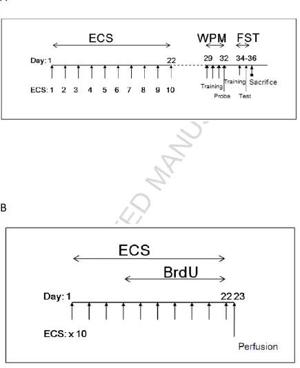
The experimental design is summarised in Figure 1A. Animals were randomly
allocated to a course of bilateral brief pulse (BP), ultrabrief pulse (UBP), or sham ECS. ECS
was delivered thrice weekly (Monday, Wednesday, Friday) for ten sessions via ear-clip
electrodes using the ECT Unit 57800 device (Ugo Basile, Italy). Parameters for BP ECS were:
0.5ms pulse-width; 100 pulses/second; 0.7s duration; 75mA current; while for UBP ECS the
pulse-width was reduced to 0.3ms. Sham animals were identically handled but received no
ACCEPTED MANUSCRIPT
5
sessions. The tonic phase of the seizure was defined as the period starting at the end of
stimulus administration during which an animal’s hind legs and forearms were held rigidly
against the body with some muscle twitching until the beginning of the clonic phase during
which the forearms and legs extended stiffly away from the body and began flexing
-extending rapidly until such motor activity was no longer apparent.
2.2. Behavioural studies
2.2.1. Water plus maze
Animals were habituated to the behavioural testing area for one week after
completing the allocated course of ECS. Hippocampal-dependent spatial reference memory
was assessed using a water plus maze (Diamond et al., 1999). The maze had four closed
arms in a plus formation with a central rectangular region and was filled with water (21±1°C)
to a depth of 45cm. A hidden platform, the same colour as the maze, was placed 2.5cm
below the level of the water and located at the distal end of one arm. Visual cues were
positioned on surrounding walls. Each animal was placed, facing away from the centre, into
in the distal end of a pre-selected entry arm on either the left or right of the hidden
platform arm for all trials on days 1-3 of testing and completed 10 such trials daily (60s/trial;
30s inter-trial intervals). On day 4, animals underwent 30 trials in which platform position
did not change but entry into arms was pseudo-randomised so that all animals started 10
trials in each of the other three different arms. Frequency of platform acquisition, as a
measure of reference memory, was calculated. Velocity (cm/s) for each treatment group
and platform acquisition on each experiment day was also measured. EthovisionXT (Noldus
Information Technology, The Netherlands) was used for data collection.
2.2.2. Forced swim test
The forced swim test, a measure of responsiveness to antidepressant treatments
(Porsolt et al., 1977), was conducted in two sessions on days 12-13 after completing the
course of ECS, consisting of a 15 minutes pre-test followed by a 5 minutes test 24 hours
later. A 50cm high, 25cm diameter, clear cylinder was filled 35cm from the base with water
ACCEPTED MANUSCRIPT
6
required to stay afloat. Animals were sacrificed 24 hours after the swim test and the
hippocampus was immediately dissected, snap-frozen in liquid nitrogen and stored at -80oC.
2.3. BDNF expression
Hippocampal BDNF protein levels were measured using a sandwich ELISA kit
according to the manufacturer’s instructions (Chemikine, Chemicon (Millipore), USA). Briefly, hippocampal samples were homogenised in the recommended lysis buffer (10%
w/v). Homogenates were centrifuged at 14,000g for thirty minutes and the supernatant was
collected for use in the BDNF assay. The amount of BDNF in each sample was analysed in
duplicate and determined from the regression line generated by BDNF standards (range
7.8pg/ml to 500pg/ml). Total protein concentrations were measured by Bradford protein
assay. BDNF results are presented as pg BDNF per mg of total protein content in each
sample.
2.4. Immunohistochemistry
To label proliferating cells in the dentate gyrus, a second set of animals was similarly
treated with ECS and administered BrdU (40 mg/kg, i.p.; Sigma-Aldrich) 2-4 hours post ECS
on treatment days three to ten (see Figure 1B). Twenty-four hours after the final ECS,
animals were anaesthetised with urethane and transcardially perfused with 0.9% saline,
followed by 4% paraformaldehyde, essentially as previously described (Kesavapany et al.,
2002).
Hippocampal coronal sections (10m) were mounted on slides for staining. Unless
specified otherwise, all procedures took place at room temperature. Endogenous
peroxidase activity was blocked using 3% hydrogen peroxide solution. Slides were washed
for 10 minutes under running water, microwaved on full power for 13 minutes in 10mM
citric acid buffer (pH6.0), washed in TBS, incubated in avidin (Vector Laboratories) for 10
minutes, washed twice in TBS and then incubated in biotin (Vector Laboratories) for 10
minutes. Following two washes in TBS and incubation in 10% normal rabbit serum (Dako) for
5 minutes, sections were incubated with rat anti-BrdU (1:10,000; ab6326, Abcam) in TBS for
1 hour. After this, slides were washed thrice in TBS and incubated in biotinylated rabbit
ACCEPTED MANUSCRIPT
7
TBS washes, StrepAB/HRP complex (Vector Laboratories) was incubated on each slide for 30
minutes. Slides were washed once in TBS and twice in distilled water before a 15 minute
DAB (SigmaFAST DAB tablets) incubation to visualise the reaction product. Following a wash
with distilled water, sections were counterstained with Hematoxylin. Sections were imaged
using an Olympus DP72 at 20x magnification. Numbers of BrdU-positive cells in dentate
gyrus subgranular zone and granule cell layer were counted blind on coded slides from
every sixth section over a range of 24 sections per animal.
2.5. Statistical analyses
Data are presented as means ± SEM and were analysed using GraphPad Prism
version 5.0 (Graphpad Software, La Jolla California, USA). A repeated measures ANOVA was
used to compare seizure durations between groups. One-way ANOVAs were used to
compare ELISA data and probe day platform acquisition for the water plus maze with
Newman Keuls post-hoc tests as required. Repeated measures ANOVA, with Day (water
plus maze Training days 1, 2, 3) or Treatment (BP ECS, UBP ECS, sham ECS) as factors, was
used to analyse platform acquisition during training on the water plus maze with Bonferroni
post tests as required. Kruskal-Wallis test with Dunn’s multiple comparison post hoc tests was used to analyse BrdU-positive cell counts. Statistical significance was set at p<0.05.
3. Results
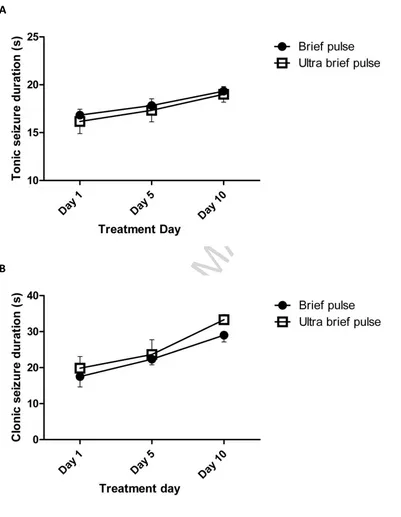
3.1. Tonic-clonic seizure durations
Tonic and clonic seizure durations were compared on ECS treatment days 1, 5 and 10
(Figure 2). Repeated measures ANOVA showed that both tonic (F(2,20)=3.88, p=0.038) and clonic (F(2,20)=18.63, p<0.0001) seizure durations lengthened with increasing number of treatments. Tonic seizure durations have previously been similarly reported to increase
over time in a study comparing the effects of ECS using pulse widths of either 0.5 ms or 10
ACCEPTED MANUSCRIPT
8
3.2. Behavioural changes after ECS
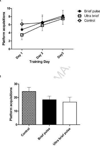
Water plus maze platform acquisition on training and probe days were investigated
to determine if animals learned the platform position and could recall this information.
Platform acquisition during the training period increased significantly over time (F(2,28)=7.23, p=0.0029) but was not affected by treatment type (F(2,28)=0.41, p=0.67; Figure 3A),
indicating that all animals learned the task, and there was no group x time interaction effect
(F(4,28)=0.48, p=0.7513). On the probe trial day, both BP and UBP ECS-treated animals obtained the platform less frequently than controls but this difference was not statistically
significant (F(2,14)=1.8, p=0.21; Figure 3B). Of note, there was no difference between the levels of platform acquisition by BP and UBP groups. The swimming velocity (cm/s) at which
the animals obtained the platform increased significantly (F(2,42)=6.47, p=0.004) over the three training days but did not differ between the three treatment groups (F(2,42)=0.16,
p=0.849), indicating no motor deficit resulted as a consequence of real ECS treatments. Similarly, no significant difference in velocity to finding the platform was found between the
three groups (F(2,9)=0.033, p=0.967) on the probe day.
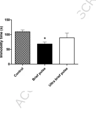
The groups differed with regards to immobility times in the Forced Swim Test
(F(2,15)=3.8, p=0.046; Figure 4). Post hoc analysis revealed that the BP group showed a significant decrease (p<0.05) in immobility time when compared to controls, whereas a
non-significant decrease in immobility time was seen in the UBP group.
3.3. BDNF protein levels after ECS
Different levels of hippocampal BDNF were found between the three groups (F(2,
15)=6.3, p=0.01) when measured two weeks after completing the allocated course of ECS with BP ECS producing significantly higher levels than found in both the control (p<0.01) and
UBP (p<0.05) groups (Figure 5). A smaller, but non-significant, increase was found in the
UBP group compared to sham-treated controls.
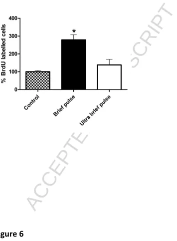
3.4. Immunohistochemistry
ECS treatment was associated with an increase in cell proliferation compared to
ACCEPTED MANUSCRIPT
9
allocated course of ECS. BP ECS induced a significant (p<0.05), almost 3-fold, increase in the
number of proliferating cells compared to sham treated control animals, whereas a much
smaller and non-significant increase in cell proliferation was seen following UBP-ECS.
4. Discussion
To our knowledge, this is the first study to systematically compare the effects of UBP
(0.3 ms) and BP (0.5 ms) width stimuli on chronic ECS and related antidepressant-related
changes in rats. We found that both forms of ECS elicited seizures that were
indistinguishable with regards to observable motor seizure activity and the durations of the
tonic and clonic phases. However, while BP ECS induced significant increases in recognised
markers of antidepressant action, UBP ECS did not have the same effect.
Compared to sham-treated animals, a large, three-fold, and significant increase in
cell proliferation was seen within the dentate gyrus one day after completing a course of BP
ECS while only a much smaller, non-significant, increase was seen following UBP ECS. This
degree of increased cell proliferation following BP ECS is similar to that previously found by
other groups (Madsen et al., 2000; Segi-Nishida et al., 2008). The majority (75-88%) of these
new cells have been reported to be neuronal (Malberg et al., 2000; Scott et al., 2000) and
such hippocampal neurogenesis is required for anti-depressant behavioural effects in both
rats and nonhuman primates (Perera et al., 2011; Santarelli et al., 2003). Additionally, two
weeks after completing a course, we found that BP ECS resulted in a significantly greater
increase in hippocampal levels of BDNF than UBP ECS. Increased BDNF expression has been
reported to mediate anti-depressant effects for a range of antidepressant drugs as well as
ECS in animals (Castren and Rantamaki, 2010).
Chronic UBP ECS was therefore not as efficient as BP ECS for inducing
antidepressant-related cellular and molecular changes in rat hippocampus. In parallel with
these differences, we found that BP ECS had a greater antidepressant behavioural effect as
it significantly reduced immobility time in the forced swim test, while immobility time
following UBP ECS did not differ significantly from sham ECS. This may be reflected in some
recent clinical studies of various forms of UBP ECT for depression that reported very low
ACCEPTED MANUSCRIPT
10
A potential benefit of UBP ECS – and UBP ECT for treating depression – is a reduction in the cognitive side effects associated with ECS treatment (Sackeim et al., 2008). In the
present study there was a non-significant trend for similar decreases in performance in both
the UBP and BP groups compared to the sham control group on hippocampal-dependant
reference memory in the water plus maze probe trial. However, we did not find any
difference between the effects of UBP and BP ECS even though it induced less cellular,
molecular and behavioural changes than BP ECS. These cognitive findings should be
interpreted with some caution because the sample sizes in our study may have been too
small to detect a relatively reduced impact of UBP ECS on memory. Additionally, the interval
of 10 days between end of treatment and cognitive assessment may have led to early post
treatment differences between UBP and BP groups no longer being apparent, reflecting
recovery of hippocampal cognitive function over time following ECS as occurs in patients
treated with ECT (Semkovska et al., 2011; Semkovska and McLoughlin, 2010). Interestingly,
some recent studies attempting to cause neuronal loss using ECS have found that repeated
brief seizures can cause specific neuronal death within hippocampal structures under
certain conditions (Cardoso et al., 2011). However, these same studies also report that cell
loss does not occur when ECS is administered on a 24-48 hour schedule (Cardoso et al.,
2008). The interval between the final seizures in the programme must be short (2 hours) to
cause the morphological and behavioural changes observed, possibly resulting from
neuronal vulnerability during the post ictal period. Of note, analyses of rodent brain
following conventionally spaced BP ECS, i.e. not designed to deliberately induce neuronal
death, have not found evidence of neuronal loss (Dalby et al., 1996; Gombos et al., 1999;
Vaidya et al., 1999; Cardoso et al., 2008; Jinno and Kosaka, 2009). Based upon the findings
of the present study, we would predict the same for UBP ECS.
For experimental purposes we altered only one parameter in the treating ECS
stimulus - the pulse width. Therefore, one possible explanation for the relatively small
antidepressant-related effects we found with UBP ECS is that the total stimulus charge per
session administered was only 60% that for BP ECS, i.e. 1.575 mC compared to 2.625 mC.
Increasing the frequency of UBP stimuli or the duration of UBP ECS administration might
result in greater anti-depressant effects. However, despite the lower total charge for UBP
ACCEPTED MANUSCRIPT
11
indistinguishable. Even though we found large and meaningful differences between UBP
and BP ECS on several different measures, some caution is warranted as noted above
because of the relatively small sample sizes and the risk of type 1 error. Our findings
therefore require further exploration and replication, possibly examining reported
differences at other timepoints. Finally, the effects of UBP ECS on animal models of
depression, in addition to normal rats, need to be studied. For example, BP ECS has been
reported to reduce forced swim test immobility time in Flinders Sensitive Line rats
(Jimenez-Vasquez et al., 2007), normalise both impaired sucrose preference (a measure for
anhedonia) and reduced hippocampal BDNF levels induced by chronic mild stress (Gersner
et al., 2010), and reverse corticosterone-mediated inhibition of neurogenesis (Hellsten et
al., 2002). It would therefore be important to study the therapeutic effects of UBP ECS in
similar models of depression.
5. Conclusions
We found UBP ECS to be much less efficient than BP ECS in normal rats for inducing a
range of cellular, molecular and behavioural changes that are believed to be important for
antidepressant mechanisms. Comparing the effects of different stimulus parameters of ECS
in animals allows us to better understand this antidepressant therapy and determine
whether modifying the existing parameters of ECT may be of benefit for patients with
depression.
Acknowledgements
This work was supported by the Health Research Board (grant no. TRA/2007/5) and the St.
Patrick’s Hospital Foundation.
References
ACCEPTED MANUSCRIPT
12 Cardoso, A., Lukoyanova, E.A., Madeira, M.D., and Lukoyanov, N.V., 2011. Seizure-induced structural and functional changes in the rat hippocampal formation: Comparison between brief seizures and status epilepticus. Behav. Brain Res. 225, 538-546.
Castren, E., and Rantamaki, T., 2010. The role of BDNF and its receptors in depression and
antidepressant drug action: Reactivation of developmental plasticity. Dev Neurobiol 70, 289-297.
Dalby, N.O., Tonder, N., Wolby, D.P.D., West, M., Finsen, B., and Bolwig, T.G., 1996. No loss of hippocampal hilar somatostatinergic neurons after repeated electroconvulsive shock: A combined stereological and in situ hybridization study. Biol. Psychiatry 40, 54-60.
Diamond, D.M., Park, C.R., Heman, K.L., and Rose, G.M., 1999. Exposing rats to a predator impairs spatial working memory in the radial arm water maze. Hippocampus 9, 542-552.
Dunne, R., and McLoughlin, D.M., 2011. Systematic review and meta-analysis of bifrontal electroconvulsive therapy versus bilateral and unilateral electroconvulsive therapy in depression. World J. Biol. Psychiatry, Nov 18, Epub ahead of print.
Eranti, S., Mogg, A., Pluck, G., Landau, S., Purvis, R., Brown, R.G., Howard, R., Knapp, M., Philpot, M., Rabe-Hesketh, S., et al., 2007. A randomized, controlled trial with 6-month follow-up of repetitive transcranial magnetic stimulation and electroconvulsive therapy for severe depression. Am. J. Psychiatry 164, 73-81.
Gersner, R., Toth, E., Isserles, M., and Zangen, A., 2010. Site-specific antidepressant effects of repeated subconvulsive electrical stimulation: potential role of brain-derived neurotrophic factor. Biol. Psychiatry 67, 125-132.
Gombos, Z., Spiller, A., Cottrell, G.A., Racine, R.J., and Burnham, W.M., 1999. Mossy fiber sprouting induced by repeated electroconvulsive shock seizures. Brain Res. 844, 28-33.
Hellsten, J., Wennstrom, M., Mohapel, P., Ekdahl, C.T., Bengzon, J., and Tingstrom, A., 2002. Electroconvulsive seizures increase hippocampal neurogenesis after chronic corticosterone treatment. Eur. J. Neurosci 16, 283-290.
Jansson, L., Wennstrom, M., Johanson, A., and Tingstrom, A., 2009. Glial cell activation in response to electroconvulsive seizures. Prog. Neuropsychopharmacol. Biol. Psychiatry 33, 1119-1128. Jimenez-Vasquez, P.A., Diaz-Cabiale, Z., Caberlotto, L., Bellido, I., Overstreet, D., Fuxe, K., and Mathe,
A.A., 2007. Electroconvulsive stimuli selectively affect behavior and neuropeptide Y (NPY) and NPYY1 receptor gene expressions in hippocampus and hypothalamus of Flinders Sensitive Line rat model of depression. Eur. Neuropsychopharmacol. 17, 298-308.
Jinno, S., and Kosaka, T., 2009. Neuronal circuit-dependent alterations in expression of two isoforms of glutamic acid decarboxylase in the hippocampus following electroconvulsive shock: a stereology-based study. Hippocampus 19, 1130-1141.
Kellner, C.H., Knapp, R., Husain, M.M., Rasmussen, K., Sampson, S., Cullum, M., McClintock, S.M., Tobias, K.G., Martino, C., Mueller, M., et al., 2010. Bifrontal, bitemporal and right unilateral electrode placement in ECT: randomised trial. Br. J. Psychiatry 196, 226-234.
Kesavapany, S., Banner, S.J., Lau, K.F., Shaw, C.E., Miller, C.C.J., Cooper, J.D., and McLoughlin, D.M., 2002. Expression of the Fe65 adapter protein in adult and developing mouse brain.
Neuroscience 115, 951-960.
Loo, C., Sheehan, P., Pigot, M., and Lyndon, W., 2007. A report on mood and cognitive outcomes with right unilateral ultrabrief pulsewidth (0.3 ms) ECT and retrospective comparison with standard pulsewidth right unilateral ECT. J. Affect. Disord. 103, 277-281.
Loo, C.K., Sainsbury, K., Sheehan, P., and Lyndon, B., 2008. A comparison of RUL ultrabrief pulse (0.3 ms) ECT and standard RUL ECT. Int. J. Neuropsychopharmacol. 11, 883-890.
ACCEPTED MANUSCRIPT
13 Malberg, J.E., Eisch, A.J., Nestler, E.J., and Duman, R.S., 2000. Chronic antidepressant treatment
increases neurogenesis in adult rat hippocampus. J. Neurosci. 20, 9104-9110. McCall, W.V., Reboussin, D.M., Weiner, R.D., and Sackeim, H.A., 2000. Titrated moderately
suprathreshold vs fixed high-dose right unilateral electroconvulsive therapy - Acute antidepressant and cognitive effects. Arch. Gen. Psychiatry 57, 438-444.
Newton, S.S., Collier, E.F., Hunsberger, J., Adams, D., Terwilliger, R., Selvanayagam, E., and Duman, R.S., 2003. Gene profile of electroconvulsive seizures: Induction of neurotrophic and
angiogenic factors. J. Neurosci. 23, 10841-10851.
Niemantsverdriet, L., Birkenhager, T.K., and van den Broek, W.W., 2011. The efficacy of ultrabrief-pulse (0.25 millisecond) versus brief-ultrabrief-pulse (0.50 millisecond) bilateral electroconvulsive therapy in major depression. J. ECT 27, 55-58.
Perera, T.D., Dwork, A.J., Keegan, K.A., Thirumangalakudi, L., Lipira, C.M., Joyce, N., Lange, C., Higley, J.D., Rosoklija, G., Hen, R., et al., 2011. Necessity of hippocampal neurogenesis for the
therapeutic action of antidepressants in adult nonhuman primates. PLoS One 6(4), e17600. Porsolt R.D., Le Pichon M., Jalfre M., 1977. Depression: A new animal model sensitive to
antidepressant treatment. Nature 266, 730-732.
Quante, A., Luborzewski, A., Brakemeier, E.L., Merkl, A., Danker-Hopfe, H., and Bajbouj, M., 2011. Effects of 3 different stimulus intensities of ultrabrief stimuli in right unilateral
electroconvulsive therapy in major depression: A randomized, double-blind pilot study. J. Psychiatr. Res. 45, 174-178.
Sackeim, H.A. (2004). Convulsant and anticonvulsant properties of electroconvulsive therapy: towards a focal form of brain stimulation. Clin. Neurosci. Res. 4, 39-57.
Sackeim, H.A., Prudic, J., Devanand, D.P., Kiersky, J.E., Fitzsimons, L., Moody, B.J., McElhiney, M.C., Coleman, E.A., and Settembrino, J.M., 1993. Effects of stimulus-intensity and electrode placement on the efficacy and cognitive effects of electroconvulsive-therapy. N. Engl. J. Med. 328, 839-846.
Sackeim, H.A., Prudic, J., Nobler, M.S., Fitzsimons, L., Lisanby, S.H., Payne, N., Berman, R.M., Brakerneier, E.L., Perera, T., and Devanand, D.P., 2008. Effects of pulse width and electrode placement on the efficacy and cognitive effects of electroconvulsive therapy. Brain Stim. 1, 71-83.
Santarelli, L., Saxe, M., Gross, C., Surget, A., Battaglia, F., Dulawa, S., Weisstaub, N., Lee, J., Duman, R., Arancio, O., et al., 2003. Requirement of hippocampal neurogenesis for the behavioral effects of antidepressants. Science 301, 805-809.
Scott, B.W., Wojtowicz, J.M., and Burnham, V.M., 2000. Neurogenesis in the dentate gyrus of the rat following electroconvulsive shock seizures. Exp. Neurol. 165, 231-236.
Segi-Nishida, E., Warner-Schmidt, J.L., and Duman, R.S., 2008. Electroconvulsive seizure and VEGF increase the proliferation of neural stem-like cells in rat hippocampus. Proc. Natl. Acad. Sci. USA 105, 11352-11357.
Semkovska, M., Keane, D., Babalola, O., and McLoughlin, D.M., 2011. Unilateral brief-pulse
electroconvulsive therapy and cognition: effects of electrode placement, stimulus dosage and time. J. Psychiatr. Res. 45, 770-780.
Semkovska, M., and McLoughlin, D.M., 2010. Objective cognitive performance associated with electroconvulsive therapy for depression: a systematic review and meta-analysis. Biol. Psychiatry 68, 568-577.
Sienaert, P., Vansteelandt, K., Demyttenaere, K., and Peuskens, J., 2009. Randomized comparison of ultra-brief bifrontal and unilateral electroconvulsive therapy for major depression: Clinical efficacy. J. Affect. Disord. 116, 106-112.
ACCEPTED MANUSCRIPT
14 Vaidya, V.A., Siuciak, J.A., Du, F., and Duman, R.S., 1999. Hippocampal mossy fiber sprouting induced
ACCEPTED MANUSCRIPT
15
Legends for figures
Figure 1 Experimental design. (A) Animals were administered real or sham ECS thrice weekly for ten sessions over 22 days. Following one week habituation to the behavioural
suite, animals were trained and tested in the water plus maze (WPM) followed by the forced
swim test (FST) two days later. The animals were sacrificed 24 hours after the swim test. (B)
Another set of animals was similarly treated with ECS and was also injected with BrdU 2-4
hours after ECS treatments 4-10. The animals were perfused 24 hours after the final ECS
session and the brains extracted to examine changes in cell proliferation.
Figure 2 Seizure durations. (A) Tonic and (B) clonic seizure durations are shown on the first, fifth and tenth treatment days of the randomly allocated ECS course. Durations of both
tonic (p=0.038) and clonic (p<0.0001) seizure phases increased significantly over time in
both BP and UBP groups but there was no significant difference between treatment groups
or significant Treatment x Time interactions. n=6 per group.
Figure 3 Water plus maze. (A) After completing the randomly allocated course of ECS, animals obtained the platform more frequently over successive training days 1-3 (p=0.024).
Treatment type had no effect on number of platform acquisitions. n = 5/6 per group. (B)
The number of platform acquisitions on probe test day 4 did not differ significantly between
groups. BP and UBP groups obtained the platform less frequently than the sham group
although this difference was not significant.
Figure 4 Forced swim test following ECS. BP animals spent significantly less time immobile than UBP or control animals. The decrease in immobility times in the UBP group was not
significantly lower than in controls. n=6 per group, *p<0.05 compared to control group.
Figure 5 BDNF protein levels in the hippocampus following ECS. BDNF was significantly increased in the BP group compared to the control and UBP ECS groups. The increase seen
in BDNF levels in the UBP group was not significantly higher than in the control group. n=6
ACCEPTED MANUSCRIPT
16
Figure 6 Cell proliferation following ECS. BP treatment significantly increased the relative number of BrdU-labelled cells in the dentate gyrus compared to sham-treated control
animals (p<0.05) (percentage of control). The increase in BrdU-labelled cells was not
ACCEPTED MANUSCRIPT
17
A
[image:18.595.72.508.88.623.2]B
ACCEPTED MANUSCRIPT
18
Figure 2
A
[image:19.595.91.492.84.601.2]ACCEPTED MANUSCRIPT
[image:20.595.94.445.78.619.2]19
Figure 3
A
ACCEPTED MANUSCRIPT
[image:21.595.105.416.206.594.2]ACCEPTED MANUSCRIPT
[image:22.595.90.456.82.611.2]ACCEPTED MANUSCRIPT
[image:23.595.102.458.132.615.2]ACCEPTED MANUSCRIPT
26
Highlights
Ultrabrief pulse electroconvulsive therapy may be as effective as brief pulse ECT We compared ultrabrief and brief pulse electroconvulsive stimulation (ECS) in rats We examined molecular, cellular and behavioural antidepressant-related changes Both forms of ECS elicited similar types and durations of seizures