ESSENTIAL GUIDES FOR
ISOLATION/PURIFICATION OF
IMMUNOGLOBULINS
A. Layer, P. Schneider, J.-D. Tissot and M. A. Duchosal, Service Re&gional Vaudois de Transfusion Sanguine, Lausanne, Switzerland
Copyright^ 2000 Academic Press
Introduction
The immunoglobulins (Igs), proteins produced by B lymphocytes, have been extensively studied both at molecular and genetic levels. They consist of two identical heavy chains and two identical light chains having therefore the same isotype and the same type, respectively. Igs are puriRed for three main purposes. (i) as therapeutic injections to patients; (ii) for use as a tool in research or clinical diagnosis; and (iii) for their biochemical analysis (speciRcity, isotype or clonal diversity). Most of these applications require that the binding activity of Igs be retained throughout all the puriRcation procedures.
PuriRcation of Igs can be performed according to their physicochemical properties, their biological activities or a combination of both. The technique used will depend on the desired degree of purity and the amount and nature of the starting mater-ial. The methods that have been described are gener-ally directly applicable to crude materials such as serum, asciticSuid or cell culture supernatant. Two-dimensional polyacrylamide gel electrophoresis (2D-PAGE) affords an efRcient way of evaluating the degree of purity reached in afRnity puriR ca-tions. Several aspects of 2D-PAGE analysis are described in detail in two other articles ‘Electro-phoresis/Two-dimensional PAGE’ and &Clinical Ap-plications of Electrophoresis/Electrophoresis’ in this Encyclopedia.
As a general rule, and independently of the tech-nique used, the starting material should always be devoid of any insoluble substances and the puriR ca-tion be preceded by centrifugaca-tion orRltration. Vis-cousSuids, such as serum, may be diluted before use, especially for chromatographic procedures. The solu-tions should contain a bacteriostatic agent, such as 0.02% sodium azide (NaN
), and be kept on ice. Ig
solutions should be handled gently, avoiding bubbl-ing or frothbubbl-ing, because such manipulations may be
accompanied by denaturing effects, and may lead to protein precipitation.
Puri
\
cation by Precipitation
Solubility of the proteins, particularly Igs, in water relies mainly on the ability to make hydrogen bonds between polar or ionic groups with water molecules (hydrophilic interactions), and on the capacity to maintain hydrophobic groups that cannot interact with water molecules buried inside the proteins. In addition, the solubility of Igs is temperature-depen-dent. Any external factor capable of modifying hy-drogen bonds or decreasing the medium hydrophilic-ity will decrease the solubilhydrophilic-ity of the proteins and may eventually lead to their precipitation. Each protein has its own physicochemical characteristic, including solubility. For this reason, several differential precipitation procedures can be developed to isolate Igs from variousSuids. These procedures are present-ed below.
Differential Ethanol Precipitation
TheRrst fractionation of plasma proteins for thera-peutic use was described in 1949 by E.J. Cohn. The basic procedure, with few modiRcations, is still widely used in industrial fractionation centres. Basically, ethanol is added progressively to the medium to a Rnal concentration varying from 8 to 40%. Subsequently, the temperature is decreased to !33C and then to !53C. Finally, the pH is decreased from 7.3 to 4.8. These steps yield pre-cipitation fractions, called Cohn’s fractions I}V. Fraction II contains the -globulins or Igs. The treatment of this fraction with caprylic acid (see be-low) allows the preparation of Igs that are enriched in IgA and IgM. This approach is used when large amounts of Igs are needed, i.e. for therapeutic pur-poses (from up to 5000 L plasma) and will not be detailed further.
Ammonium Sulfate Precipitation
solubility and, when the ammonium sulfate concen-tration is increased stepwise, a sequential precipita-tion of proteins may be obtained.
Practically, a saturated solution of ammonium sul-fate (761 g L\1; 4.1 mol L\1) is slowly added to a stirred (in order to avoid local over-increase in concentration) solution of Igs, until theRnal desired concentration, usually expressed as a percentage of ammonium sulfate saturation, is reached. Although some interspecies variability is observed for Ig solu-bility, a 50% ammonium sulfate saturation is usually appropriate for most Ig precipitation procedures. Pre-precipitation at 40% ammonium sulfate saturation may be useful to remove large protein aggregates and proteins that may precipitate at low ammonium ion concentration. The precipitation is allowed to occur at 43C for 6}12 h. The precipitate is recovered by centrifugation at 2000}5000gfor 20}30 min. In gen-eral, the pellet is then gently solubilized in a minimal volume of a physiological buffer, and dialysed to remove the residual ammonium sulfate. Alterna-tively, Ig purity can be increased by washing the pellet with a 50% saturated solution and solubilization in PBS, followed by another round of ammonium sul-fate Ig precipitation. When starting with serum or cell supernatant containing serum, this method allows the removal of most albumin and haemoglobin, but the precipitated fraction still contains several serum pro-teins in addition to Igs. In this case this approach must be coupled with another puriRcation technique if pure material is needed. When starting with a serum-free cell culture supernatant, this method is convenient to isolate, and to concentrate, monoclonal Igs in one step. In addition, the method is easy, cheap and can be applied to large volumes.
Caprylic Acid Precipitation
The solubility of proteins is altered by the presence of some short chain fatty acids, such as octanoic acid (caprylic acid) at mildly acidic pH. Basically, caprylic acid increases medium hydrophobicity. In practice the pH of the starting solution must be adjusted to 4 by the addition of about 2 vol of a 60 mmol L\1 sodium acetate buffer. Then, 0.04}0.07 vol of caprylic acid (depending on the starting material as well as on the animal species of the Igs) is added drop by drop while stirring, and the solution is incubated at room temperature for 30 min. Under these condi-tions, most of the serum proteins are precipitated, with the exception of IgG, which is recovered in the supernatant after centrifugation at 5000gfor 10 min. This method bears similarities with that of am-monium sulfate precipitation. In particular, it needs to be coupled with another one to yield highly puri-Red Ig fractions.
Chromatographic Methods
In chromatographic procedures, compounds in solu-tion are separated by allowing them toSow through a selective medium poured in a column. Differen-tial interactions between molecules and matrix are responsible for them migrating at various speeds, or even completely immobilizing them. Separated mol-ecules are recovered in the efSuent of the column. Several commercially available preparations allow separation of proteins according to their various physicochemical properties. Detailed information about the use of such media is furnished by the manufacturers or can be found in the literature for particular applications.
Ion Exchange Chromatography
An ion exchanger consists of a positively or negative-ly charged group covalentnegative-ly bound to an insoluble matrix. Charged molecules with complementary po-larity to that of the immobilized groups bind to the matrix through electrostatic interactions, whereas uncharged or similarly charged molecules pass freely through the matrix. Since the net charge of a pro-tein depends on the pH, the starting experimental conditions must be carefully chosen. The bound pro-teins may be desorbed either by change in pH, or by change in the ionic strength. The former modiRes the charge of the protein, whereas salts compete with the binding of the protein to the resin. Addition of NaCl is most frequently used for elution. As the strength of the protein}matrix interaction depends on the net protein charge, a sequential elution can be performed by gradually increasing salt concentration. Because Igs have a more basic isoelectric point than most other serum proteins, ion exchange chromatography can be used for their puriRcation. Practically, the matrix should be extensively washed with 0.5 mol L\1 HCl or 0.5 mol L\1 NaOH before use, and then equilibrated with the binding buffer. In addition, the sample must be dialysed against the binding buffer before being loaded on the resin.
differentially puriRed using this method. Rela-tively pure IgG is usually recovered in theRrst eluted fraction; IgM, the last eluted Ig isotype, may also be recovered in quite a pure form, whereas IgD and IgA, with intermediate elution properties, are only poorly resolved with this method. Ion exchange chromatog-raphy can yield sufRciently pure antibodies if the starting material is a cell culture supernatant or an asciticSuid, but it must be coupled to an additional puriRcation step when samples such as serum are used. The method is also cheap and is convenient for large initial volumes.
Hydroxyapatite Chromatography
Immobilized hydroxyapatite (calcium phosphate hy-droxide) is used for another kind of adsorption chromatography. At pH 6.8, Igs bind to the matrix, and are eluted when a linear gradient of phosphate buffer from 120 to 300 mmol L\1 is applied. When highly puriRed Ig fractions are needed, this technique must be coupled to another one, again depending on the starting material.
Gel Filtration Chromatography
A gel Rltration matrix consists of beads containing pores of various sizes. As the sample Sows through the matrix, the largest molecules are excluded from the beads. They stay only in the mobile phase, and move fast. The smaller molecules, depending princi-pally on their sizes but also on their shapes, dif-fuse more or less inside the pores, and move more slowly within the column. Thus, this system allows the separation of the proteins according to their sizes and shapes. Various matrices with particular structures, pore sizes and excluding limits (theMrat which the proteins are no more able to enter into the beads) are available commercially. GelRltration can be used within broad pH ranges, with or without detergents such as 1% sodium dodecyl sulfate (SDS), or dissociating agents such as urea or guanidine. Igs puriRcation is usually performed without sophisti-cated conditions, and allows the separation of IgM molecules that are considerably larger than IgG as well as many other serum proteins. Using an exclu-sion limit of 300}500 kDa, and a column volume of at least 20 times larger than that of the starting solution, IgM is easily recovered in the excluded peak. Due to the time needed to allow a complete passage, the use of a fraction collector is highly rec-ommended. Fractions corresponding to 1}3 initial volumes should be collected. GelRltration must usu-ally be coupled with other methods to yield sufR -cient Ig purity, and is mainly limited to the puriR ca-tion of IgM.
Precipitation of Immune Complexes
The Precipitin Reaction
When antigens are added in adequate proportions to a mixture of antibodies, speciRc Igs and antigens will form a lattice which is susceptible to precipitate (the precipitin reaction). This macromolecular complex can then be easily recovered by centrifugation. The required amount of antigens to be added must be determined by establishing a precipitation curve. This implies that one should also have a procedure allowing the precipitate to be quantiRed. The use of radiolabel-led antigens is particularly suitable for this when restricted amounts of antigens are available. Comp-lement activation, which occurs in normal fresh serum, is able to inhibit the precipitation strongly. Therefore, it is necessary to make either a pre-puriRcation of the Igs or to inhibit the complement cascade by the addi-tion of ethylenediaminetetraacetic acid before per-forming a precipitin reaction. The precipitated lattice is subsequently re-solubilized either by incubation with serum or with an excess of free antigen, or by digestion with papain, which generates Fc fragments.
Polyethylene Glycol Precipitation
Soluble (nonlattice) immune complexes, either pres-ent naturally, or generated by adding a corresponding antigen, precipitate from serum in the presence of 3}4% polyethylene glycol (PEG; Mr 6000) after 2}12 h incubation at 43C. This method has been widely used to isolate circulating immune complexes in various pathological situations. Other high mo-lecular weight proteins, as well as aggregated Igs, can also precipitate using above-mentioned conditions. Therefore, additional steps of puriRcation, using pro-tein G or A (see below), are generally warranted before sufRciently pure material can be used. Solubilization of most PEG-precipitated immune complexes can easily be performed using most buf-fers that do not contain PEG. In contrast, dissociation of immune complexes requires quite harsh condi-tions, that are frequently not compatible with tech-niques allowing the further puriRcation of free Igs devoid of antigens. Immunoprecipitation of speciRc antibodies is therefore mainly limited to analytical purposes that do not need biological activity. Ig light and heavy chains from immune complexes are easily solubilized in buffers containing SDS, and can be analysed by SDS-PAGE or 2D-PAGE.
Af
\
nity Chromatography
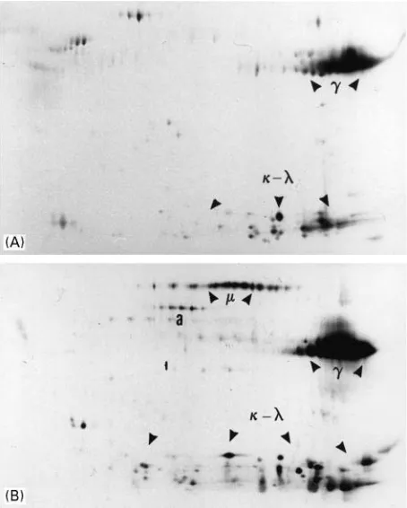
consist-Figure 1 (A) 2D pattern of IgG purified over protein G-Sepharose. Human serum was incubated with commercially available protein G-Sepharose (Pharmacia), and IgG eluted as indicated in the text.heavy chains migrate within pIs ranging from 6 to more than 10, with a size of about 50 kDa. Light chains display pIs ranging from 5 to 10 and size between 21 and 26 kDa.
, IgG heavy chain;,, light chains. (B) 2D pattern of IgA affinity purified over a homemade anti- chain-Sepharose resin. The affinity resin was prepared from commercially available CNBr-activated Sepharose (Pharmacia), according to the manufac-turer’s recommendations, and commercially available goat anti-humanchain antibodies.chains migrate with a pI ranging from 4.9 to 6.1 and a size of about 58 kDa., IgA heavy chain;,, light chains.
ing of an Ig-binding molecule covalently coupled to a bead support. Unbound molecules are removed by washing, and speciRcally bound Igs are then eluted using an appropriate buffer. The method is high-ly speciRc and high Ig purity is usually reached in a single step. Ig-binding molecules belong to three major groups: (i) bacterial protein A or protein G; (ii) speciRc antigens; and (iii) monospeciRc antibodies directed to epitopes on Igs (such as goat anti-human Ig antibody). Various bead supports and coupling procedures have been studied. The use of commer-cially available activated beads has now allowed preparation of afRnity media within most labor-atories. In particular cyanogen bromide (CNBr)-activated Sepharose beads can easily be used for most applications and detailed instructions are furnished by the manufacturers. After coupling, the resin should be extensively washed to remove all un-coupled Ig-binding molecule, and equilibrated in binding buffer. The binding capacity of each resin should be determined experimentally before use. If sample binding is indifferently performed in a container (batch procedure) Rxed on a rotating wheel or through a column, washing and elution are best performed through a column. The use of a peri-staltic pump is highly recommended to ensure aRxed Sow.
Washing and elution steps are best followed online with a UV detector set to monitor at 280 nm. Alterna-tively, fractions may be collected and tested individ-ually using either UV absorption or more speciRc procedures. When necessary (see elution conditions below), the eluate has to be collected in a neutralizing solution, and the resin immediately re-equilibrated in the binding buffer. Unused resins should be stored at 43C in the presence of a bacteriostatic agent, such as 0.02% sodium azide or 20% ethanol.
Af\nity Chromatography using Immobilized Protein G and A
Protein A and protein G are present within the bacter-ial cell walls of Staphylococcus aureusand of group Gstreptococci, respectively. Both proteins have high afRnity for the Fc region of IgG, but bind dif-ferentially IgG subclasses from various species. Whereas protein G bind all human and mouse IgG subclasses, protein A presents only low binding capa-city for human IgG3 and mouse IgG1. Ready-to-use matrix-immobilized protein G or A is commercially available, and detailed information about the binding properties of these two proteins can be found in the literature or furnished by the manufacturers. Binding buffers usually contain 100 mmol L\1 Tris or 10 mmol L\1phosphate with 0.15 mol L\1NaCl, at pH 8 for protein A and pH 7 for protein G. After Ig
[image:4.568.291.519.59.403.2]Figure 2 (A) 2D pattern of a mixture of affinity purified IgM and IgA; (B) 2D pattern of a mixture of affinity purified IgM and IgG. Immunoglobulins were purified over homemade anti- chain-Sepharose resin, anti-chain-Sepharose resin and anti -chain-Sepharose resin and mixed as indicated before electrophoresis. Resins were prepared according to the manufacturer’s instruc-tions, from CNBr-activated Sepharose and commercially avail-able goat anit-human,andchains. Immunoglobulins were prepared from serum as indicated in the text.chains migrate with pIs between 5.6 and 6.4, and size of 72 kDa., IgM heavy chain;, IgA heavy chain;, IgG heavy chain;,, light chains.
interactions, can be increased by raising salt concen-trations of the binding and washing buffers to 3.3 and 3 mol L\1, respectively. This high salt con-centration method was initially described for the puriRcation of mouse IgG1on protein A, but is now obsolete, due to the availability of protein G. The use of an excess of protein A allows IgG1, IgG2and IgG4 to be depleted from samples and IgG3 to be puriRed using protein G in a second step. A sequential elution of all four IgG subclasses from protein A using a pH gradient has been described, but the resolution is quite low and subclass-speciRc puriRcation of IgG1\4 is now best performed using immobilized mono-speciRc anti-Ig raised against either 1, 2, 3 or
4heavy chains of the IgG molecule.
The elution yield from protein G or A using stan-dard procedures is never 100% (see below) and resid-ual IgG can be eluted during a second run procedure and may contaminate the new IgG being puriRed with IgG from the previous run. In order to avoid any Ig contamination from a previous experiment, it is therefore highly advisable to use either the same batch of protein G or A for the same initial IgG preparation or to use a new batch of resin for each procedure.
Af\nity Chromatography using Immobilized anti-Igs
In this method, the immobilized binding molecules are Igs (mouse, rabbit, goat, sheep) directed against Ig heavy and/or light chains. Using antibodies of various speciRcities, it is possible to isolate either total Igs (using anti- and - chains), particular Ig isotopes (using anti-,-,-,-or-chain) or IgG subclasses (using anti-1, -2, -3 or-4chains). The interaction between immobilized and targeted immunoglobulins is just a particular type of antibody}antigen interac-tion. Binding and elution are therefore basically per-formed using the same conditions as those used for immobilized antigen supports (see below). Figure 1B shows that IgA puriRed from a human serum sample over an anti-chain resin does not display any other heavy butchain isotope. The resolution obtained by 2D-PAGE in separating various Ig heavy chains is illustrated inFigure 2.
IgG subclasses can be puriRed using two dif-ferent procedures named positive and negative isola-tions. In positive isolation, the desired subclass is immobilized on a resin, washed and recovered by elution, whereas in the negative isolation, all un-wanted subclasses are bound on resin and the desired subclass is recovered in the Sow-through. The ad-vantage of this latter approach is that theRnal prep-aration is not exposed to strong nonphysiological conditions which may denature the puriRed IgG
sub-class. Disadvantages are that the methodology re-quires larger amounts of resin and several immobi-lized antibodies and that, when biologicalSuids are processed, theRnal preparation still contains proteins other than IgG.
Af\nity Chromatography using Immobilized Antigens
Figure 3 2D patterns of affinity purified tetanus toxoid anti-bodies. Immunoglobulins were purified from two severe combined immunodeficient (SCID) mice (A and B) previously injected with human lymphocytes and boosted with tetenus toxoid. Homemade tetanus toxoid-Sepharose resin was prepared according to manu-facturer’s recommendations, from commercially available (Phar-macia) CNBr-activated Sepharose and a home preparation of tetanus toxoid. Igs were SDS-eluted, as indicated in the text., IgG heavy chain;, IgM heavy chain;,, light chains, a, albumin.
(10 mmol L\1 Tris or phosphate buffer saline (PBS) 0.15 mol L\1 NaCl, pH 7.5). The unbound material is washed away with about 20 resin-volumes of binding buffer. Elution of bound antibodies is commonly performed by successive washes with 100 mmol L\1 glycine, pH 2}3 and 100 mmol L\1 triethylamine, pH 11}12. The eluted fractions are subsequently recovered into tubes containing neu-tralizing buffer. Elution solutions such as 5 mol L\1 LiCl/PBS, 3.5 mol L\1 MgCl
2/PBS, 1% SDS, 2}8 mol L\1urea, 3 mol L\1thiocyanate, 10% dioxane, or 50% ethylene glycol can also be used.
The puriRcation of anti-tetanus toxoid Igs over a toxoid-coated resin is presented in Figure 3. Both 2D-PAGE light chain patterns shown depict a limited number of easily distinguishable spots, typical of oligoclonal Igs. These patterns are clearly different from that of total Igs (and puriRed total IgG, not shown) from the same serum, indicating that a subpopulation of Igs was puriRed. Whereas the anti-tetanus toxoid antibodies shown in Figure 3A
consist only of IgG, some IgM anti-tetanus toxoid antibodies were also present in the case shown in Figure 3B.
Af\nity Chromatography Using Jacalin or Complement
Jacalin (a carbohydrate-binding molecule) allows the separation of both subclasses of pre-puriRed IgA (jacalin binds IgA1 but not IgA2). Complement C1q will bind antigen-complexed Igs. Anti-complement Igs will bind immune complexes bound to compo-nents of the complement system.
Recovery from Af\nity Resins
As already mentioned, elution from protein G or A-Sepharose may be incomplete. In our hands, puriR -cation of 6}12 mg batches of IgG from various sour-ces resulted in a recovery of about 50%, using acidic elution. Similar recovery yields have also been re-ported by others, using similar elution. PuriRcation of anti-tetanus toxoid antibodies on tetanus toxoid-Sepharose resulted likewise in a 50% loss of antibody activity. We further investigated antibody recovery yield using afRnity-puriRed radiolabelled anti-bodies. When puriRcation was scaled down to 10g Ig (an amount that allows enzyme-linked immunosor-bent assay or electrophoresis techniques), recovery of bound material from protein G or tetanus toxoid-Sepharose was only about 10%. The percentage of lost Igs was roughly inversely proportional to the initial Ig amount. Some loss is acceptable when purifying large batches of monoclonal antibodies. On the other hand, when purifying antibodies for analyti-cal purposes, one should keep in mind that antibody losses may skew the Rnal results; the composition (isotype, subclass and diversity) of the eluted fraction may indeed no longer reSect the composition of Igs that were initially loaded on the resin. The problem of low recovery could be solved by heating Ig-loaded protein G- or tetanus toxoid-Sepharose in the pres-ence of SDS and dithioerythritol; more than 97% of bound Igs could be recovered by this way. Of course, such treatment does limit further analysis to methods that do not require biological activity of Igs, such as electrophoresis, since Igs are denatured under such conditions.
Conclusion
Table 1 Summary of the major approaches for purifying immunoglobulins
Starting material Methods Purposes
Plasma, asciitis Affinity chromatography on protein G or A Isolation of pure IgG
Plasma, asciitis, pre-purified immunoglobulin fractions
Affinity chromatography on purified antigens
Purification of monospecific antibodies
Plasma, asciitis, pre-purified immunoglobulin fractions
Affinity chromatography using mono-specific antibodies (anti-, -, -, -or -)
Isolation of immunoglobulins of a single isotype
Plasma, asciitis, pre-purified immunoglobulin fractions
(NH4)2SO4/DEAE Sepharose Preparation of large amounts of relatively
pure immunoglobulin fractions Plasma, asciitis, pre-purified immunoglobulin
fractions
(NH4)2SO4-Hydroxyapatite Preparation of large amounts of relatively
pure immunoglobulin fractions Plasma, asciitis, pre-purified immunoglobulin
fractions
Gel filtration/DEAE Sepharose Preparation of relatively pure IgM
frac-tions
variations and/or combinations of methods may be used to satisfy a particular need, depending on the starting material, as well as for the purpose of the puriRcation. However, for most current applications, afRnity puriRcation procedures appear to be the most elegant and selective methods. The binding ca-pacities of afRnity resins are usually high (up to 20 mg of immunoglobulins per mL resin), and their reusability allows the puriRcation of quite large amounts of pure immunoglobulins in relatively short times.
Table 1 summarizes the most efRcient methods of purifying Igs.
Further Reading
AkerstroKm B and BjoKrck L (1986) A physicochemical study of protein G: a molecule with unique immunoglobulin G-binding properties.Journal of Biological Chemistry
261: 10240}10247.
AkerstroKm B, Brodin T, Reis K and BjoKrck L (1985) Protein G: a powerful tool for binding and detection of mon-oclonal and polyclonal antibodies.Journal of Immunol-ogy135: 2589}2592.
Bukovsky J and Kennett RH (1987) Simple and rapid puriR -cation of monoclonal antibodies from cell culture super-natants and ascitesSuids by hydroxyapatite chromatog-raphy on analytical and preparative scales.Hybridoma
6: 219}228.
Burnouf T (1994) New trends in plasma fractionation and plasma products (review).Vox Sanguinis67 (suppl 3): 251}253.
Crowley-Nowick PA, Campbell E, Schrohenloher REet al. (1996) Polyethylene glycol precipitates of serum con-tains large proportion of uncomplexed immunog-lobulins and C3. Immunological Investigations 25: 91}101.
Harlow E and Lane D (1988)Antibodies: A Laboratory Manual, pp. 283}318. Cold Spring: Cold Spring Harbor Laboratories.
Labrou N and Clonis YD (1994) The afRnity techno-logy in downstream processing.Journal of Biotechnol-ogy36: 95}119.
Langone JJ (1982) Applications of immobilized protein A in immunochemical techniques.Journal of Immunolo-gical Methods55: 277}296.
Langone JJ (1982) Protein A ofStaphylococcus aureusand related immunoglobulin receptors produced by strepto-cocci and pneumostrepto-cocci.Advances in Immunology32: 157}252.
Page M, Baines MG and Thorpe R (1994) Preparation of puriRed immunoglobulin G (IgG).Methods in Molecu-lar Biology32: 407}432.
Perosa F, Carbone R, Ferrone S and Dammacco F (1990) PuriRcation of human immunoglobulins by sequential precipitation with caprylic acid and ammonium sulfate.
Journal of Immunological Methods128: 9}16.
Rojas G, Jimenez JM and Gutierrez JM (1994)
Caprylic acid fractionation of hyperimmune horse plasma: description of a simple procedure for antivenom production.Toxicon32: 351}363.
Scholz GH, Vieweg S, Leistner S et al. (1998) A simpliRed procedure for the isolation of immuno-globulins from human serum using a novel type of thiophilic gel at law salt concentration.