Thesis by
David Michael Chenoweth
In Partial Fulfi
llment of the Requirements
for the Degree of
Doctor of Philosophy
California Institute of Technology
Pasadena, California
2009
© 2009
Acknowledgments
This thesis is dedicated to my wife Kim for her constant love, support, and friendship
through the years. I can’t thank her enough for her love and support, which has guided me through our
time at Caltech. Our scientifi c collaborations and discussions have been another source of constant
enjoyment for me. This thesis would not have been possible without her help and support and I am
fi lled with excitement as I think about the arrival of our fi rst child and our future together.
I would like to thank my Ph.D. committee Peter Dervan, Linda Hsieh-Wilson, Doug Rees,
and Brian Stoltz for all the time and effort they have invested in me while at Caltech. They have all
been fantastic role models and their dedication and passion for science has been truly inspiring. I
would like to express my sincere gratitude to my advisor Peter B. Dervan for his guidance, support,
and constant willingness to let me pursue all my crazy ideas and projects while in his group. I have
truly valued his guidance and mentoring as I navigated my way through research and I thank him
for the privilege to work in his research group. I sincerely thank the chair of my committee Linda
Hsieh-Wilson for all of her guidance and support during graduate school. Teaching Chem 145 with
her has been one of the highlights of my time at Caltech. I will miss the enjoyment of trying to
make the best possible problem sets and test questions together. I think often times I learned more
than the students during our endeavors. Doug Rees has been a fantastic mentor and I am grateful for
his support throughout my Ph.D, his excitement about science, and his mentorship has profoundly
infl uenced me and will continue to be an inspiration. My interactions with Doug and his research
group have been one of the highlights of my graduate school experience at Caltech. I thank Brian
Stoltz for serving on my committee. His passion for science, intellectual curiosity, and hard work
are truly inspiring. The time he has devoted and his comments, honest criticisms, and mentoring
during my proposal exams and thesis defense are greatly appreciated.
Many other professors have made an incredible impression on me while at Caltech and
I thank them all. Bill Goddard has been a fantastic collaborator and mentor and his excitement
about science is truly infectious. I will miss our discussions about science and collaborations on
projects orthogonal to my Ph.D research. His generosity and willingness to give me access to his
computers and group resources has been very valuable and is greatly appreciated. I thank Harry
Gray for his mentorship and advice and for being another inspirational professor to me. I especially
valued our discussions about research (fl uorescent polyamides) and his willingness to share his
interactions and electrostatic potential map analysis). Dennis has also been an inspirational professor
and his Physical Organic Chemistry textbook has served as a constant source of enjoyment to me,
often occupying my night stand and keeping me reading late into the night. Jack Roberts is thanked
for our many discussions about NMR and his constant willingness to talk to students and help them
with problems. Just walking down the hall between Church and Crellin every day and seeing Jack
intensely pounding away on his computer has been very motivating. I always knew he was thinking
deeply about science and this was very comforting for me.
I have had the privilege to work with many collaborators while at Caltech and I thank them
all. Dan Harki has been a great friend and collaborator and I greatly enjoyed our time at Caltech
and look forward to future collaborations. I would also like to thank Christian Dose for many great
times in the lab and the rest of my collaborators that I have had the privilege of working with (Justin
Cohen, John Phillips, Anne Viger, Mike Marques, Mike Brochu, and Julie P.). I also thank the rest
of the Dervan group for making the lab a wonderful place.
I sincerely thank Mike Day and Jens Kaiser for putting up with my constant questions and
nagging day in and day out during my adventures in crystallography. I thank both of them for their
guidance and friendship during my time at Caltech. I will defi nitely remember the Friday night
plasma party with Jens in the Rees group microwave and the many fun synchrotron trips. I thank
Bill Scott at UCSB for help with crystallography software setup on my mac and the Chimera team
at UCSF for their help with new software features and script writing.
I sincerely thank all of my friends and family outside Caltech for their constant love and
support through the years: Mom, Dad, Dannielle, Ed and Sandy Shuler, and Kieth Shuler, Angela
Harki, Sam Harki, and Tom Britton. We really miss all of you! Angela and Sam Harki are thanked
for being great friends and listening to all the complaints during Grad school. We will defi nitely
miss the two of you (and Dan too)! We will have to fi nd some way to visit each year.
I would especially like to thank Tom Britton for his friendship and mentoring during
my time at Eli Lilly. He has made a lasting impression on me and taught me more than I could
Abstract
The work presented in this thesis is focused on the molecular recognition of DNA by minor
groove binding polyamides. Methods and strategies for the solution-phase synthesis of hairpin and
cyclic pyrrole-imidazole polyamides are presented with optimized protocols requiring little to no
chromatography. These synthetic strategies have led to the design of cyclic polyamides targeted to
the androgen response element and are shown to be biologically active and cell permeable in cell
culture experiments in addition their binding affi nities rival that of most polyamide architectures.
The structural elucidation of an α-amino-turn-linked cyclic polyamide is presented at 1.17 Å resolution providing insight into the detailed molecular recognition process and allosteric modulation
responsible for the inhibition of transcription factor-DNA binding. Additionally, structural
elucidation of a β-amino-turn-linked cyclic polyamide, highlighting the conformational differences compared to the α-amino-turn linked structure is presented. A structural basis for the inability of polyamides to bind dsRNA is also proposed based on biophysical, structural, and modeling data.
In addition to these studies a new class of programmable oligomers targeting the DNA sequence
5’-WGGGGW-3’ were shown to inhibit DNA binding of the Nf-kB transcription factor by EMSA
gel shift. Compounds synthesized in this study were found to possess unique fl uorescent properties
with the ability to modulate their fl uorescence by binding their targeted dsDNA, leading to sequence
specifi c fl uorescent detection reagents. Efforts toward the templated-assembly of polyamides using
higher-order DNA structure (NCP) are also reported and the development of a new profl uorescent
class of heterocycle, which has the potential to be used as a chemical reporter of ligation events is
Dedication... ... iii
Acknowledgments ...iv
Abstract ...vi
Table of Contents ... vii
List of Figures ... xii
List of Schemes ...xx
List of Tables ...xxi
Nomenclature and Symbology ... xxiii
Chapter 1: Introduction to Molecular Recognition of DNA ...1
1.1 Background and Signifi cance ...2
1.2 Nucleic Acid Structure ...3
1.3 Molecular Recognition of DNA ...5
1.5 Scope of this work ...23
1.6 Notes and Reference ...25
Chapter 2: Solution-Phase Synthesis of Pyrrole–Imidazole Polyamides ...30
Abstract ...31
2.1 Introduction ...32
2.2 Results and Discussion ...33
2.3 Conclusion ...37
2.4 Experimental Section ...37
2.4.1 General ...37
2.4.2 HCl•H2N-Py-CO2Me (10) ...39
2.4.3 BocHN-PyPy-CO2Me (11) ...39
2.4.4 HCl•H2N-PyPy-CO2Me (12) ...39
2.4.5 BocHN-PyPyPy-CO2Me (13) ...39
2.4.6 HCl•H2N-PyPyPy-CO2Me (4) ...40
2.4.7 ImPyPyPy-CO2Me (14) ...40
2.4.8 ImPyPyPy-CO2H (3) ...41
2.4.9 BocHN-(R)β-CbzHNγ-Im-CO 2Et (15) ...41
2.4.10 HCl•H2N-(R)β-CbzHNγ-Im-CO 2Et (5) ...42
2.4.11 ImPyPyPy-(R)β-CbzHNγ-Im-CO 2Et (16) ...42
2.4.12 ImPyPyPy-(R)β-CbzHNγ-Im-CO 2H (17) ...43
2.4.13 ImPyPyPy-(R)β-CbzHNγ-ImPyPyPy-CO 2Me (2) ...43
2.4.14 ImPyPyPy-(R)β-CbzHNγ-ImPyPyPy-CO 2H (22) ...44
2.4.15 BocHN-(+)-BnOIPA (20) ...44
2.4.16 ImPyPyPy-(R)β-CbzHNγ-ImPyPyPy-(+)-BnOIPA (23)...45
2.4.17 ImPyPyPy-(R)β-H2Nγ-ImPyPyPy-(+)-IPA (1) ...46
2.4.18 Calculation of Molar Extinction Coeffi cients ...47
2.5 Notes and References ...48
2.6 Spectra and Supplemental Information ...50
Chapter 3: Cyclic Pyrrole–Imidazole Polyamides Targeted to the Androgen Response Element ..82
Abstract ...83
3.1 Introduction ...84
3.2 Results and Discussion ...85
3.2.1 Solution-Phase Synthesis of Cyclic Polyamides ...85
3.2.2 Thermal Stabilization of DNA duplexes by Polyamides ...87
3.2.3 Biological Assay for Cell Permeability ...89
3.3 Conclusion ...90
3.4 Experimental Section ...90
3.4.1 General ...90
3.4.2 UV Absorption Spectrophotometry ...91
3.4.3 Measurement of Androgen-Induced PSA mRNA ...91
3.4.4 BocHN-(R)β-CbzHNγ-Im-CO 2H (8) ...91
3.4.5 BocHN-(R)β-CbzHNγ-ImPyPyPy-CO 2Me (9) ...92
3.4.6 HCl•H2N-(R)β-CbzHNγ-ImPyPyPy-CO 2Me (10) ...92
3.4.7 BocHN-(R)β-CbzHNγ-ImPyPyPy-CO 2H (11)...93
3.4.8 BocHN-(R)β-CbzHNγ-ImPyPyPy-(R)β-CbzHNγ-ImPyPyPy-CO 2Me (12) ...93
3.4.9 BocHN-(R)β-CbzHNγ-ImPyPyPy-(R)β-CbzHNγ-ImPyPyPy-CO 2H (13) ...94
3.4.10 BocHN-(R)β-CbzHNγ-ImPyPyPy-(R)β-CbzHNγ-ImPyPyPy-CO 2Pfp (14) ...95
3.4.11 cyclo-(-ImPyPyPy-(R)β-H2Nγ-ImPyPyPy-(R)β-H2Nγ-) (1) ...95
3.4.12 cyclo-(-ImPyPyPy-(R)β-AcHNγ-ImPyPyPy-(R)β-H2Nγ-) (3) and cyclo-(-ImPyPyPy-(R) β-AcHNγ-ImPyPyPy-(R)β-AcHNγ-) (2) ...96
3.4.13 ImPyPyPy-(R)β-H2Nγ-ImPyPyPy-(+)-IPA (4) ...96
3.4.14 ImPyPyPy-(R)β-AcHNγ-ImPyPyPy-(+)-IPA (5) ...96
3.5 Notes and References ...97
3.6 Spectra and Supplemental Information ...99
Chapter 4: Oligomerization Route to Polyamide Macrocycles ...117
Abstract ...118
4.1 Introduction ...119
4.2 Results and Discussion ...120
4.3 Conclusion ...121
4.4 Experimental Section ...122
4.4.1 General ...122
4.4.2 BocHN-(R)β-CbzHNγ-ImPyPyPy-CO 2H ...123
4.4.3 BocHN-(R)β-CbzHNγ-ImPyPyPy-CO 2Pfp (4) ...123
4.4.4 Oligomerization procedure ...123
4.4.5 UV Absorption Spectrophotometry ...124
4.5 References and Notes ...124
4.6 Spectra and Supplemental Information ...126
Chapter 5: Allosteric Modulation of DNA by Small Molecules ...127
Abstract ...128
5.1 Introduction ...129
5.2 Results and Discussion ...130
5.3 Conclusion ...134
5.4 Experimental Section ...135
5.4.1 General ...135
5.4.2 Synthesis ...135
5.4.3 Cyclo-(-ImImPyPy-(R)α-BocHNγ-ImImPyPy-(R)α-H2Nγ-) (6) ...135
5.4.4 Cyclo-(-ImImPyPy-(R)α-H2Nγ-ImImPyPy-(R)α-H2Nγ-) (1) ...136
5.4.5 Oligonucleotide Purifi cation and Crystallization ...137
5.4.6 UV-visible analysis ...137
5.4.7 Data collection, Structure Determination, and Refi nement ...137
5.4.8 Structure Analysis and Figure Preparation ...138
5.5 Notes and References ...138
5.6 Spectra, Data Statistics, and Supplemental Information ...141
Abstract ...154
6.1 Introduction ...155
6.2 Overall Structure ...157
6.3 Overall structure of DNA-polyamide complex ...158
6.4 Turn conformation ...159
6.5 Allosteric Perturbations ...160
6.6 Solvation ...161
6.7 RNA Binding Studies ...162
6.8 Conclusion ...163
6.9 Experimental ...164
6.9.1 Synthesis ...164
6.9.2 Oligonucleotide purifi cation and Crystallization ...164
6.9.3 Data collection, Structure determination, and refi nement ...164
6.9.4 Structure Analysis ...165
6.10 Notes and References ...165
6.11 Supplemental Information ...168
Chapter 7: Programmable Oligomers Targeting 5’-GGGG-3’ in the Minor Groove of DNA and NF-κB Binding Inhibition ...171
Abstract ...172
7.1 Introduction ...173
7.2 Results and Discussion ...175
7.2.1 Heterocycle Synthesis ...175
7.2.2 Oligomer Synthesis ...175
7.2.3 DNA affi nity and sequence specifi city ...177
7.2.4 NF-κB electrophoretic mobility gel shift assay ...178
7.3 Conclusion ...183
7.4 Experimental ...183
7.4.1 General ...183
7.4.2 Heterocycle Synthesis ...186
7.4.3 Oligomer Synthesis ...189
7.4.4 Resin Cleavage Procedure ...191
7.4.5 Footprinting Experiments ...192
7.4.6 NF-κB Electrophoretic Mobility Shift Assay ...192
7.5 Notes and References ...194
Chapter 8: Fluorescent Sequence-Specifi c dsDNA Binding Oligomers ...198
Abstract ...199
8.1 Introduction ...200
8.2 Results and Discussion ...200
8.3 Conclusion ...202
8.4 Experimental ...202
8.5 Notes and References ...203
8.6 Spectra and Supplemental Information ...205
Chapter 9: Polyamide/NCP Ligation and Profl uorescent Azido-Carbostyrils ...209
Abstract ...210
9.1 Introduction ...211
9.1.1 Templated Dimerization of Polyamides ...211
9.2 Results and Discussion ...214
9.3 Conclusion ...219
9.4 Experimental ...220
9.4.2 Plasmids ...223
9.4.3 Polyamide Synthesis ...223
9.4.4 Reconstitution of NCP ...224
9.4.5 NCP Templated Ligation Reactions ...224
9.5 Notes and References ...224
Appendix A: Next Generation Hairpin Polyamides with (R)-3,4-Diaminobutyric Acid Turn Unit ... 226 Abstract ...227
A.1 Introduction ...228
A.2 Results and Discussion ...228
A.2.1 Thermal stabilization of DNA duplexes by hairpin polyamides ...228
A.2.2 Sequence apecifi city at the turn position ...232
A.2.3 Acetylated chiral hairpin polyamides ...234
A.2.4 Biological assay for cell permeability ...235
A.3 Conclusion ...237
A.4 Experimental ...237
A.4.1 General ...237
A.4.2 Synthesis of polyamides ...238
A.4.3 UV Absorption Spectrophotometry ...239
A.4.4 Molecular Modeling ...240
A.4.5 Measurement of Androgen-Induced PSA mRNA ...240
A.5 Notes and References ...240
A.6 Supplemental Information ...243
Appendix B: Apredica ADMET Report ...251
Appendix C: Programmable Oligomers for Minor Groove DNA Recognition ...287
Abstract ...288
C.1 Introduction ...289
C.2 Experimental ...290
C.2.1 Polyamide Synthesis ...290
C.3 Results ...292
C.3.1 DNA Affi nity and Sequence Specifi city of Dimer Caps ...292
C.3.2 Design of a Programmable Oligomer for 5’-GTAC-3’ ...292
C.4 Discussion ...293
C.5 Conclusion ...297
C.6 Notes and References ...297
C.7 Supplemental Information ...299
C.7.1 General ...299
C.7.2 Heterocycle Synthesis ...300
C.7.3 Polyamide Synthesis ...305
C.7.4 Deprotection of the O-Methyl-Protected Polyamides ...306
C.7.5 Oligomer 9 Synthesis ...307
C.7.6 O-Methyl Deprotection ...309
C.7.7 Cleavage From Resin ...309
Appendix D: Peptoid Cell Uptake Studies ...310
Abstract ...311
D.1 Summary ...312
D.2 Experimental ...315
D.2.1 Materials ...315
D.2.2 General bromoacetic acid addition procedure ...316
D.2.4 Peptoid synthesis procedure (Synthesis of Resin PR-1) ...316
D.2.5 Procedure for fi rst mini-PEG coupling (Synthesis of Resin PR-2) ...317
D.2.6 Preparation of peptoid 5-FAM (1) ...318
D.2.7 Preparation of peptoid 6-FAM (2) ...318
Appendix E: Cell Uptake Studies of 4G Targeting Polyamides ...320
Abstract ...321
E.1 Introduction ...322
E.2 Results and Discussion ...322
E.3 Conclusion ...334
E.4 Experimental ...335
E.5 Notes and References ...335
List of Figures
CHAPTER 1: Introduction to Molecular Recognition of DNAFigure 1.1
Chemical structure of DNA. ...4Figure 1.2
DNA base pairs ...5Figure 1.3
DNA sugar phosphate backbone...6Figure 1.4
DNA polymorphs and A-form RNA. ...7Figure 1.5
Nucleosome core particle structures. ...9Figure 1.6
Anatomy of the DNA base pair edges ...10Figure 1.7
X-ray structures of DNA binding transcription factors ...11Figure 1.8
Atomic model of the interferon-β enhancesome ...12Figure 1.9
DNA-binding natural products. ...13Figure 1.10
DNA recognition by netropsin and distamycin ...14Figure 1.11
Crystal structure of a 2:1 binding single strand Py/Im polyamide ...15Figure 1.12
Crystal structure of a 2:1 binding single strand ImHpPyPy-β-Dp polyamide ...16Figure 1.13
Polyamide pairing rules ...17Figure 1.14
Consequence of covalent attachment of two polyamide strands ...18Figure 1.15
Polyamide GABA-based turns ...19Figure 1.16
NMR models of 1:1 and 6-ring cyclic polyamide DNA complexes ...20Figure 1.17
Polyamide 2:1 DNA crystal structures colored by B-factor ...21Figure 1.18
X-ray crystal structures of polyamide-NCP complexes ...22Figure 1.19
Polyamide clamp bound to the NCP ...22Figure 1.20
Current state of macromolecular crystallography: A DNA-drug perspective ....24CHAPTER 2: Solution-Phase Synthesis of Pyrrole–Imidazole Polyamides
Figure 2.1
Structure of Py-Im hairpin polyamide 1 ...33Figure 2.3
Analysis of polyamide 2 purity by analytical HPLC ...37Figure 2.4
UV properties of polyamide 1 ...38Figure 2.5
1H NMR of HCl•H2N-Py-CO2Me (10) ...50
Figure 2.6
13C NMR of HCl•H2N-Py-CO2Me (10) ...51
Figure 2.7
1H NMR of BocHN-PyPy-CO2Me (11) ...52
Figure 2.8
13C NMR of BocHN-PyPy-CO2Me (11) ...53
Figure 2.9
1H NMR of HCl•H2N-PyPy-CO2Me (12) ...54
Figure 2.10
13C NMR of HCl•H2N-PyPy-CO2Me (12) ...55
Figure 2.11
1H NMR of BocHN-PyPyPy-CO2Me (13) ...56
Figure 2.12
13C NMR of BocHN-PyPyPy-CO2Me (13) ...57
Figure 2.13
1H NMR of HCl•H2N-PyPyPy-CO2Me (4) ...58
Figure 2.14
13C NMR of HCl•H2N-PyPyPy-CO2Me (4) ...59
Figure 2.15
1H NMR of ImPyPyPy-CO2Me (14) ...60
Figure 2.16
13C NMR of ImPyPyPy-CO2Me (14) ...61
Figure 2.17
1H NMR of ImPyPyPy-CO2H (3) ...62
Figure 2.18
13C NMR of ImPyPyPy-CO2H (3) ...63
Figure 2.19
1H NMR of BocHN-(R)β-CbzHNγ-Im-CO2Et (15) ...64
Figure 2.20
13C NMR of BocHN-(R)β-CbzHNγ-Im-CO2Et (15) ...65
Figure 2.21
1H NMR of HCl•H2N-(R)
β-CbzHNγ-Im-CO
2Et (5) ...66
Figure 2.22
13C NMR of HCl•H2N-(R)
β-CbzHNγ-Im-CO
2Et (5) ...67
Figure 2.23
1H NMR of ImPyPyPy-(R)β-CbzHNγ-Im-CO2Et (16) ...68
Figure 2.24
13C NMR of ImPyPyPy-(R)β-CbzHNγ-Im-CO2Et (16) ...69
Figure 2.25
1H NMR of ImPyPyPy-(R)β-CbzHNγ-Im-CO2H (17) ...70
Figure 2.26
13C NMR of ImPyPyPy-(R)β-CbzHNγ-Im-CO2H (17) ...71
Figure 2.27
1H NMR of ImPyPyPy-(R)β-CbzHNγ-ImPyPyPy-COFigure 2.28
13C NMR of ImPyPyPy-(R)β-CbzHNγ-ImPyPyPy-CO2Me (2) ...73
Figure 2.29
1H NMR of ImPyPyPy-(R)β-CbzHNγ-ImPyPyPy-CO 2H (22) ...74Figure 2.30
13C NMR of ImPyPyPy-(R)β-CbzHNγ-ImPyPyPy-CO 2H (22) ...75Figure 2.31
1H NMR of BocHN-(+)-BnOIPA (20) ...76Figure 2.32
13C NMR of BocHN-(+)-BnOIPA (20) ...77Figure 2.33
1H NMR of ImPyPyPy-(R)β-CbzHNγ-ImPyPyPy-(+)-BnOIPA (23) ...78Figure 2.34
13C NMR of ImPyPyPy-(R)β-CbzHNγ-ImPyPyPy-(+)-BnOIPA (23) ...79Figure 2.35
1H NMR of ImPyPyPy-(R)β-H2Nγ-ImPyPyPy-(+)-IPA (1) ...80Figure 2.36
13C NMR of ImPyPyPy-(R)β-H2Nγ-ImPyPyPy-(+)-IPA (1) ...81CHAPTER 3: Cyclic Pyrrole–Imidazole Polyamides Targeted to the Androgen Response Element
Figure 3.1
Structures of cyclic and hairpin polyamides 1–5...84Figure 3.2
Targeting the ARE with DNA-binding polyamides ...88Figure 3.3
Polyamides 1 and 5 ADMET testing ...99Figure 3.4
1H NMR BocHN-(R)β-CbzHNγ-Im-CO 2H (8) ...101Figure 3.5
13C NMR BocHN-(R)β-CbzHNγ-Im-CO 2H (8) ...102Figure 3.6
1H NMR BocHN-(R)β-CbzHNγ-ImPyPyPy-CO 2Me (9) ...103Figure 3.7
13C NMR BocHN-(R)β-CbzHNγ-ImPyPyPy-CO 2Me (9) ...104Figure 3.8
1H NMR HCl•H 2N-(R) β-CbzHNγ-ImPyPyPy-CO 2Me (10) ...105Figure 3.9
13C NMR HCl•H 2N-(R) β-CbzHNγ-ImPyPyPy-CO 2Me (10) ...106Figure 3.10
1H NMR BocHN-(R)β-CbzHNγ-ImPyPyPy-CO 2H (11) ...107Figure 3.11
13C NMR BocHN-(R)β-CbzHNγ-ImPyPyPy-CO 2H (11) ...108Figure 3.15
13C NMR BocHN-(R)β-CbzHNγ-ImPyPyPy-(R)β-CbzHNγ-ImPyPyPy-CO2H (13) ...112
Figure 3.16
1H NMR BocHN-(R)β-CbzHNγ-ImPyPyPy-(R)β-CbzHNγ-ImPyPyPy-CO 2Pfp (14) .113Figure 3.17
1H NMR cyclo-(-ImPyPyPy-(R)β-H2Nγ-ImPyPyPy-(R)β-H2Nγ-) (1) ...114Figure 3.18
Analytical HPLC characterization of cyclic polyamide 15. ...115Figure 3.19
Analytical HPLC characterization of cyclic polyamide 1. ...115Figure 3.20
Analytical HPLC characterization of cyclic polyamide 3. ...115Figure 3.21
Analytical HPLC characterization of cyclic polyamide 2. ...116Figure 3.22
Analytical HPLC characterization of cyclic polyamide 5. ...116CHAPTER 4: Oligomerization Route to Polyamide Macrocycles
Figure 4.1
Structures of macrocyclic polyamides 1z–3z and 1–3 ...119Figure 4.2
Synthesis of macrocyclic polyamides 1–3 ...120Figure 4.3
Reverse phase HPLC analysis of the oligomerization reaction ...121Figure 4.4
1H NMR BocHN-(R)β-CbzHNγ-ImPyPyPy-CO 2Pfp (4) ...126CHAPTER 5: Allosteric Modulation of DNA by Small Molecules
Figure 5.1
Chemical structure of the cyclic polyamide 1 ...129Figure 5.2
Comparison of native DNA to polyamide/DNA complex ...130Figure 5.3
Analysis of groove width for native DNA and polyamide complexed DNA ...131Figure 5.4
Conformation of the α-amino substituted GABA turn ...132Figure 5.5
Direct and water-mediated non-covalent molecular recognition interactions. ...134Figure 5.6
Solid-phase synthesis of cyclic polyamide 1 ...141Figure 5.7
Polyamide analytical data ...142Figure 5.8
Single crystal of the polyamide-DNA complex and UV-Vis. ...144Figure 5.9
Hydrogen bond map of polyamide-DNA complex...145Figure 5.11
DNA structure map ...147Figure 5.12
Hydrogen bond map of DNA crystal structure ...148Figure 5.13
Molecular electrostatic potential maps of compounds 7-10 and complex 11 ..149Figure 5.14
Comparison of Local base-pair step parameters ...150Figure 5.15
Comparison of Local base-pair parameters ...151Figure 5.16
Comparison of Local base-pair helical parameters ...152CHAPTER 6: Structural Elucidation of a β-amino-γ-linked Cyclic Polyamide-DNA Complex and RNA Binding Studies
Figure 6.1
Structure of cyclic polyamide–DNA complex at 0.95 Å resolution. ...156Figure 6.2
Molecular recognition details from the X-ray structure ...158Figure 6.3
DNA minor and major groove dimensions. ...159Figure 6.4
Allosteric distortion upon polyamide binding ...160Figure 6.5
Polyamide–RNA binding. ...162Figure 6.6
Comparison of Local base-pair step parameters...168Figure 6.7
Comparison of Local base-pair parameters ...169Figure 6.8
Comparison of Local base-pair helical parameters ...170CHAPTER 7: Programmable Oligomers Targeting 5’-GGGG-3’ in the Minor Groove of DNA and NF-κB Binding Inhibition
Figure 7.1
Structures of pyrrole-benzimidazole dimers ...174Figure 7.2
Crystal structure of the NF-κB heterodimer bound to the DNA ...174Figure 7.3
Postulated hydrogen-bonding models for the polyamide–DNA complexes ...176Figure 7.4
Illustration of NF-κB:DNA binding inhibition by oligomer 2. ...177Figure 7.5
DNaseI footprinting for polyamides 1 and 2 ...180Figure 7.6
DNaseI footprinting for polyamides 3 and 4 ...181Figure 7.8
Gel shift screen for compounds 1–6 ...184Figure 7.9
EC50 value for compound 2 from gel shift experiment ...185Figure 7.10
Antibody supershift on match DNA with anti-p50 or anti-p65 antibody ...185CHAPTER 8: Fluorescent Sequence-Specifi c dsDNA Binding Oligomers
Figure 8.1
Structure of oligomers O1 and O2 ...200Figure 8.2
Design of dsDNA library ...201Figure 8.3
Fluorescence emission spectra of O1 and O2 ...202Figure 8.4
Plot of dsDNA concentration versus fl uorescence ...203Figure 8.5
UV spectra for compounds O1 and O2. ...205Figure 8.6
Flourescence emission as a function of DNA concentration. ...206Figure 8.7
Flourescence emission spectra compound O1...207Figure 8.8
Flourescence emission spectra compound O2...208CHAPTER 9: Polyamide/NCP Ligation and Profl uorescent Azido-Carbostyrils
Figure 9.1
Crystal structure of an NCP bound polyamide clamp ...211Figure 9.2
Illustration of the clamp dimerization strategy ...212Figure 9.3
Sequence of the 146 base pair fragment of α-satellite DNA ...213Figure 9.4
Analysis of the linker dependence ...214Figure 9.5
Synthesis of alkyl azido linkers ...214Figure 9.6
Synthesis of azido-polyamides. ...215Figure 9.7
Synthesis of alkynyl-polyamides. ...216Figure 9.8
DNA templated ligation on the NCP ...217Figure 9.9
Control reactions for the NCP templated ligation ...218Figure 9.10
Linker distances for incorporation of the profl uorescent coumarin. ...219Figure 9.12
Synthesis of profl uorescent azide 17 ...221Figure 9.13
Photophysical properties of compound 17 ...222Figure 9.14
Representative gels of the NCP reconstitution ...224APPENDIX A: Next Generation Hairpin Polyamides with (R)-3,4-Diaminobutyric Acid Turn Unit
Figure A.1
Increased DNA-binding affi nity caused by different γ-turn units ...229Figure A.2
Chemical structures for hairpins 1-16. ...230Figure A.3
Normalized UV denaturation profi les. ...231Figure A.4
Models of different turn conformations ...233Figure A.5
Chemical structures polyamides 17-19 ...234Figure A.6
Androgen receptor (AR)-mediated transcription ...235Figure A.7
Chemical structures polyamides 20-23 ...236Figure A.8
DNase I footprint titration experiments (polyamides 2–4) ...245Figure A.9
DNase I footprint titration experiments (polyamides 6–8) ...246Figure A.10
DNase I footprint titration experiments (polyamides 10–12) ...247Figure A.11
DNase I footprint titration experiments (polyamides 14–16) ...248Figure A.12
A) Chemical structures of polyamides 20-23 ...250APPENDIX B: Apredica ADMET Report
Figure B.1
Polyamides 1 and 5 ...252APPENDIX C: Programmable Oligomers for Minor Groove DNA Recognition
Figure C.1
Structures of dimers ...290Figure C.2
Postulated hydrogen-bonding models for polyamide-DNA complexes. ...291Figure C.3
Quantitative DNase I footprinting experiments for polyamides 1, 2, and 3 ...294APPENDIX D: Peptoid Cell Uptake Studies
Figure D.1
Compound 1, 5-FAM, and 2, 6-FAM. ...312Figure D.2
Synthesis of compound 1 and 2. ...313Figure D.3
HeLa cell uptake studies for compound 1 ...314
Figure D.4
HeLa cell uptake studies for compound 2 ...315
APPENDIX E: Cell Uptake Studies of 4G Targeting Polyamides
Figure E.1
Fluorescent polyamide 2 ...322Figure E.2
Tail and turn modifi cations for 23 compound polyamide library. ...323Figure E.3
Fluorescent polyamide cell uptake studies (Compounds 1-4) ...326Figure E.4
Fluorescent polyamide cell uptake studies (Compounds 5-8) ...327Figure E.5
Fluorescent polyamide cell uptake studies (Compounds 9-11) ...327Figure E.6
Fluorescent polyamide cell uptake studies (Compounds 12-15) ...328Figure E.7
Fluorescent polyamide cell uptake studies (Compounds 16-19) ...329Figure E.8
Fluorescent polyamide cell uptake studies (Compounds 20–23) ...330Figure E.9
Mechanism of mitochondrial staining using MitoTracker. ...331Figure E.10
Results of HeLa cell uptake study using MitoTracker and compound 4 ...331Figure E.11
Results of HeLa cell uptake study using Lysotracker and compound 4 ...332Figure E.12
Cell uptake with (±)-verapamil (Compound 23) ...333CHAPTER 2: Solution-Phase Synthesis of Pyrrole–Imidazole Polyamides
Scheme 2.1
Preparation of 3 and 4 ...35Scheme 2.2
Preparation of 5 and assembly of core polyamide 2 ...36Scheme 2.3
Preparation of 21 ...37Scheme 2.4
Final steps for the synthesis of Py-Im polyamide 1 ...37CHAPTER 3: Cyclic Pyrrole–Imidazole Polyamides Targeted to the Androgen Response Element
Scheme 3.1
Preparation of 10 and 11 ...85Scheme 3.2
Preparation of 1, 2, and 3 ...86CHAPTER 7: Programmable Oligomers Targeting 5’-GGGG-3’ in the Minor Groove of DNA and NF-κB Binding Inhibition
Scheme 7.1.
Synthesis of imidazopyridine–imidazole dimers...178Scheme 7.2.
Representative solid-phase synthesis of polyamide 6. ...179APPENDIX C: Programmable Oligomers for Minor Groove DNA Recognition
Scheme C.1
Representative solid-phase synthesis of polyamide 2 and 3 ...293Scheme C.2
Synthesis of 15...301Scheme C.3
Synthesis of 7...302Scheme C.4
Synthesis of 8...303Scheme C.5
Synthesis of 1...304Scheme C.6
Compound 2. ...305Scheme C.7
Compound 3. ...305Scheme C.8
Synthesis of 9-13. ...307APPENDIX E: Cell Uptake Studies of 4G Targeting Polyamides
Scheme E.1.
Synthesis of R4 ...324CHAPTER 1: Introduction to Molecular Recognition of DNA
Table 1.1
Typical nucleic acid structural parameters. ...8CHAPTER 3: Cyclic Pyrrole–Imidazole Polyamides Targeted to the Androgen Response Element
Table 3.1
Tm values for polyamides for 1–5 ...87Table 3.2
Caco-2 permeability summary...99Table 3.3
Cytotoxicity summary ...99Table 3.4
Fluorescent Cyp IC50 summary ...100Table 3.5
hERG FastPatch summary ...100Table 3.6
Plasma half-life summary ...100Table 3.7
Plasma protein binding summary ...100CHAPTER 4: Oligomerization Route to Polyamide Macrocycles
Table 4.1
Tm values for cycles 1–3 in the presence of DNA.a ...122CHAPTER 5: Allosteric Modulation of DNA by Small Molecules
Table 5.1
Data collection and refi nement statistics. ...143CHAPTER 6: Structural Elucidation of a β-amino-γ-linked Cyclic Polyamide-DNA Complex and RNA Binding Studies
Table 6.1
Data collection and refi nement statistics. ...157Table 6.2
Buckle and opening values. ...161Table 6.3
Polyamide-DNA and Polyamide-RNA melting temperatures. ...162CHAPTER 7: Programmable Oligomers Targeting 5’-GGGG-3’ in the Minor Groove of DNA and NF-κB Binding Inhibition
Table 7.1
Affi nities of 5’-GGGG-3’ binding oligomers. ...183APPENDIX A: Next Generation Hairpin Polyamides with (R)-3,4-Diaminobutyric Acid Turn Unit
Table A.1
Melting temperatures of DNA/polyamide complexes ...232Table A.2
Melting temperatures of DNA/polyamide complexes ...235Table A.3
Melting temperatures of DNA/polyamide complexes ...243Table A.4
Equilibrium association constants for hairpin polyamides ...244Table A.5
Melting temperatures of polyamides ...249Appendix C: Programmable Oligomers for Minor Groove DNA Recognition ...287
Nomenclature and Symbology
A adenine
Å angstrom
A•T adenine Watson-Crick hydrogen bonded to thymine Ac2O acetic anhydride
ADMET absorption, distribution, metabolism, excretion, and toxicity AP activating protein
AR androgen receptor
ARE androgen response element atm atmosphere
Bi benzimidazole Boc tert-butyloxycarbonyl bp base pair
ºC degrees Celsius
C cytosine
calc’d calculated Cbz carbobenzyloxy
CCDC Cambridge Crystallographic Data Centre Ct 2-carboxy-3-chlorothiophene
Dbu diazabicycloundecane DCM dichloromethane DHT dihydrotestosterone
DIEA N,N-diisopropylethylamine DMF N,N-dimethylformamide DMSO dimethylsulfoxide DNA deoxyribonucleic acid
Dp N,N-dimethylaminopropylamine ds double strand
Em emission
ESI electrospray ionization
Et ethyl
Ex excitation
FAB fast-atom bombardment Fmoc fl uorenylmethyloxycarbonyl
G guanine
g grams
G•C guanine Watson-Crick hydrogen bonded to cytosine GABA gamma-aminobutyric acid
h hour(s)
HBTU 2-(1H-benzotriazole-1-yl)-1,1,3,3-tetramethyluronium hexafl uorophosphate HF hartree fock
Hp 3-hydroxypyrrole
hν light
Hz hydroxybenzimidazole
IC50 median inhibition concentration (50%) Im N-methylimidazole
Ip imidazopyridine Ka association constant Kd dissociation constant λ wavelength
m/z mass to charge ratio μ micro
M molar
max maximum
MALDI Matrix-assisted LASER desorption/ionization min minute(s)
mol mole(s)
mmol millimole(s) MS mass spectrometry
N normal
N A, T, G, or C
NCP nucleosome core particle
No oxazole
NOESY nuclear Overhauser enhancement spectroscopy PCR polymerase chain reaction
Py-Im pyrrole-imidazole PNA peptide nucleic acid PSA prostate specifi c antigen Py-Im pyrrole-imidazole
RT-PCR reverse transcriptase PCR Py N-methylpyrrole
PyBOP (benzotriazol-1-yloxy)tripyrrolidinophosphonium hexafl uorophosphate OBt hydroxytriazole ester
Rf retention factor RNA ribonucleic acid
RP-HPLC reverse-phase high performance liquid chromatography sat. saturated
satd. saturated ss single strand
T thymine
TFA trifl uoroacetic acid TMR tetramethyl rhodamine TO thiazole orange TOF time-of-fl ight
U uracil
UV ultraviolet Vis visible
1.1 Background and Signifi cance
The fi eld of molecular recognition has come a long way since the organic solvent based
host-guest chemistry of Lehn and Cram (crown ether cation complexes).1-3 Understanding in a
predictive and mechanistic sense the molecular recognition between synthetic ligands and biological
macromolecules in water is fundamental to understanding biochemical processes and cellular
composition.4 The overall free energy of these complexes often includes a superposition of
non-covalent forces such as hydrogen bonding interactions, dipole-dipole, induced dipole, cation-π, lone pair-π, and van der Waals interactions in addition to hydrophobic effects. Understanding the intimate interplay of these forces and their contributions to the overall free energy of a
host-guest system has remained one of the ultimate challenges in chemistry and biology. The molecular
recognition processes involved in nucleic acid-drug and nucleic acid-protein interations are similar
with both being driven by the hydrophobic effect, a phenomenon which is still not well understood.
As ligand-receptor recognition proceeds, the optimization of multiple forces ensues including
minimization of water exposed hydrophobic surfaces and simultaneous maximization of van der
Waals interactions. Additionally, optimization of all buried hydrogren-bond donor and acceptor
pairings including solvent-assisted and counterion charge neutralization contribute to the complex
recognition event. Intimate structural and biophysical knowledge of these processes is fundamental
to the understanding of nature at the molecular level.
The DNA double helix, in addition to being the molecular storage unit of genetic information,
represents one of the ultimate challenges in aqueous based molecular recognition. Over billions of
years, nature has used selection to evolve protein surfaces that recognize DNA in a cooperative and
combinatorial fashion allowing for the stringent regulation of the molecular processes crucial to all
living organisms on earth. Prior to the 1960s, histologists and cell biologists realized that certain
small molecules could interact specifi cally with cell nuclei.5 Dye molecules such as aminoacridines
were regularly used for staining tissues and cells and it was recognized that specifi city for different
nucleic acid structures could be obtained using different dyes. However it was not until the 1960s
that a formal DNA drug binding hypothesis would be formulated. The “intercalation hypothesis”
formulated by Leonard Lerman (a graduate student of Linus Pauling at Caltech) in 1961, working
at the Cambridge MRC laboratory, provided the pivotal turning point in the fi eld of drug-nucleic
acid recognition.6 Since the intercalation hypothesis, a plethora of biophysical, biochemical, and
biostructural investigations have unveiled the detailed chemistry and biology of many DNA binding
intercalating natural product actinomycin D remained one of the most potent chemotherapeutics
throughout the 1950s and 1960s along with other nucleic acid binding drugs including
cross-linking agents and powerful alkylators, however the fi rst minor groove binding agents would not
be discovered until the mid 1960s.5 Even though Lerman himself relied upon X-ray fi ber diffraction
data for his intercalation hypothesis in the 1960s, it took another 15-20 years before the fi rst single
crystal X-ray structures of drug-nucleic acid complexes (intercalators) would emerge with the
seminal work of Sobell, Rich, and Neidle.9-11 The fi rst X-ray structure of a minor groove binder
would not appear until Dickerson’s report on the 1:1 structure of netropsin complexed with DNA
in 1985.12 This was soon followed by the 1:1 structure of the distamycin-DNA complex by Rich in
1987.13 In a seminal study, structural evidence using NMR for the 2:1 binding motif of distamycin
was provided by Wemmer in 1989,14 however the fi rst single crystal X-ray structure of a 2:1 minor
groove binding ligand-DNA complex was not realized until the work of Ramakrishnan in 1994 on
distamycin A.15 Since this work many advances have been made in the fi eld of DNA molecular
recognition, with minor groove binders representing one of the most promising classes of
DNA-binding molecules for targeted transcriptional therapy.
The modulation of gene expression using small molecules has been one of the ultimate
goals of nucleic acid molecular recognition. Complex natural products such as actinomycin D,
netropsin, and distamycin A have served as inspiration to chemists for the construction of molecular
architectures capable of nucleic acid recognition with specifi city and affi nities equivalent to and
rivaling that of endogenous proteins. Initially inspired by the 2:1 binding natural product distamycin,
pyrrole-imidazole polyamides have evolved into a modular programmable molecular recognition
system capable of specifi cities and affi nities rivaling that of endogenous transcription factors.16,17
Modulation of transcription factor-DNA interfaces with small molecules such as pyrrole-imidazole
polyamides provides a powerful strategy for controlling regulation of the genetic material and
could eventually impact human medicine. The future of molecular recognition is poised to benefi t
greatly from advances in biochemical, biophysical, computational, and structural (X-ray, NMR,
EM, Cryo-EM, etc.) methods along with the new tools of physical biology leading to ever increasing
resolution and a quantitative understanding of molecular level processes.4,18
1.2 Nucleic Acid Structure
Deoxyribose nucleic acid (DNA) is the fundamental storage material of genetic information
through their sugar-phosphate backbones.19,20 The 5’ and 3’ hydroxyl groups of the deoxyribose
sugar defi ne the directionality of the DNA strand while a set of four nucleobases [adenine (A),
guanine (G), cytosine (C), and thymine (T)] distinguish the nucleotide monomers, providing the
fundamental building blocks of the genetic code. Figure 1.1 shows the chemical structure of a
short DNA strand containing all four bases and Figure 1.2 shows the atom numbering conventions.
Early studies by Chargaff demonstrated that A and T occurred in similar molar ratios as did G
and C, which in combination with fi ber diffraction data from Rosalind Franklin and Maurice
Wilkins would eventually lead to Watson and Crick’s base-paired helical model of B-DNA.21 The
Watson-Crick base paired model of DNA contains a set of rules for which A prefers to bind T
through two hydrogen bonds and G prefers to bind C through three hydrogen bonds on opposite
strands as shown in Figure 1.2 (U replaces T in RNA).22 The strands are oriented in an antiparallel
fashion as they base pair and wind around a central axis. These opposite strands form a double
helical structure where the Watson-Crick base pairs are stacked and stabilized by a combination of
favorable hydrophobic effects and hydrogen bonding between paired bases. Due to the length of
the sugar-phosphate backbone, a helical twist is required to minimize the distance between adjacent
base pairs and maximize their hydrophobic stacking.19,20
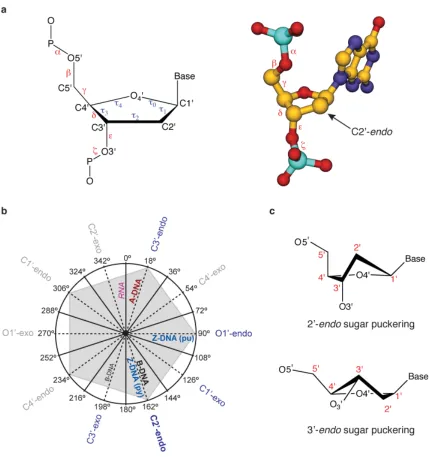
The sugar-phosphate backbone of DNA is highly dynamic allowing for a diverse range
of higher order structures depending on
environmental conditions. The torsion
angles for the sugar-phosphate backbone
are defi ned in Figure 1.3 and typically vary
with ionic strength, pH, sequence, and
many other factors.19,20 In constrast to RNA,
where the 2’-hydroxyl of the sugar locks the
A-form helix into a fairly rigid structure, the
sugar-phosphate backbone of DNA is highly
mobile.19,20 DNA conformation can often
be defi ned by the sugar puckering modes,
which by convention are named after the
ring atom and either endo or exo referring
to the 5’ side of the furanose ring or the 3’
[image:31.612.115.372.422.701.2]side, respectively. Figure 1.3 shows typical
sugar pucker conformations along with their preference in
nucleic acid structures. In addition to the sugars displaying
conformational preferences, the phosphodiester bond exhibits
conformational rigidity analogous to a peptide bond. This
conformation rigidity, known as the gauche effect, is a result
of stereoelectronic effects from lone pair hyperconjugation/
donation of the O3' and O5' oxygen atoms into the σ* orbital of the P-O5' and P-O3' bonds, respectively.23 Double helical
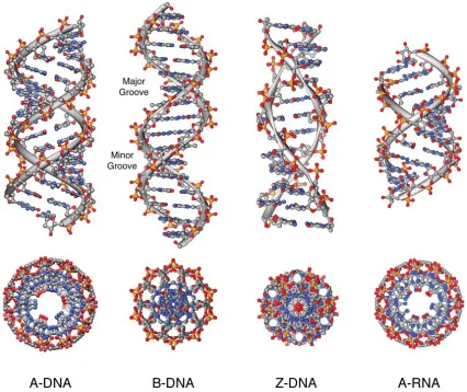
DNA is a dynamic structure which is capable of forming
three primary double strand conformations known as A, B,
and Z forms. In contrast to this, double helical RNA is far less
fl exible with the 2'-OH locking its sugar ring conformation
into a C3'-endo pucker resulting in a preference for an A form
helix similar to that of A-form DNA. A structural comparison
of these ideal DNA polymorphs along with A-form RNA is
shown in Figure 1.4 and Table 1.1.19,20,23,24,
In biological systems, especially eukaryotic cells, DNA is assembled around octameric
proteins called histones and compacted into macrohelical fi bers forming the high-order structure of
chromatin. This DNA-histone complex is called the nucleosome core particle (NCP) and represents
the fundamental repeating unit of chromatin consisting of 147 base pairs of DNA forming two
super helical turns around the histone octomer with 20-80 base pairs of linker DNA separating
one NCP from the next. The Richmond group25-27 at ETH Zurich has made seminal contributions
to elucidate biologically relevant higher-order DNA structures such as the NCP25,26 and the
tetra-NCP27 presented Figure 1.5. In addition, a theoretical model of four tetra-NCPs assembled into a
super-helical chromatin fi ber is presented in Figure 1.5. Chromatin architecture and accessibility in
biological systems represents a higher-order level of regulation and a profoundly important problem
for the fi eld of DNA recognition.
1.3 Molecular Recognition of DNA
One of the largest projects in modern science, the human genome project,28-30 is poised to
deliver detailed information and make major impacts in biotechnology and medicine through the
[image:32.612.110.268.87.287.2]physical and functional characterization of the approximately 20,000 to 25,000 genes in the human
genome. These genes are tightly regulated in higher organisms by transcription factor assemblies
that function in a concerted cooperative and combinatorial fashion to modulate eukaryotic gene
expression. The molecular recognition processes involved in nucleic acid-protein interations
are completely analogous to those of nucleic acid-drug interactions where initial complexation
is often driven by the hydrophobic effect. Optimization of the same forces is also required,
involving minimization of water exposed hydrophobic surfaces and maximization of van der
Waals interactions in conjuction with the optimization of all buried hydrogren bond donor and
acceptor pairings (solvent-assisted or counterion charge neutralization).31 The recognition of the
B-DNA interface by proteins and small molecules can occur at the major groove, minor groove,
and phosphate backbone, or any combination, with interactions mediated through electrostatics,
hydrogen bonding, and van der Waals interactions along with base pair stacking for the case of
[image:34.612.115.541.72.430.2]intercalators. The DNA base pair edges in the major groove and minor groove provide an array
of functionality for hydrogen bonding, hydrophobic interaction, and steric complementarity with
proteins and small molecule binders.5,8,16,17,31 The molecule electrostatic potential surfaces for the
minor and major groove base pair edges are shown in Figure 1.6.32 In addition, primary driving
forces such as the hydrophobic effect and shape complementarity are common to both proteins and
small molecules.
The regulation of gene transcription is controlled by the sequence specifi c cooperative
assembly of transcription factors, which form regulatory switches and networks in the cell
[image:35.612.109.531.101.540.2]DNA provide distinct surfaces for the interaction of transcription factors through specifi c and
nonspecifi c interactions such as (hydrogen bonding, electrostatics, van der Waals, etc.). Several
DNA-binding transcription factors are presented in Figure 1.7 to highlight the diverse architectures
used for recognizing DNA, ranging from homodimeric coiled coils interacting with the major
groove to monomeric beta-sheet containing proteins interacting with the minor groove. In addition
to homodimeric motifs, heterodimeric motifs are utilized along with metal ion coordinated
assemblies (i.e. Nf-κB p65-p50 and androgen receptor).33 Transcriptional co-activating proteins
serve to integrate information from transcription factor assemblies and modulate gene expression
through communication with RNA polymerase II leading to the transcription of protein-coding
regions in the eukaryotic genome.33,34
Transcription factors (TF) can communicate indirectly through allosteric modulation of DNA
resulting in cooperative assembly with very little direct protein-protein interaction. Transcription
factor binding can cause DNA-sequence dependent structural perturbations which modulate the
binding of the next TF. TF's can also interact directly through protein-protein interactions to increase
cooperativity. The β-enhancesome (Figure 1.8) is one such example of a cooperative assembly with cooperativity most likely arising at the DNA and coactivator levels. A conserved stretch of 55 bp's
(160 Å long) in a nucleosome free region of the IFN-β promoter serves as a regulatory element for the cooperative assembly of 8 proteins into a continuous surface, burying 72% of the DNA
solvent accessible area with very little protein-protein interaction.35 Transcriptional co-activating
proteins serve to integrate information from the assembly to modulate gene expression through
communication with RNA polymerase II leading to transcription.33,34
A diverse range of natural products and secondary metabolites have been shown to bind
DNA with interaction modes consisting primarily of either intercalation or groove binding.5,7,8
In addition, some ligands rely on a combination of intercalation and groove binding that can
also be augment by covalent modifying chemical domains, as in the case of anthramycin and
neocarzinostatin. A collection of diverse DNA binding natural products are shown in Figure 1.9
with echinomycin and daunomycin representing intercalators and anthramycin and dystamicin A
representing minor groove binders. The natural product distamycin (Figure 1.9) binds to A,T tracks
in the minor groove of DNA, four to fi ve base pairs in size, in both a 2:1 and a 1:1 ligand:DNA
stoichiometry.12-14 The affi nity and specifi city of dystamycin is controlled by a superposition of
shape complementarity, hydrophobic effects, and specifi c hydrogen bonding to the minor groove
of B-form DNA. Due to it's modular design of repeating pyrrole amino acids and ammenability
to rational modifi cation, distamycin has served as the inspiration for the design of several classes
sequence specifi c DNA minor-groove binders, with the ultimate goal of designing highly specifi c
targeted gene regulation agents.
1.4 DNA Recognition by Minor-Groove Binders
Prior to the fi rst structure of a molecule bound to DNA, specifi c recognition of B-form
DNA was predicted to occur in major groove rather than minor groove.5 An observation that was
based on the fact that the hydrogen bond acceptors at N3 of adenine and O2 of thyamine A/T base
pairs are similarly placed and lack any prominent distinguishing features.36 With the combination of
biophysical and structural data from NMR and X-ray studies, it was verifi ed that the minor groove of
B-form DNA was a legitimate target for specifi c recognition.12-15 (For crystal structures of netropsin
and distamycin A, see Figure 1.10.) Building upon inspiration from the natural products, netropsin
and dystamycin, minor groove binders have progressed to a modular molecular recognition platform
with high affi nity and specifi city for many different sequences of DNA.5,7,8,16,17
Over the past two decades, the development of minor groove DNA binders has evolved from
the initial discovery of the natural product dystamycin to a new class of programmable heterocyclic
oligomers demonstrating high affi nity and sequence specifi city.16,17 In addition to the incorporation
of alternative heterocycles such as imidazole that have enabled specifi city for guanine recognition
using the Im-Py pair, much research has gone into linking the two heterocyclic strands in a dimeric
motif.37-39 Covalent linkage of the two anti-parallel heterocyclic strands by a gamma amino butyric
acid (GABA) linkage results in increases in affi nity of 100–3600 fold relative to the unlinked
homodimeric motif.40,41 The incorporation of the turn linkage in the form of a GABA or substituted
GABA turn represented a major technological advance allowing for the fi rst time the incorporation
of unsymmetrical ring pairs for the targeting of non-palindromic DNA sequences.37 In addition,
covalent linkage of the two strands has led to sub-nanomolar increases in affi nity competing with
and often rivaling that of andogenous DNA binding proteins.16,17,37-39 This high affi nity modular
dimeric motif has allowed for the regulation of gene expression by direct interaction with the
protein interface.16,17
The four Watson-Crick base pairs can be differentiated by their molecular shape, electrostatic
potential, and positions of hydrogen bond donors and acceptors in the DNA minor groove fl oor.
The minor groove edge of a G•C base pair contains a steric hydrogen bond donating “bump” in the
form of the exocyclic amine of guanine. The steric properties of the exocyclic amine of guanine
form the basis for the A•T selectivity observed for netropsin and dystamycin binding due to steric
interaction with the edge of the pyrrole ring. It was discovered in a key study in the early 1990s that
imidazole in place of pyrrole in a three ring polyamide analogous to dystamycin could bind the 5
base pair sequence 5’-WGWCW-3’ (where W=A or T) resulting in a 2:1 polyamide-DNA complex
where the imidazole ring is stacked against a pyrrole ring allowing differentiation of G•C base pairs
from C•G, A•T, or T•A.42 The Im/Py pair has been used extensively in unlinked polyamides and in
turn linked polyamides culminating in the recent publication of a polyamide library, that represents
the solutions for targeting various 6 base pair sequences with high affi nity and specifi city.16,17,43
Thermodynamic studies have revealed that the Im/Py pair sequence selectivity is primarily driven
by favorable enthalpic factors44,45 and X-ray crystallographic studies in collaboration with the Rees
group provided structural insight based on a specifi c hydrogen bond between the imidazole lone
pair and the exocyclic amine of guanine in unlinked 2:1 homodimeric polyamides (Figure 1.11).46
Discrimination of T•A from A•T base pairs represents a much greater challenge due to the
ability of thyamine and adenine to both accept a hydrogen bond and the lack of unsymmetrical
steric features as in the G•C case.16,17 Despite this challenge, a small asymmetric cleft between the
C2 of adenine and the O2 of thyamine has been exploited for specific targeting by the N
-methyl-3-hydroxypyrrole/N-methylpyrrole (Hp/Py) pair, however affinities of these molecules are slightly
lower than their Py/Py containing counterparts.16,17 In another seminal structural study with the
Rees group on Hp containing 2:1 binders, it was revealed that a combination of shape selective
recognition of the asymmetric cleft along with a specific hydrogen bond between the Hp hydroxyl
[image:43.612.129.512.359.645.2]and the thymine O2 was responsible for the A•T specificity (Figure 1.12).47,48 The combination of
Py, Im, and Hp combined as unsymmetrical pairs in opposite strands of a unlinked homodimeric
or turn linked polyamide can be used to specifically recognize the four Watson-Crick base pairs
(Figure 1.13).16,17 These interactions can be described as a set of guidelines or pairing rules for the
design of sequence specific B-form DNA targeted polyamides where Im/Py specifies G•C and
Hp/Py specifies A•T. Some limitations do exist for certain sequences such as homopurine tracts,
certain G-rich sequences, and sequences beyond 6 base pairs due to the sequence-dependent DNA
microstructure and overcurvature of longer polyamides, however unique solutions to some of these
problems have been developed (i.e. incorporation of a flexible β-Ala residue in 1:1 and hairpin polyamide motifs).16,17
In addition to the 2:1 and hairpin polyamide architectures many other strand linkage strategies
have been explored such as linking through the N-methyl groups on the central heterocycles (H-pin
motif)49-50 or in the terminal heterocycles (U-pin motif).51 However, one of the highest affi nity and
in some cases most specifi c polyamide architectures has been the covalent linking of the C- to
N-termini at both ends of the polyamide into a macrocycle, eliminating all possibility of extended
binding modes.41,52-55 Macrocyclic γ-turn linked polyamides were fi rst explored as 6 ring systems
targeting a 5 base pair sequence of DNA in 1995 and were shown to have signifi cantly higher
affi nity, however their specifi city versus mismatch DNA was only 3-fold compared to 40-fold
for their hairpin counterparts.52 Mainly due to limitations in synthetic methodology and initial
discouraging thermodynamic results the cyclic polyamide motif was not investigated further
until 1999.41 After improvements in solid-phase synthetic methodology, nanomole to micromole
quantities of polyamides could be readily synthesized although cyclic polyamides still remained
challenging.53-55 Using solid-phase methods cyclic polyamides were reinvestigated with two major
architectural changes.41,53-55 The fi rst being the use of an 8 rings system as oppose to 6 in the original
studies and the second major change was moving the charge from the pyrrole N-methyl group to the
alpha position of the γ-turn in the form of (R)-2,4-diaminobutyric acid, that had been discovered to increase the affi nity, sequence specifi city, and orientational preference of hairpin polyamides. This
second generation cyclic 8 ring polyamide motif was found to have greatly improved specifi city
and affi nity over its hairpin and unlinked counterparts targeting the sequence 5'-AGTACT-3'.41
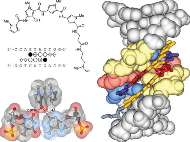
The results of covalent attachment of the two polyamide strands can be seen in Figure 1.14. In
a second study, multiple Hp/Py pairs were introduced into the 8-ring cyclic polyamide motif,
which resulted in increased affi nity and specifi city relative to hairpin polyamides targeted to the
sequences 5'-TGAACT-3' and 5'-TGATCT-3'.53 Despite these advances the cyclic polyamide motif
has received little attention relative to its hairpin counterpart mainly due to synthetic limitations.
The chiral (R)-2,4-diaminobutyric acid turn (α-turn) was a major advance in polyamide design not only for the cyclic polyamides but primarily for the hairpin motif.38 The addition of
an amino substituent to the alpha position of the γ-turn helps to disfavor extended 1:1 binding
modes and reverse binding due to a steric clash with the minor groove fl oor. In addition, the chiral
amino turn helps to increase the overall affi nity of polyamides while maintaining specifi city and
improving water solubility.38 The chiral α-turn was proposed to increase affi nity through electrostatic
interactions between the protonated cationic amine group and the anionic DNA backbone however
addition to substitution of the γ-turn at the alpha position, many other variations have been studied with shorter turns, longer turns, and conformationally constrained variations.16,17,56 The most recent
and one of the most successful advances in polyamide turn technology is the β-amino γ-linked
turn.39 Recent studies have demonstrated that this turn can provide substantial increases in affi nity
for certain polyamide sequences, however the effect is less pronounced as the imidazole content of
the polyamide is increased (Figure 1.15).39
NMR structural studies using NOESY-restrained molecular dynamics models have also
provided insight into 1:1 and 6-ring cyclic polyamides complexed with DNA.55,57 Figure 1.16 shows
an NMR model of a 1:1 polyamide with 11 out of 40 of the best calculated models overlayed.57
This shows very small coordinate deviation towards the center of the DNA helix and bound
polyamide with increasing conformational mobility at the ends. The 6-ring cyclic polyamide model
represented in Figure 1.16 shows an overlay of 21 of the best calculated models.55 This structure
shows signifi cant conformational mobility in the 6-ring cyclic complex with a highly fl exible DNA
sugar-phosphate backbone.
X-ray crystallographic studies resulting from collaborations between the Rees and Dervan
groups have provided valuable insight into the polyamide-DNA molecular recognition process by
elucidating the structure of fi ve 2:1 polyamide-DNA complexes at a resolution ranging from 2.00
to 2.27 Å and a summary comparing specifi c structural parameters is shown in Figure 1.17.46-48
These crystallographic studies revealed that the DNA rise per base pair matches the polyamide rise
Figure 1.18
X-ray crystal structures of polyamide-NCP complexes. a) Five polyamides in com-plex with the NCP at 2.30 Å resolution. b) Two polyamides in comcom-plex with the NCP at 2.45 Å resolution. c) One polyamides in complex with the NCP at 2.65 Å resolution.per residue, however the polyamide structure is over-curved with respect to the DNA minor groove
and shape complementarity is lost beyond a sequence of 5 contiguous base pairs.46 In addition,
these crystallographic studies elucidated the basis for GC recognition by the Im/Py pair46 and TA
recognition by the Hp/Py pair47,48 providing fundamental insight into polyamide binding. A variety
of space groups were observed including monoclinic, orthorhombic, and trigonal with resolutions
ranging from 2.00 to 2.27 Å. Average R-factors were in the mid-20s and polyamide B-factors
averaged 47 to 86 Å2 whereas DNA B-factors averaged 43 to 67 Å2 for all structures presented in
Figure 1.17. The 2:1 binding polyamide crystal structures also frequently exhibited disorder in the
polyamide tail region and was usually modeled in alterate conformations refl ecting the dynamic
nature of the β-alanine linked dimethylamino propylamine terminus.
DNA binding polyamides are also able to bind sequence specifi cally to DNA on the
nucleosome core particle.58 Hairpin polyamide-NCP crystal structures have been solved at
resolutions ranging from 2.05 to 2.65 Å providing structural proof that polyamides can bind
biologically relevant higher-order DNA structure however a combination of resolution limits and
high B-factors for the polyamide prevented a detailed picture beyond confi rmation of the polyamide
binding location (Figures 1.18 and 1.19).58,59 The current state of macromolecular crystallography,
with regard to minor groove binding DNA-drug structures, was assessed prior to beginning the
structural work presented in Chapter 5 and 6 of this thesis and is presented in Figure 1.20. This
survey demonstrates the lack of high resolution structures of DNA minor groove binders and the
notable absence of linked dimeric minor groove binder structures. This survey underscores the
pressing need for atomic resolution X-ray crystal structures of DNA minor groove binders to truly
understand the molecular basis of recognition.
1.5 Scope of this work
The work presented in this thesis is focused on the molecular recognition of DNA by
minor groove binding polyamides. In Chapter 2 of this thesis, a solution-phase synthesis of
pyrrole-imidazole polyamides is presented with optimized protocols utilizing little to no chromatography.
Chapter 3 builds on synthetic methodology in Chapter 2 allowing the effi cient synthesis of cyclic
polyamides targeted to the androgen response element. This chapter demonstrates that cyclic
polyamides can be synthesized in an effi cient manner, are biologically active and cell permeable
in cell culture experiments, and rival the binding affi nity of most other polyamide architectures.
Figure 1.20
Current state of macromolecular crystallography: A DNA-drug perspective. Data compiled from the PDB on 11/04/2007.60 (The number of structures solved is designated inbinding ability of higher order macrocycles. The structural elucidation of an α-amino-turn-linked cyclic polyamide is presented in Chapter 5 at 1.18 Å resolution providing insight into the detailed
molecular recognition processes. Chaper 6 details the structural elucidation of a β -amino-turn-linked cyclic p