The Effects of Acute and
Chronic Exercise on
Brain Derived Neurotrophic Factor and
Cognitive Performance in
Mild Cognitive Impairment
Kate Elizabeth Devenney
Bsc. Physiotherapy
Supervised by Professor Brian Lawlor and Dr Emer Guinan
Submitted for the degree of Doctor of Philosophy
Trinity College Dublin, the University of Dublin
Discipline of Physiotherapy
School of Medicine
i
Declaration
This thesis is submitted by the undersigned to Trinity College for the examination of the degree in Doctor of Philosophy.
All work described herein is entirely my own work except where otherwise stated and has not been submitted as an exercise for a degree at this or any other University. I hereby agree to deposit this thesis in the University’s open access institutional repository or allow the library to do so on my behalf, subject to Irish Copyright Legislation and Trinity College Library conditions of use and acknowledgement.
_________________________________
ii
Summary
A growing body of literature suggests that structured exercise interventions may help slow the progression of cognitive decline in people with Mild Cognitive Impairment (MCI), yet little is known about the effects of acute (a single bout of exercise) and chronic exercise on brain derived neurotrophic factor (BDNF) concentration and cognitive performance in this patient population. BDNF, a neurotrophin widely expressed in the brain is upregulated through exercise and believed to enhance neuronal survival, brain plasticity and learning and memory. Exploring the BDNF response to acute and chronic exercise may offer new insights into the underlying mechanism that mediates the exercise-cognitive relationship in a cognitively impaired population. Accordingly, the overarching aim of this thesis was to investigate the effect of acute (varying modality, duration or intensity) and chronic exercise on serum BDNF concentration and cognitive performance in a group of sedentary older adults with MCI.
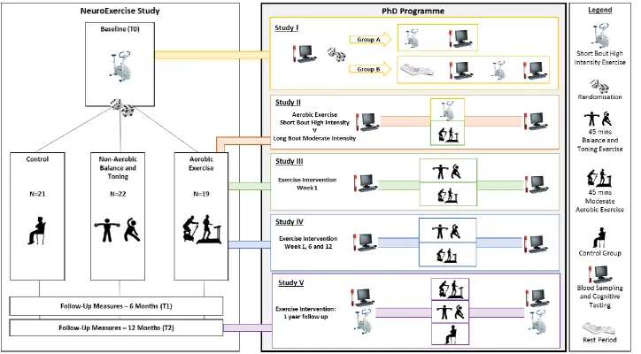
Work on this study began in September 2015 when I commenced a full-time research post as a PhD candidate on the NeuroExercise study. The NeuroExercise study is a European multi-centre Randomised Controlled Trial (RCT) examining ‘The Effects of an Extensive Exercise Programme on the Progression of Mild Cognitive Impairment’. Funding for the NeuroExercise study was awarded by the EU Joint Programme – Neurodegenerative Disease (JPND) Research grant with funding for the Dublin arm of the trial supported by the Health Research Board (HRB). I had responsibility for overseeing study implementation and coordination of the NeuroExercise study at the Dublin site which ran for a 32-month duration. Duties included ethics applications, participant screening and recruitment, study assessments, implementation of the 12-month NeuroExercise exercise protocol, data management and liaising with local referring sites and international partners. The NeuroExercise study recruited from the German Sports University in Cologne, Radboud University Medical Centre in Nijmegen, the Netherlands and Trinity College Dublin. In this study, participants (n=62 at the Dublin site) were randomised to either a yearlong aerobic exercise intervention, balance and toning (non-aerobic) exercise intervention or control group. The primary outcome was change in cognitive performance as measured by a neuropsychological test battery.
iii
this, specific research questions and a study protocol was formulated. Participants with MCI, recruited to participate in the NeuroExercise study also consented to participate in all studies within this PhD thesis. Studies I - III were designed to address gaps within the literature and answer specific research questions around acute exercise, BDNF and cognitive performance in an MCI cohort. Results in Study I demonstrated that a short bout of high intensity exercise increased serum BDNF (sBDNF) concentration compared to a resting control condition. While high intensity interventions were feasible in this patient group, the increase in sBDNF did not result in improvements in cognitive performance. Study II and III examined the effect of manipulating acute exercise variables (intensity, duration and mode) on sBDNF concentration and cognitive performance. Results found no difference between varying aerobic exercise conditions (short bout high intensity versus longer bout moderate intensity) or between two distinct exercise modalities (aerobic versus balance and toning) on study outcomes.
Study IV and V examined the effects of purposeful structured exercise training. Study IV investigated whether 12 weeks of exercise training: 1) altered basal sBDNF concentration and cognitive performance and 2) augmented the acute exercise BDNF and cognitive response. Results revealed significant increases in basal sBDNF concentration following 6 weeks of supervised aerobic or balance and toning exercise training. Finally, Study V provided insights into the relationship between sBDNF concentration, episodic memory and cardiorespiratory fitness following participation in a 1-year exercise intervention. Results showed a 1-year exercise intervention with poor levels of adherence did not impact BDNF or cognition.
iv
Acknowledgement
First and foremost to my supervisors. Making the move from a clinical physiotherapy post to a research position was a daunting process and would not have been possible without your support and guidance. To Professor Brian Lawlor, thank you for welcoming me to the memory clinic team in St. James’s Hospital and for your leadership and support over the last few years. Your advocacy work, and tireless pursuit in research will leave a lasting impact on the lives of people living with dementia in Ireland. To Dr Emer Guinan, who was always a voice of reason, a wealth of wisdom and provided sound advice throughout. Thank you for your patience - no problem was ever too big or too small and you continually gave selflessly of your time, energy and expertise. I wish to thank you unreservedly for your unwavering support and endless reading of chapters (even when you were supposed to be off the clock!). I couldn’t have done this without you.
To all the staff past and present at the memory clinic, particularly Rachel Farley, Irene Bruce and Dr Robert Coen – thank you for giving of your time and assisting me in the study design and with study recruitment. I greatly admire the work, care, compassion, and commitment you demonstrate to patients attending the memory clinic service.
The work on this thesis would not have been possible without a cohesive team effort. To both local and international colleagues who collaborated on the NeuroExercise study. Special thanks to Dr Damien Ferguson, who provided exceptional medical cover to study participants. To Leona Connolly – thank you for assisting with NeuroExercise study assessments. To all the research assistants that worked on the NeuroExercise study – Andrew Brooks, Louise O’ Connor, Sophie Reynolds and Megan O’Grady. A special big thanks to Gemma Foley, research assistant extraordinaire who seamlessly helped with day-to-day operations of the NeuroExercise study to allow me time to work on my PhD. Thank you so much for sticking it out until the bitter end!
To all the participants in the NeuroExercise study – thank you for giving so generously of your time and energy to this study. I thoroughly enjoyed getting to know you all and chatting over the occasional coffee and croissant after classes.
v
assistance and expertise in the laboratory analysis of over 350 biological samples which was greatly appreciated and of huge added value to the project. To Cathal Walsh – thank you for providing advice on statistical approach used across studies contained within this thesis.
To Professor John Gormley, thank you for allowing me to expand my horizons beyond PhD work by giving me the opportunity to grow my teaching practice over the years. To Professor Juliette Hussey, many thanks for taking time to read and provide valuable feedback on thesis chapters.
Thank you to the Physiotherapy Department in St. James’s Hospital particularly to the manager, Niamh Murphy who has been a constant support and mentor in my professional development throughout my career.
To my friends in the post grad room who understand the many ups and downs of the PhD process. To my wide and varied groups of friends outside of the research world - Girl Crew Centra, Sound People and LHC – thanks for the wine and cheese, coffee and chocolate but above all the chats and laughter that I needed to get through the last few years. To my family, particularly my parents Mo and Declan, thank you for your love and support. From a young age, you instilled the mantra “you can do anything you want to do”, although I think the PhD has pushed the limits of this statement! Final mention has to go to Dr Emma Dunne, my girlfriend and best friend. Thank you for being my number one supporter always.
vi
Table of Contents
Declaration ... i
Summary ... ii
Acknowledgement ... iv
Table of Contents ... vi
List of Figures ... xiii
List of Tables ... xiv
List of Abbreviations ... xvi
List of Appendices ... xviii
Dissemination of Research ... xix
1 Chapter 1: Introduction... 1
1.1 Mild Cognitive Impairment ... 1
1.2 The aging brain ... 2
1.3 Protective factors against cognitive decline ... 3
1.4 Cognitive function, physical activity and exercise ... 5
1.5 The impact of acute and chronic exercise on cognitive function ... 7
1.6 Brain Derived Neurotrophic Factor ... 8
1.6.1 BDNF in aging, MCI and AD ... 9
1.6.2 BDNF response to exercise ... 10
1.7 Acute exercise, BDNF and cognitive performance ... 12
1.8 Summary ... 13
2 Chapter 2: Methodology ... 17
2.1 Introduction ... 17
2.2 Background research methods ... 18
2.2.1 Study designs ... 18
2.2.2 Sampling ... 19
2.2.3 Reliability and validity ... 20
vii
2.2.5 Statistical approach in this thesis ... 22
2.3 Overview of the NeuroExercise Study ... 23
2.3.1 Ethics approval ... 23
2.3.2 Recruitment and screening ... 23
2.3.3 Diagnostic criteria... 24
2.3.4 Inclusion criteria ... 24
2.3.5 Exclusion criteria ... 25
2.3.6 NeuroExercise intervention ... 26
2.3.7 Relationship between the NeuroExercise Study and this thesis. ... 26
2.4 Exercise conditions ... 26
2.4.1 Exercise condition 1: Short bout high intensity aerobic exercise ... 26
2.4.2 Exercise condition 2: 45 minutes moderate intensity aerobic exercise .. 32
2.4.3 Exercise condition 3: 45 minutes balance and toning exercise (non-aerobic) 33 2.5 Materials ... 36
2.5.1 Blood sampling ... 36
2.5.2 Blood collection ... 36
2.5.3 BDNF analysis ... 36
2.5.4 Enzyme linked immunosorbent assay (ELISA) ... 36
2.5.5 Cognitive testing ... 38
2.5.6 Cambridge Neuropsychological Test Automated Batteries (CANTAB): Paired Associate Learning Task ... 38
2.5.7 Sustained Attention to Response Task (SART) ... 41
2.5.8 Stroop Neuropsychological Screening Test (SNST) ... 45
2.6 Conclusion ... 48
3 Chapter 3: The effect of structured aerobic exercise on cognitive performance in individuals with MCI: a systematic review ... 49
3.1 Introduction ... 49
3.2 Methods ... 50
viii
3.2.2 Inclusion criteria ... 50
3.3 Results ... 51
3.3.1 Identification of studies ... 51
3.3.2 Study quality ... 52
3.3.3 Study design and subject characteristics ... 55
3.3.4 The effect of exercise on measures of cognitive performance ... 55
3.3.5 Biomarkers ... 67
3.3.6 Cardiorespiratory fitness ... 68
3.4 Discussion ... 70
3.5 Conclusion ... 73
4 Chapter 4: Study I: High intensity acute aerobic exercise increases brain derived neurotrophic factor in mild cognitive impairment: a randomised controlled study ... 74
4.1 Introduction ... 74
4.2 Aims and objectives ... 74
4.3 Methods ... 75
4.3.1 Study design ... 75
4.3.2 Participants ... 75
4.3.3 Study outcomes ... 76
4.3.4 Blood sampling for BDNF measurement ... 76
4.3.5 Cognitive performance ... 76
4.3.6 Intervention ... 76
4.3.7 Sample size ... 76
4.3.8 Statistical analysis ... 76
4.4 Results ... 77
4.4.1 Participant characteristics ... 77
4.4.2 Primary analysis: between group difference in BDNF and cognitive performance ... 80
4.4.3 Secondary analysis: change over time within group B ... 81
4.4.4 Sex differences in acute exercise BDNF and cognitive response ... 83
ix
4.6 Conclusion ... 86
5 Chapter 5: Study II: A comparison of contrasting aerobic exercise protocols on serum brain derived neurotrophic factor concentration and cognitive performance in mild cognitive impairment ... 87
5.1 Introduction ... 87
5.2 Aims and objectives ... 88
5.3 Methods ... 89
5.3.1 Participant recruitment ... 89
5.3.2 Study design ... 89
5.3.3 Outcomes ... 90
5.3.4 Blood sampling ... 90
5.3.5 Cognitive performance ... 90
5.4 Intervention ... 90
5.4.1 Condition A: Short duration high intensity ... 90
5.4.2 Condition B: Long duration moderate intensity ... 90
5.4.3 Statistical analysis ... 91
5.5 Results ... 91
5.5.1 Exercise parameters ... 92
5.5.2 Participant characteristics ... 92
5.5.3 Difference between conditions: BDNF ... 93
5.5.4 Difference between conditions: cognitive performance ... 93
5.6 Discussion ... 97
5.7 Conclusion ... 99
6 Chapter 6: Study III: An investigation of two distinct acute exercise paradigms on serum brain derived neurotrophic factor concentration and cognitive performance in mild cognitive impairment ... 100
6.1 Introduction ... 100
6.2 Aims and objectives ... 100
6.3 Methods ... 101
x
6.3.2 Study design ... 101
6.3.3 Outcomes ... 102
6.3.4 Blood sampling ... 102
6.3.5 Cognitive performance ... 102
6.3.6 Exercise conditions ... 102
6.3.7 Statistical analysis ... 103
6.4 Results ... 103
6.4.1 Participant characteristics ... 103
6.4.2 Brain Derived Neurotrophic Factor ... 105
6.4.3 Cognitive performance ... 105
6.5 Discussion ... 108
6.6 Conclusion ... 109
7 Chapter 7: Study IV: The impact of acute and chronic exercise on serum brain derived neurotrophic factor concentration and cognitive performance following 6 and 12 weeks of structured exercise training in mild cognitive impairment ... 110
7.1 Introduction ... 110
7.2 Aims and objectives ... 111
7.3 Methods ... 111
7.3.1 Participant recruitment ... 111
7.3.2 Study design ... 111
7.3.3 Blood sampling ... 112
7.3.4 Cognitive performance ... 112
7.3.5 Intervention ... 113
7.3.6 Statistical analysis ... 113
7.4 Results ... 113
7.4.1 Adherence to exercise intervention ... 113
7.4.2 Participant characteristics ... 114
7.4.3 Brain Derived Neurotropic Factor ... 115
7.4.4 Cognitive performance ... 119
xi
7.6 Conclusion ... 125
8 Chapter 8: Study V: Peripheral brain derived neurotrophic factor and episodic memory in mild cognitive impairment: results from a one year exercise intervention study 126 8.1 Introduction ... 126
8.2 Aims and objectives ... 127
8.3 Methods ... 127
8.3.1 Participant recruitment and randomisation ... 127
8.3.2 Study design ... 128
8.3.3 Outcomes ... 128
8.3.4 Blood sampling ... 128
8.3.5 Cognitive measures ... 128
8.3.6 Episodic memory... 128
8.3.7 Global cognition ... 129
8.3.8 Cardiorespiratory fitness ... 129
8.3.9 Intervention ... 130
8.3.10 Statistical analysis ... 130
8.4 Results ... 131
8.4.1 Adherence to exercise intervention ... 131
8.4.2 Participant characteristics ... 131
8.4.3 Recruitment (memory clinic versus community participants) ... 134
8.4.4 Change in BDNF, cognitive function and cardiorespiratory fitness ... 134
8.4.5 Brain Derived Neurotrophic Factor ... 134
8.4.6 Cognitive function... 135
8.4.7 Cardiorespiratory fitness ... 135
8.4.8 Correlation ... 138
8.5 Discussion ... 138
8.6 Conclusion ... 141
9 Chapter 9: General Discussion ... 142
xii
9.2 Analysis of key points ... 143
9.3 Acute exercise and BDNF ... 143
9.4 Chronic exercise and BDNF ... 144
9.5 Cardiorespiratory fitness and BDNF in MCI... 146
9.6 Acute exercise and cognitive performance ... 147
9.7 Chronic exercise and cognitive performance ... 148
9.8 Critical Analysis ... 150
9.8.1 Study sample ... 151
9.9 Adherence ... 152
9.10 Considerations in BDNF analysis ... 153
9.10.1 Blood sampling ... 155
9.11 Cognitive testing ... 155
9.12 Exercise conditions ... 156
9.13 Potential bias ... 156
9.14 Future Directions ... 157
9.15 Summary ... 157
10 References ... 159
xiii
List of Figures
Figure 1:1 Annual rates of conversion from mild cognitive impairment (MCI) to dementia
over 48 months ... 2
Figure 1:2 The timeline of risk factors and protective factors for dementia. ... 4
Figure 1:3 Potential brain mechanisms for preventive strategies in dementia ... 5
Figure 2:1 Recruitment, screening and selection of participants to the NeuroExercise Study ... 24
Figure 2:2 Cosmed bike and K4B2 equipment ... 29
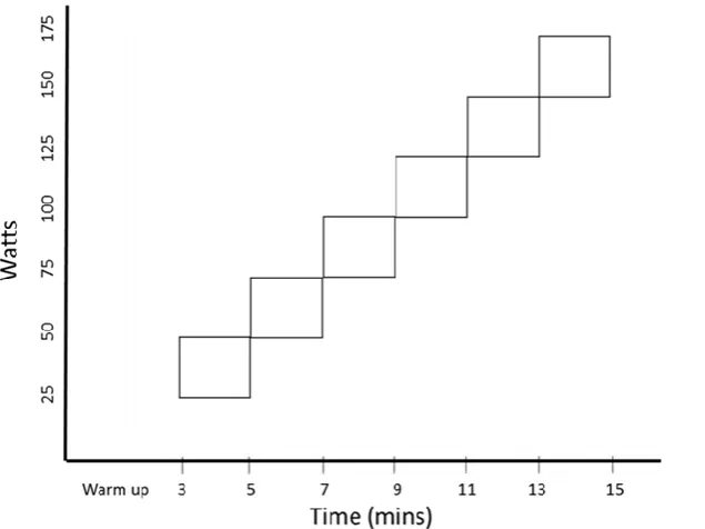
Figure 2:3 WHO exercise test progression ... 31
Figure 2:4 Timeline outlining PhD studies relative to the NeuroExercise Study ... 35
Figure 2:5 iPad interface showing PAL task ... 39
Figure 2:6 Laptop displaying SART programme ... 42
Figure 2:7 Procedure fixed version SART ... 43
Figure 2:8 Stroop Neuropsychological Screening Test ... 46
Figure 3:1 PRISMA diagram showing flow of studies into review ... 52
Figure 4:1 Study design and schedule of outcome assessments in Study I ... 75
Figure 4:2 Boxplot showing the change in serum BDNF in response to a high intensity exercise or resting control condition ... 80
Figure 5:1 Outline of study design, intervention and outcome assessments in Study II ... 89
Figure 6:1 Study design showing study outcome measures and intervention in Study III ... 102
Figure 7:1 Study design outlining study outcomes measures and intervention in Study IV ... 112
xiv
List of Tables
Table 1-1 Overview of the studies in the PhD programme outlining study title, aims and
study type. ... 15
Table 2-1 American College of Sports Medicine indications for stopping exercise testing ... 28
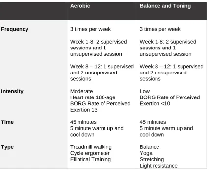
Table 2-2 Components of exercise prescription are described using the FITT principle: Frequency, Intensity, Time (duration) and Type. ... 32
Table 2-3 Capture Antibody Concentration in BDNF analysis ... 37
Table 2-4 PAL task outcome measured ... 40
Table 2-5 SART outcomes ... 44
Table 2-6 Stroop task score and description ... 47
Table 3-1 Risk of bias for RCTs assessed using the Cochrane collaboration risk of bias tool ... 53
Table 3-2 ROBINS-I: Risk Of Bias In Non-randomised Studies of Interventions ... 54
Table 3-3 Summary of study design, intervention and outcomes of included studies. Exercise interventions described in line with the FITT (frequency, intensity, time and type) principle... 56
Table 3-4 Studies that included an objective measurement of cardiorespiratory fitness pre-post structured aerobic exercise intervention ... 69
Table 4-1 Baseline participant characteristics Study IV. ... 78
Table 4-2 Objective measurements of exercise intensity and duration during performance of the exercise condition (short bout high intensity) in group A and group B. . ... 78
Table 4-3 Effect of intervention (exercise v control) on BDNF and cognitive outcomes. ... 79
Table 4-4 Secondary analysis measuring change over time in study outcomes within group B. ... 82
Table 5-1 Exercise prescription parameters collected during condition A (short bout high intensity) and condition B (long bout moderate intensity). ... 92
Table 5-2 Baseline participant characteristics Study II. ... 93
Table 5-3 Effects of contrasting aerobic exercise protocols on BDNF and cognitive outcomes. ... 95
xv
xvi
List of Abbreviations
ACSM American College of Sports Medicine
AD Alzheimer’s disease
ADAS-cog Alzheimer's Disease Assessment Scale-cognitive subscale
aMCI Amnestic Mild Cognitive Impairment
AAN American Academy of Neurology
ANOVA Analysis of Variance
ApoE Apolipoprotein E
AT Aerobic Exercise Training
AVLT Auditory Verbal Learning Test
BAT Balance and Toning Exercise
BBB Blood Brain Barrier
BDNF Brain Derived Neurotrophic Factor
BP Blood Pressure
CANTAB Cambridge Neuropsychological Test Automated Batteries
CE Commission Errors
CFQ Cognitive Failures Questionnaire
CI Confidence Interval
CPET Cardiopulmonary Exercise Testing
CR Cardiorespiratory
CRF Clinical Research Facility
CRT Choice Reaction Time
DSST Digit Symbol Substitution Test
ECG Electrocardiogram
EDTA Ethylenediamine Tetraacetic Acid
EG Emer Guinan
ES Effect Size
FITT Frequency Intensity Time Type
HRB Health Research Board
HR Heart Rate
HRmax Heart Rate Max
HRR Heart Rate Reserve
ICCs Intraclass correlation coefficients
IGF-1 Insulin Growth Factor-1
xvii
KD Kate Devenney
M Mean
Md Median
MCI Mild Cognitive Impairment
MDC Minimal Detectable Change
METs Metabolic Equivalent
MMSE Mini–Mental State Examination
MoCA Montreal Cognitive Assessment
MRI Magnetic Resonance Imaging
PA Physical Activity
PAL Paired Associate Learning
PALTEA Paired Associate Learning Total Errors Adjusted PPMCC Pearson Product Moment Correlation Coefficients
RAVLT Rey Auditory Verbal Learning Test
RCT Randomised Controlled Trial
REC Research and Ethics Committee
ROCF Rey–Osterrieth Complex Figure
RPE Rate of Perceived Exertion
RT Reaction Time
SART Sustained Attention to Response Task
sBDNF Serum Brain Derived Neurotrophic Factor
SCWT Stroop Colour Word Task
SDMT Symbol Digit Modalities Test
SJH St. James’s Hospital
SNST Stroop Neuropsychological Screening Test
SD Standard Deviation
SRT Simple Reaction Time
TBI Traumatic Brain Injury
TMT Trail Making Test
VO2max Maximal Oxygen Uptake
VO2peak Peak Oxygen Uptake
WAIS Wechsler Adult Intelligence Scale
xviii
List of Appendices
Appendix I: The Effects of an Extensive Exercise Programme on the Progression of Mild Cognitive Impairment (MCI): Study Protocol for a Randomised Controlled Trial
Appendix II: Real World Recruiting of Older Subjects with Mild Cognitive Impairment for Exercise Trials: Community Readiness is Pivotal
Appendix III: Exercise and Alzheimer’s disease: Current Perspectives
Appendix IV: Joint Research and Ethics Committee Approval from St. James’s Hospital / Tallaght Hospital
Appendix V: Ethics Committee Approvals from Mater Misericordiae University Hospital, St. Patrick’s University Hospital, St. Vincent’s University Hospital
Appendix VI: Study PIL and Consent Form
Appendix VII: Study Advertisement
Appendix VIII: Study Poster
Appendix IX: Home Exercise Diary
Appendix X: Absolute and Relative Indications for Stopping Exercise
Appendix XI: Borg’s Rating of Perceived Exertion
Appendix XII: Case Report Form Baseline Assessment
Appendix XIII: Case Report Forms Exercise Classes
xix
Dissemination of Research
Bursary award:
• Recipient of the 2017 Irish Society of Chartered Physiotherapists Glennon AIG Bursary (€2500) for my project entitled “The effects of acute exercise on cognitive performance and brain-derived neurotrophic factor (BDNF) in individuals with Mild Cognitive Impairment (MCI)
Published papers:
• Devenney, K.E., Sanders, M.L., Lawlor, B., Rikkert, M.G.O. and Schneider, S.,
2017. The effects of an extensive exercise programme on the progression of Mild Cognitive Impairment (MCI): study protocol for a randomised controlled trial. BMC geriatrics, 17(1), p.75. (Appendix I)
• Sanders, M.L., Stuckenschneider, T., Devenney, K.E., Lawlor, B., Schneider, S., Rikkert, O., Marcel, G.M. and NeuroExercise Study Group, 2018. Real World Recruiting of Older Subjects with Mild Cognitive Impairment for Exercise Trials: Community Readiness is Pivotal. Journal of Alzheimer's Disease, 62(2), pp.579-581. (Appendix II)
Papers under review:
• High intensity acute aerobic exercise effects Brain Derived Neurotrophic Factor (BDNF) in Mild Cognitive Impairment (MCI): a randomised controlled study. 2018. Devenney, K.E, Guinan, E.M., Mota, B.C, Kelly, A.M., Olde Rikkert, M.G.M., Schneider S., and Lawlor, B.
Currently submitted for British Journal of Sports Medicine for review
Conference oral presentations:
Irish Gerontological Society - Postgraduate Study Day in Ageing Research - Friday 27th April 2018, St Vincent's Hospital, Dublin
xx
Marcel Olde Rikkert (Nijmegen/Netherlands), Stefan Schneider (Cologne/Germany), Emer Guinan (Dublin/Ireland)
Health Across Lifespan (HAL) - International conference on healthiness and fitness across the lifespan. September 12–15, 2018, Otto von Guericke University Magdeburg, Germany. Winner of 3rd prize for Best Oral Presentation / Young Researcher Award of HAL 2018. Presentation title as above.
Public and patient dissemination:
• Sharing of recruitment experiences with CANDI research investigators meeting April 4th 2018, Trinity College Dublin
• Public talk on ‘Exercise and Brain Health’ at the Alzheimer’s Café Glasnevin April 2018
• Education session ‘Exercise and Brain Health’ given to the cognitive rehabilitation MCI group at the memory clinic, St. James’s Hospital
• Key note speaker at the Physiotherapy Department Research Presentations, St. James’s Hospital July 2018
1
1 Chapter 1: Introduction
1.1 Mild Cognitive Impairment
Mild Cognitive Impairment (MCI) is a clinical syndrome with a continuum of symptoms and is regarded as an intermediate stage between the cognitive change associated with normal aging and dementia (Petersen, 2004). MCI is marked by either focal or multifocal cognitive impairment, with minimal impairment of instrumental activities of daily living, which is a defining threshold for a dementia diagnosis (Dubois et al., 2010). MCI can be broadly classified into two subtypes: 1) amnestic MCI is a syndrome in which memory dysfunction is the predominant cognitive impairment and 2) non-amnestic MCI in which the primary cognitive impairment is established within another cognitive domain (e.g., language, visuospatial, executive) (Gauthier et al., 2006, Petersen and Negash, 2008).
2
Figure 1:1 Annual rates of conversion from mild cognitive impairment (MCI) to
dementia over 48 months (Petersen et al., 1999)
As our ageing population increases, the number of people living with dementia in Ireland is expected to rise to over 130,000 by 2041 (Pierce et al., 2014), which is consistent with the rising occurrence of dementia worldwide (Ferri et al., 2005). The estimated annual cost of dementia care in Ireland in 2010 was over €1.69 billion (Cahill et al., 2012). At the 2013 summit on dementia convened by G8 in London, a range of initiatives were announced in which leading nations made commitments to actively work towards a cure or disease modifying treatment for dementia by 2025 (Dementia, 2014). With the ongoing difficulties’ in establishing new disease modifying drugs, research studies into non-pharmacological strategies to prevent dementia, and to optimise cognitive function in older people, particularly individuals at risk of developing dementia, have been increasing in number (Pieramico et al., 2014).
1.2 The aging brain
As people age, there is a decline in a number of physiological functions including reduced immune function, loss of muscle mass and strength, a decrease in bone mineral density and an increase in fat mass (Lamberts et al., 1997, Rudman and Rao, 1992). Furthermore, increases in blood pressure and inflammatory load are also known to occur during the aging process, with both independently linked to risk for dementia and cognitive decline (Qiu et al., 2005, Singh and Newman, 2011, Yaffe et al., 2004).
3
working memory), and appear especially sensitive to age-related change (Rypma and D'Esposito, 2000, Salthouse, 1996, Kausler, 1994, De Luca et al., 2003, Singh-Manoux et al., 2012, Burke and Barnes, 2006, Glisky, 2007). The susceptibility to learning and memory impairments is generally attributed to a decrease in neuronal plasticity of the cortex and hippocampus, which is considered vulnerable to the aging process and has been directly related to the decline of cognitive performance (Barrientos et al., 2015, Morrison and Baxter, 2012).
In aMCI, memory impairments (recognition memory and free recall) arise early in the clinical presentation because they rely heavily on areas affected in the early stages of Alzheimer’s neuropathology i.e. the spread of neurofibrillary tangles and neuropil threads in the medial temporal lobe, initially in the transentorhinal cortex and then in the entorhinal cortex and hippocampus (Braak and Braak, 1991, Petersen et al., 2006). Longitudinal Magnetic Resonance Imaging studies have reported significantly greater grey matter loss in the hippocampal area, inferior and middle temporal gyrus, posterior cingulate, and precuneus in those with MCI who transition to AD (Chetelat et al., 2005, Fennema-Notestine et al., 2009). However, an autopsy study found those clinically diagnosed with MCI (including aMCI) presented as pathologically heterogeneous, with a number of people exhibiting mixed pathologies (Schneider et al., 2009).
1.3 Protective factors against cognitive decline
4
Figure 1:2 The timeline of risk factors and protective factors for dementia. SES:
Socio-Economic Status. NSAID: Non-Steroidal Anti-Inflammatory Drug. HRT: Hormone Replacement Therapy (Fratiglioni et al., 2004)
5
Figure 1:3 Potential brain mechanisms for preventive strategies in dementia
(Livingston et al., 2017)
It must also be noted that a number of non-modifiable risk factors for dementia and cognitive decline have also been identified which include higher age (Tervo et al., 2004); female sex (Fratiglioni et al., 2000), a genetic predisposition (apolipoprotein E (ApoE) Ε4 allele) (Tervo et al., 2004); and a lower socio-economic status (Sattler et al., 2012).
1.4 Cognitive function, physical activity and exercise
Physical Activity (PA) is defined as any bodily movement produced by skeletal muscles that results in energy expenditure. Exercise is considered a subset of PA and involves planned, structured, and repetitive movement that has an intermediate objective of improving or maintaining physical or cardiorespiratory (CR) fitness (Caspersen et al., 1985). These definitions are offered as an interpretational framework for discussing PA, exercise, and cognitive function.
[image:27.595.138.485.71.404.2]6
1994). However, the emergence of PA as a mediator of cognitive function is of particular interest due to its modifiable nature (Kramer et al., 2006). Converging evidence from both animal and human studies, suggest that a physically active lifestyle acts as a promoter of brain health mediating neural homeostasis via neuroprotective and neurorestorative mechanisms, thereby counteracting brain ageing and preserving cognitive function (Hill et al., 1993, Rowe and Kahn, 1997, Cotman and Berchtold, 2002, Colcombe and Kramer, 2003b, Palleschi et al., 1996). In older adults, associations have been demonstrated between PA and global cognition (Colcombe and Kramer, 2003b), executive function (Angevaren et al., 2008b, Erickson and Kramer, 2009b) and memory (Floel et al., 2010). Historically a relatively consistent relationship between higher PA levels and a reduced risk of developing dementia has been established (Buchman et al., 2012, Sattler et al., 2011, Yaffe et al., 2001, Cotman and Berchtold, 2002). However, a small number of observational studies failed to report a positive relationship between PA and cognition or dementia (Sturman et al., 2005, Wilson et al., 2002, Yamada et al., 2003), and recent epidemiological data has not replicated the positive associations reported previously (Brasure et al., 2018, Sabia et al., 2017). Furthermore, a Cochrane review found that PA interventions of varying intensity, duration or frequency, including those that successfully improved CR fitness, presented no cognitive benefit to cognitively healthy older adults (Angevaren et al., 2015).
One issue with PA epidemiological study designs, is that results may distort observational associations and impede the distinction between correlation and causation. Another criticism of a number of the observational PA studies is the broad use of only self-report activity without objective measurements of PA, which may be prone to social desirability biases with artificially inflated rates of participation (Erickson et al., 2012b). Clear definition between the various modalities and quantities of PA behaviour is necessary to clarify the relationship between PA and cognitive function including the distinction between aerobic and nonaerobic physical activities (Barnes et al., 2003).
7
(RCTs) by Heyn et al. (2004) reported that PA (defined as any exercise program or form of rehabilitative exercises, physical activity, fitness, or recreational therapy) increased physical fitness and improved cognitive function in adults with cognitive impairment, with a large effect size (Effect Size (ES)=0.57). The most recent meta-analytic review by Ströhle et al. (2015) found that exercise interventions had a large pooled ES in AD (SMCR=0.83, 95% CI 0.59 to 1.07) and small ES in MCI (SMCR=0.20, 95% CI 0.11 to 0.28), suggesting that physical exercise has larger effects on cognition than drug therapy.
In recent years, a broad range of exercise intervention studies have demonstrated cognitive benefits can be achieved with varying exercise modalities in populations with MCI (Baker et al., 2010a, Nagamatsu et al., 2013b, Nascimento et al., 2014a, Lautenschlager et al., 2008). Aerobic exercise in particular has received the most attention within the literature and interventional studies have reported significant improvements in global cognitive scores (Mini-Mental State Examination (MMSE) scores: 0.98, 95% CI 0.5 to 1.45, p<0.0001; Montreal Cognitive Assessment (MoCA) scores: 2.7, 95% CI 1.11 to 4.29, p=0.0009) with a weak but significant effect on memory in older adults with (immediately recall: 0.29, 95% CI 0.13 to 0.46, p=0.0005; delay recall: 0.22, 95% CI 0.09 to 0.34, p=0.0005) (Zheng et al., 2016), and small positive effects for tasks measuring verbal fluency (ES=0.17) in people with MCI (Gates et al., 2013). To further examine the effect of structured aerobic exercise interventions on cognitive performance in MCI, a systematic review of this literature will be presented in Chapter 3.
1.5 The impact of acute and chronic exercise on cognitive function
8
(van Praag et al., 1999a) in areas of the brain that support cognitive process. Changes in cardiovascular health, production of Brain Derived Neurotrophic Factor (BDNF), insulin sensitivity, stress, and inflammation have all been explored as potential mechanisms underlying the exercise-cognitive relationship (Kennedy et al., 2017).
Acute exercise refers to the practice of a single bout of exercise lasting from a few seconds to perhaps several hours (Dietrich and Audiffren, 2011). Behavioural and psychological changes in cognitive performance induced as a result of acute exercise paradigms are assumed to be transient in nature, generally occurring at some point during or quite rapidly after the beginning of exercise (seconds to minutes) and disappearing relatively quickly following exercise cessation (minutes to hours). Rationale for using acute exercise to improve memory is attributed to biological and psychological theories (McMorris and Hale, 2012). Psychological theories propose that a single bout of exercise optimises the level of arousal and therefore facilitates cognitive processing (Audiffren et al., 2008) and memory consolidation (Berlau and McGaugh, 2006). Biological mechanisms implicated in these processes include increased cerebral blood flow (Erickson et al., 2011); reduced neuroinflammation (Parachikova et al., 2008); increased circulation of neurotrophins (Ferris et al., 2007) and neurotransmitters, which may augment cognition (Brisswalter et al., 2002).
1.6 Brain Derived Neurotrophic Factor
Brain-derived neurotrophic factor (BDNF) is a basic protein and a member of the neurotrophin family of growth factors that plays an important role in neuronal regulation, survival, repair, differentiation and plasticity by activating the receptor tyrosine kinase TrkB and p75 low-affinity neurotrophin receptor (Huang and Reichardt, 2001, Poo, 2001, McAllister et al., 1999, Yang et al., 2014, Egan et al., 2003). Among the neurotrophin family, BDNF has emerged as crucial mediator of neuronal plasticity, since it is highly concentrated in brain regions particularly relevant for plasticity and responsible for higher cognition (the hippocampus and cortex), but also because it plays a vital role in learning and memory and has demonstrated activity-dependent regulation of expression and secretion (Bramham and Messaoudi, 2005, Poo, 2001, Tyler et al., 2002, Cowansage et al., 2010, Bekinschtein et al., 2014).
9
the BBB in both directions, it has been suggested that a substantial proportion of the circulating BDNF may originate from neurons and glial cells within the central nervous system (Pan et al., 1998). In the peripheral system, BDNF is stored in platelets and released during clotting processes, which leads to concentrations of serum BDNF being approximately 200-fold higher relative to the concentration of plasma BDNF (Fujimura et al., 2002). However, BDNF also has the capacity to be produced within tissues in the periphery, leading some authors to question whether BDNF changes in serum levels are the result from changes in central or peripheral BDNF processes (Erickson et al., 2012a, Murer et al., 2001). Furthermore, it should be noted that a large degree of variability and individual differences in BDNF are not unexpected and a number of influencing factors have been identified including; age, gender, weight status, diurnal fluctuations, diet and disorders of the metabolic or immunological systems (Lommatzsch et al., 2005, Szuhany et al., 2015).
1.6.1 BDNF in aging, MCI and AD
10
no differences were found between AD and MCI subjects in BDNF levels (11 studies, Hedges' g=0.058, 95% CI=−0.120 to 0.236, P=0.522) (Qin et al., 2017). While a number of studies have found peripheral BDNF to be significantly reduced in MCI and AD patients compared to healthy controls (Forlenza et al., 2015, Lee et al., 2009), increased peripheral BDNF has been reported among a number of other studies (Angelucci et al., 2010, Laske et al., 2006). In addition to MCI, decreased peripheral levels of BDNF have been observed in a number of psychiatric and neurodegenerative disorders including anxiety, depression and AD and dementia (Phillips et al., 1991, Yasutake et al., 2006, Brunoni et al., 2008, Chen et al., 2006). However, consensus opinion on the role BDNF plays in the neuropathology of these disease processes, including AD and MCI is equivocal.
Although the above-mentioned studies comparing diagnosed MCI and AD patients with healthy controls failed to find consistent differences in serum BDNF levels between groups, Laske et al. (2006) suggest that peripheral levels of BDNF may increase in the early stages of the disease as a compensatory repair mechanism and then decrease according to the severity of the neurodegeneration and Aβ amyloid accumulation. Another plausible explanation for inconsistent results regarding the levels of BDNF for individuals with MCI is the differences in the methodology used to classify MCI patients and the inclusion different of different MCI subgroups (Borba, 2012).
1.6.2 BDNF response to exercise
11
A recent meta-analysis found that exercise interventions led to higher resting concentrations of peripheral BDNF in healthy adult populations (0.39, 95% CI: 0.17– 0.60, p < 0.001), with results indicating a significant effect in aerobic (0.66, 95% CI: 0.33– 0.99, p < 0.001) but not resistance training (0.07, 95% CI: -0.15–0.30, p = 0.52) (Dinoff et al., 2016). Similarly, a single session of exercise has been shown to increase concentrations of peripheral BDNF in an intensity-dependent and transient nature (Schmidt-Kassow et al., 2014, Ferris et al., 2007, Tang et al., 2008, Saucedo Marquez et al., 2015, Schmolesky et al., 2013, Knaepen et al., 2010). Szuhany et al. (2015) reported pre-post exercise change in BDNF levels across a single exercise session reflected a moderate ES (Hedges' g = 0.46, SE = 0.08, 95% CI = 0.30–0.63, z = 5.54, p < 0.001). The most recent meta-analysis examining the effect of acute exercise on peripheral blood BDNF concentration in healthy adults also found a significant increase after a single session of exercise (SMD = 0.59, 95% CI: 0.46–0.72, P < 0.001), with longer duration of exercise associated with greater increases in BDNF (Dinoff et al., 2017).
12
Exercise-driven increases in BDNF levels are controlled by neuronal activity, neurotransmitters and interactions with peripheral factors that include oestrogen, corticosterone and possibly Insulin Growth Factor-1 (IGF-1) (Cotman and Berchtold, 2002). It is known that BDNF expression is modulated by increased oestrogen levels (Singh et al., 1995). In that way, the decline in oestrogen levels in women in the menopause (Simpkins et al., 1997) may result in lower expression of BDNF. Meta-analytic reviews have found a gender-based response of BDNF to exercise with men presenting a greater BDNF response to exercise compared to women (Dinoff et al., 2017, Szuhany et al., 2015). In females, the benefits of exercise appear to depend on the presence of oestrogen (Berchtold et al., 2001), as reduced levels of oestrogen compromise neuronal function, survival and synaptogenesis in animal models (Wise et al., 2001), and decrease hippocampal availability of BDNF (Berchtold et al., 2001, Singh et al., 1995).
While evidence supports strong associations between higher levels of CR fitness and cognitive function (Wong et al., 2015, Smith et al., 2010), the relationship between BDNF and CR fitness is unclear. The majority of observational studies have suggested an inverse relationship between the two variables (Huang et al., 2014, Babaei et al., 2014, Currie et al., 2009, Jung et al., 2011, Cho et al., 2012), while others have shown negligible effects (Swift et al., 2012, Whiteman et al., 2014). One proposed theory underlying this negative association is that highly fit subjects present with elevated resting cortisol levels (Luger et al., 1987, Mastorakos et al., 2005) and high levels of cortisol (induced by high fitness or stress) inhibits hippocampal BDNF production in animal models (Schaaf et al., 2000, Murakami et al., 2005, Grønli et al., 2006). Another proposed theory, is that reduced levels of serum BDNF in more physically active individuals reflects a more efficient uptake mechanism of peripheral BDNF into the central nervous system, hence lowering basal resting levels. However, despite the inverse relationship described between BDNF and CR fitness, a recent meta-analytic review by Dinoff et al. (2017) reported significant associations between VO2peak and effect sizes in studies measuring the effects of acute exercise on BDNF (β = 3.548, P = 0.002, df = 23), indicating greater increases in peripheral BDNF after acute exercise in those with greater CR fitness. Given the conflicting reports, further research is needed to elucidate the relationship between BDNF and CR fitness.
1.7 Acute exercise, BDNF and cognitive performance
13
2002, Vaynman et al., 2004, Van Praag et al., 2005, Griffin et al., 2009). A meta-analytic review examining the effect of exercise on BDNF levels in three exercise paradigms (Szuhany et al., 2015), indicated a moderate ES for increases in BDNF following a single session of exercise (Hedges’ g = 0.46, p < 0.001). Interestingly, chronic exercise training intensified the effect of a single session of exercise on BDNF levels (Hedges’ g = 0.58, p = 0.02). Finally, results indicated a small effect of chronic exercise on resting BDNF levels (Hedges’ g = 0.28, p = 0.005). Increased BDNF in response to exercise has been demonstrated with some studies also reporting simultaneous improvements in cognitive performance (Ferris et al., 2007, Griffin et al., 2011, Tsai et al., 2014b, Winter et al., 2007). Meta-analytic reviews support the beneficial effects of acute exercise on cognitive performance, with reasonably robust effects that are generally small in magnitude (ES = 0.11–0.20) (Chang et al., 2012, Lambourne and Tomporowski, 2010). Studies exploring the acute exercise–BDNF–cognitive performance relationship have had mixed findings, but this may be more reflective of methodological differences between studies than it is a statement about the role of BDNF. For example, significant associations have been observed between acute exercise-induced change in peripheral BDNF concentration and cognitive performance in studies assessing memory while non-significant associations were found in studies assessing non-memory cognitive domains (Piepmeier and Etnier, 2015). Given that memory tasks tend to rely on hippocampal activity, executive functions are frontal lobe-dependent and that the hippocampus is a major area of BDNF expression, it may be that BDNF is only critical to the performance of memory tasks and is not implicated more broadly in explaining the effects of acute exercise on other types of cognitive performance (Piepmeier and Etnier, 2015).
The acute exercise-BDNF response had previously not been studied in an MCI cohort. However, a recent study by Tsai et al. (2018) found an acute bout of 30 minutes aerobic exercise significantly increased serum levels of BDNF and IGF-1 in elderly aMCI individuals, with levels returning to baseline resting levels approximately 20 minutes after exercise. This PhD thesis will provide a more comprehensive analysis on the acute exercise-BDNF and cognitive relationship in an MCI cohort.
1.8 Summary
14
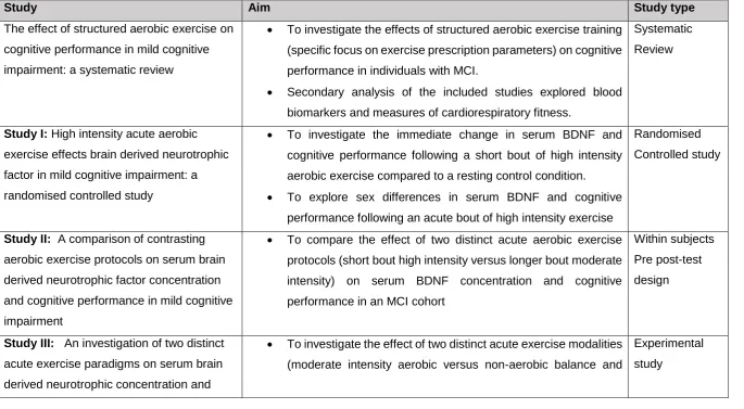
examining the relationship between acute exercise, BDNF and cognition is in its infancy, and while positive effects have been demonstrated in healthy young adult populations, this research is notably lacking in cognitively impaired populations. Exploring the BDNF response to acute and chronic exercise may offer new insights into a mechanism of action underlying the exercise-cognitive relationship in an MCI cohort. Across studies, one component of exercise prescription (modality, duration or intensity) was manipulated to test the impact of manipulating these variables on levels of BDNF concentration and cognitive performance. The overarching aim of this thesis was to investigate the effect of acute (varying modality, duration or intensity) and chronic exercise on serum BDNF concentration and cognitive performance (visuospatial learning and memory, attention and executive function) in a group of sedentary older adults with amnestic type MCI (Table 1-1).
15
Table 1-1 Overview of the studies in the PhD programme outlining study title, aims and study type. MCI: Mild Cognitive Impairment. BDNF:
Brain Derived Neurotrophic Factor.
Study Aim Study type
The effect of structured aerobic exercise on cognitive performance in mild cognitive impairment: a systematic review
• To investigate the effects of structured aerobic exercise training (specific focus on exercise prescription parameters) on cognitive performance in individuals with MCI.
• Secondary analysis of the included studies explored blood biomarkers and measures of cardiorespiratory fitness.
Systematic Review
Study I: High intensity acute aerobic
exercise effects brain derived neurotrophic factor in mild cognitive impairment: a randomised controlled study
• To investigate the immediate change in serum BDNF and cognitive performance following a short bout of high intensity aerobic exercise compared to a resting control condition.
• To explore sex differences in serum BDNF and cognitive performance following an acute bout of high intensity exercise
Randomised Controlled study
Study II: A comparison of contrasting
aerobic exercise protocols on serum brain derived neurotrophic factor concentration and cognitive performance in mild cognitive impairment
• To compare the effect of two distinct acute aerobic exercise protocols (short bout high intensity versus longer bout moderate intensity) on serum BDNF concentration and cognitive performance in an MCI cohort
Within subjects Pre post-test design
Study III: An investigation of two distinct
acute exercise paradigms on serum brain derived neurotrophic concentration and
• To investigate the effect of two distinct acute exercise modalities (moderate intensity aerobic versus non-aerobic balance and
16 cognitive performance in mild cognitive
impairment
toning) on serum BDNF concentration and cognitive performance in individuals with MCI.
Pre post-test design
Study IV: The impact of acute and chronic
exercise on serum brain derived neurotrophic factor concentration and cognitive performance following 6 and 12 weeks of structured exercise training in mild cognitive impairment
• To examine whether 12 weeks of structured aerobic training had a cumulative effect on resting measures of serum BDNF concentration and cognitive performance compared to a balance and toning group.
• To explore whether chronic aerobic training increases the magnitude of acute exercise BDNF and cognitive response.
Experimental Study
Pre post-test design
Study V: Peripheral brain derived
neurotrophic factor and episodic memory in a mild cognitive impairment population: results from a one-year exercise intervention study
• To examine serum BDNF concentration and episodic memory in a group of older adults with amnestic MCI over a 1-year period and to investigate whether there was a difference between three intervention groups (aerobic exercise v balance and toning v control).
• To investigate change in cardiorespiratory fitness (VO2max) in an MCI cohort over a one-year period and explore whether participation in a structured exercise intervention lead to changes in cardiorespiratory fitness compared to a control group
17
2 Chapter 2: Methodology
2.1 Introduction
This chapter will describe study design, sampling methods, data analysis and the background to assessment procedures used across studies I-V, presented in Chapters 4-8 in this thesis. Several assessments are common to all studies and subsequent chapters will refer back to the relevant sections in this chapter when describing individual study methods.
The studies discussed in this thesis were designed as a number of sub-studies to the NeuroExercise study. As such, participant selection criteria was uniform across studies and will be discussed in this chapter to limit duplication in later chapters. The NeuroExercise study is a European multi-centre Randomised Controlled Trial (RCT) examining ‘The effects of an extensive exercise programme on the progression of Mild Cognitive Impairment (MCI): study protocol for a randomised controlled trial’ (Devenney et al., 2017). The protocol paper for the NeuroExercise study, which was written by the author of this thesis (KD), was published in BMC Geriatrics and is available in Appendix I. I was study coordinator at the Dublin site and had responsibility for overseeing study implementation.
The NeuroExercise study recruited from the German Sports University in Cologne, Radboud University Medical Centre in Nijmegen, the Netherlands and Trinity College Dublin. In the NeuroExercise study, participants (target accrual of 75 at each site) were randomised to either a yearlong aerobic exercise (AT) intervention, balance and toning (BAT) (non-aerobic) exercise intervention or control group. The primary outcome was change in cognitive performance as measured by a neuropsychological test battery. Secondary outcomes included measures of cardiovascular fitness, physical activity, epigenetics, quality of life and measures of frailty.
18
2.2 Background research methods
2.2.1 Study designs
Two broadly defined types of studies are reported in health research: observational and experimental. There are three main types of observational studies that are distinguished by the study objective, sampling methods and the timeline of data collection. These are cross-sectional, case-control, and cohort studies which have been widely utilised in clinical research to answer observational research questions.
In observational studies, the inability to control for confounding variables is a considerable limitation. A confounding variable is independently associated with both the variable of interest and the outcome and may provide an explanation for the result (Mann, 2003). In addition, because the exposure and outcome are simultaneously assessed, there is generally no evidence of a temporal relationship between exposure and outcome. Observational studies possess an ability to address a broader range of questions with results more likely to include a broad representation of the population at risk.
Experimental design (randomised or non-randomised) refers to studies in which the investigator assigns an exposure to a group and may further compare the outcome of this exposure to a control group. Experimental studies can be subdivided into randomised and non-randomised trials (Grimes and Schulz, 2002). In experimental studies, the randomised controlled trial (RCT) is considered the most reliable research design to test interventions (Riffenburgh, 2012, Stel et al., 2007). Randomised controlled trials (RCTs) were first introduced into clinical medicine in 1948 when streptomycin was evaluated in the treatment of tuberculosis. Since then, RCTs are widely regarded as the gold standard in clinical research as the randomisation procedure controls for confounding variables and help to eliminate selection bias, both important methodological issues observed in trials.
19
2.2.2 Sampling
An adequate sampling method and sufficient sample size are necessary in quantitative research to ensure that statistical results can be detected, findings have external validity and can be generally applied to the population of interest (Fox et al., 2007). Statistical work in health research typically involves using a sample to draw conclusions from a larger population. To this end, it is necessary to utilise a means of reducing the number of participants in a study without increasing the risk of bias. Random or probability sampling are sampling methods used in a specific populations with the aim of reducing selection bias (Bland, 2015, Riffenburgh, 2012). The probability that a member of the population will be chosen for the sample is purely by chance. The list from which the sample is to be drawn is referred to as the sampling frame (Bland, 2015, Carter and Lubinsky, 2016). Random sampling implies that all people within the sampling frame have an equal probability of being selected for the study (Fox et al., 2007).
When random sampling is not possible, non-probability sampling methods are used as an alternative sampling method. As non-probability sampling methods remove the element of random selection, they cannot be fully reflective of a population and are inherently biased. However, these methods can be useful in exploratory research such as pilot studies and are widely used in rehabilitative research (Carter and Lubinsky, 2016). Common non-probability sampling methods include convenience sampling, quota sampling, purposive sampling, and self-selection sampling. In this PhD thesis, purposive criterion and self-selection sampling were used as sampling methods and these will be discussed in later chapters.
When planning a study, a key consideration is the number of participants and observations required to adequately answer the research question. Too many unnecessary observations wastes resource, while too few will reduce the power of the study (Suresh and Chandrashekara, 2012). The objective during sample size calculation is to find a balance between the risk of a type I or type II error occurring. A type I error (α) occurs when the null hypothesis is rejected when it is true. A type II error (β) occurs when the null hypothesis is accepted when it is in fact false (Bland, 2015).
Calculation of sample size involves specification of a number of parameters including:
• An estimate of the standard deviation is required to give indication of magnitude of chance variation.
20
• A significance level (α) i.e. risk of type I error must be defined. Conventionally 0.05 and 0.01 are used.
• The minimal important difference (∆) of the outcome of interest must be specified.
• The power (1- β) of the test must be determined. A powerful test is one which has a high probability of rejecting the null hypothesis when it is false. Most studies aim to have a power of 80% or higher (Riffenburgh, 2012, Mullins, 2003).
2.2.3 Reliability and validity
In quantitative research, reliability and validity are often used when assessing research rigour. Reliability relates to the stability of findings while validity is concerned with the truthfulness of findings (Carpenter and Suto, 2008).
2.2.3.1 Reliability
Reliability is regarded as the credibility and consistency of research findings and is often related to whether findings are free from measurement error when repeated under identical conditions (McDowell and Newell, 1996). Reliability can be described in term of relative and absolute reliability. Relative reliability, reported as inter-rater and intra-rater reliability examines the relationship between two or more sets of repeated measures and informs us about the error that may exist in an outcome measure (Huang et al., 2011). Intra-rater reliability describes the extent to which the same person can rate the same performance consistently. Inter-rater reliability describes the extent to which different people rate the same performance consistently.
2.2.3.2 Validity
Validity is concerned with the integrity of measurement and refers to the degree to which a scale measures what it is intended to measure (Bryman, 2015). Evaluating the validity of a tool is especially important when measuring constructs that are not directly observable (Liamputtong, 2013). Validity is not inherent to an instrument and is best evaluated within the context of a test’s intended use and specific population (Streiner et al., 2015). Methods and types of validity that are commonly evaluated include: content, construct and criterion validity (Liamputtong, 2013).
2.2.4 Principles of data analysis in quantitative research
21
analyse the data. Descriptive statistics include measures of central tendency (the mean (M) and the median (Md)), and measures of dispersion (the standard deviation (SD) and interquartile range (IQR)). Categorical variables (derived from nominal or ordinal data) are analysed using frequencies or the percentage of frequencies. Descriptive statistics for categorical variables are presented in pie-charts and bar charts. Continuous variables (scale data) are described by a measure of central tendency (mean, median, and mode) and a measure of variation around the mean / median (standard deviation / interquartile range). Graphically descriptive statistics for continuous data can be presented in histograms or boxplots. Descriptive statistics for continuous variables provide information regarding the distribution of scores. Skewness refers to the symmetry of the distribution and kurtosis gives an indication of the peak of the distribution (Pallant, 2016).
Data normality is an underlying assumption in parametric testing and it illustrated by a normal curve around the mean. An assessment of normality of data us a prerequisite for many statistical tests. Normality can be identified through the visual interpretation of the descriptive data including normality Q-Q plots and histograms. Normality can also be assessed through numerical methods such as the Shapiro-Wilk test. The Shapiro-Wilk test assesses if data is normally distributed for each independent variable and is recommended in small sample sizes of (<50 participants). The statistical result of the test indicates whether the data is normally distributed (p >0.05) or not normally distributed (p <0.05). In the case of non-normally distributed data, log transformations can be applied to achieve log-normality or non-parametric tests can be used. Normally distributed data is described as mean (standard deviation) and non-normally distributed data is described as median (inter-quartile range).
22
is the value below which the difference between groups is not likely due to chance. Alpha is most commonly set at 0.05. A p-value of >0.05 indicates a non-significant result and that the null hypothesis should not be rejected. A p-value of <0.05 indicates a statistically significant result and that the null hypothesis should be rejected. Alternatively, confidence intervals which represent the spread of the data can be used to determine statistical significance. Confidence limits are typically set at 0.95 (95% CI). In statistical testing 95% CIs which do not contain zero are deemed to be statistically significant at the 5% level (Gardner and Altman, 1986).
Relationships between variables can be analysed using correlations and regression analysis. Correlations coefficients are used to summarise the degree of relationship (correlation) between variables. The value of correlation coefficient ranges from + 1 (perfect positive correlation), through 0 (no correlation), to -1 (a perfect negative correlation). In health research, the Pearson correlation coefficient (interval or ratio) and the Spearman’s correlation coefficient (non-parametric, mixed levels of measurement) are commonly used. Multiple regression analysis involves a multivariate procedure that assesses the degree to which scores for a subset of variables predict scores for another variable in the set.
2.2.5 Statistical approach in this thesis
Data analysis was performed in all studies in this thesis using IBM SPSS Statistics version 24 (SPSS Inc, Chicago, IL). Preliminary analyses were performed to ensure no violation of the assumptions of normality by visual inspection of histogram and Q-Q plots and using Kolmogorov-Smirnov test (p>.05). Normally distributed data are presented as means (SD) and non-normally distributed data as medians (IQR). Categorical variables are presented as frequency (percentage). Change in BDNF concentrations and cognitive performance from pre-post each exercise bout was examined using paired sample t-tests for repeated measures on each patient and independent samples t-test for comparison between two exercise states. Non-parametric tests were used if data was not normally distributed. Correlations between BDNF and cognition were examined using Pearson Product Moment correlations or Spearman’s rho correlations as appropriate. Significance is taken at p<0.05.
23
reported as appropriate, consistent with published reports (Knaepen et al., 2010, Saucedo Marquez et al., 2015).
A power analysis on the primary outcome (BDNF) was performed in study I, Chapter 4. As all other studies within this thesis were considered exploratory work and nested within the larger NeuroExercise trial, a power calculation was not performed. Full details of statistical analysis for each study is presented in the relevant chapters.
2.3 Overview of the NeuroExercise Study
Within the NeuroExercise study, the author (KD) had the responsibility for overseeing study implementation and coordination at the Dublin site. Duties included ethics submission at each site, participant screening and recruitment, study assessments, implementation of the 12-month NeuroExercise exercise protocol, data management, data analysis and liaising with local referring sites and international partners. Recruitment began in January 2016 and the final study assessment was complete in August 2018.
2.3.1 Ethics approval
Ethical approval for the NeuroExercise Study and all studies within the PhD programme was obtained from the joint St. James’s / Tallaght Hospital Joint Research and Ethics Committee (REC) (Appendix IV). In addition, REC approval was obtained from Mater Misericordiae University Hospital, St. Vincent’s University Hospital and St. Patrick’s University Hospital which were referral sites to the NeuroExercise study (Appendix V). All procedures were performed in accordance with the 1964 Helsinki declaration and its later amendments. All researchers completed training in Good Clinical Practice and General Data Protection Regulation provided by Wellcome Trust Health Research Board Clinical Research Facility in St. James’s Hospital. All participants provided written informed consent (Appendix VI).
2.3.2 Recruitment and screening
24
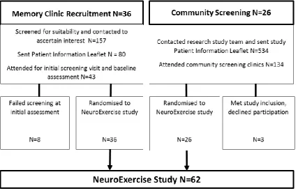
newsletters and GP practices around the Dublin area. In both recruitment strategies, initial screening was completed over the telephone to determine eligibility. Participants were then invited to a face-to-face screening appointment and those who met the defined diagnostic, inclusion and exclusion criteria and successfully completed all baseline measures were enrolled. Using these methods, 62 previously sedentary adults aged 50 years or older meeting MCI diagnostic criteria were recruited to the NeuroExercise study at St. James’s Hospital Dublin (Table 2:1).
Figure 2:1 Recruitment, screening and selection of participants to the
NeuroExercise Study
2.3.3 Diagnostic criteria
Participants had a diagnosis of MCI due to AD according to the National Institute on Aging and Alzheimer’s Association (NIA-AA) criteria (Albert et al., 2011) criteria. All enrolled participants with MCI were classified as having memory decline but not dementia (Clinical Dementia Rating global score = 0.5), consistent with established MCI classification (Albert et al., 2011, Petersen, 2004).
2.3.4 Inclusion criteria
[image:46.595.72.500.222.499.2]25
stable medical condition for more than six months; (3) stable medication for more than three months; (4) adequate visual and auditory acuity to complete neuropsychological testing; (5) electrocardiogram without significant abnormalities that might interfere with the study; (6) physical ability sufficient to allow performance of endurance exercise training; (7) capacity to provide written and dated informed consent form; (8) medical clearance to undergo a symptom-limited cardiopulmonary exercise test and extensive aerobic exercise training.
Participants recruited from the community via newspaper articles and community advertisements completed additional testing to determine MCI status. To distinguish between amnestic and non-amnestic MCI, agreed education adjusted cut-offs of -2 SD for low education (<10 years of education), -1.5 SD for the middle group (10-13 years of education) and -1 SD for the highly educated (>13 years of education) were taken from the delayed recall portion of an age adjusted episodic memory test using the Logical Memory (story recall) subtest of the Wechsler Memory Scale (WMS-IV) (Wechsler, 2014).
2.3.5 Exclusion criteria
26
2.3.6 NeuroExercise intervention
Participants in the NeuroExercise study were randomised to one of three groups. Following baseline assessment (T0), participants were randomised to one of three arms using a centrally controlled computer-generated randomisation list generated by an independent statistician. Investigators were blinded to allocation order and the treatment was assigned using sealed envelopes based on order of recruitment. Study outcome assessors were not blin