1 BRCA1/2 MUTATION ANALYSIS IN 41 OVARIAN CELL LINES REVEALS ONLY
ONE FUNCTIONALLY DELETERIOUS BRCA1 MUTATION
Britta Stordal1, Kirsten Timms2, Angela Farrelly3, Danielle Gallagher3, Steven
Busschots1, Mickaël Renaud3, Julien Thery1, Deborah Williams2, Jennifer Potter2, Thanh Tran2, Greg Korpanty4, Mattia Cremona3, Mark Carey5, Jie Li5, Yang Li5, Ozlem Aslan3, John J. O’Leary1, Gordon B. Mills5# and Bryan T. Hennessy 3,4,5#*
1) Department of Histopathology, St James’ Hospital and Trinity College Dublin, Dublin 8, IRELAND.
2) Myriad Genetics, Salt Lake City, Utah, USA.
3) Department of Medical Oncology, Royal College of Surgeons Ireland, Dublin 2, IRELAND
4) Department of Medical Oncology, Beaumont Hospital, Dublin 9, IRELAND. 5) Department of Systems Biology, The University of Texas M. D. Anderson Cancer
Center, Houston, Texas, USA.
1) Britta Stordal PhD – [email protected] 2) Kirsten Timms PhD - [email protected] 3) Angela Farrelly PhD- [email protected] 4) Danielle Gallagher BSc - [email protected] 5) Steven Busschots BSc - [email protected]
2 7) Julien Thery BSc- [email protected]
8) Deborah Williams MSc - [email protected] 9) Jennifer Potter BSc- [email protected] 10)Thanh Tran MSc – [email protected]
11)Greg Korpanty MD – [email protected] 12)Mattia Cremona PhD - [email protected] 13)Mark Carey PhD - [email protected]
14)Jie Li PhD - [email protected] 15)Yang Li PhD - [email protected] 16)Ozlem Aslan PhD - [email protected] 17)John O’Leary MD PhD - [email protected]
18)Gordon B. Mills MD PhD - [email protected] 19)Bryan T. Hennessy MD - [email protected] # Joint Senior Authors
*Corresponding Author Dr. Bryan T. Hennessy
Department of Medical Oncology, Beaumont Hospital,
Beaumont Road, Dublin 9.
IRELAND.
Tel: +353 01-8092010 Fax: +353 041-9874731
3 Abstract
Mutations in BRCA1/2 increase the risk of developing breast and ovarian cancer. Germline BRCA1/2 mutations occur in 8.6-13.7% of unselected epithelial ovarian cancers, somatic mutations are also frequent. BRCA1/2 mutated or dysfunctional cells may be sensitive to PARP inhibition by synthetic lethality. The aim of this study is to comprehensively characterise the BRCA1/2 status of a large panel of ovarian cancer cell lines available to the research community to assist in biomarker studies of novel drugs and in particular of PARP inhibitors.
The BRCA1/2 genes were sequenced in 41 ovarian cell lines, mRNA expression of BRCA1/2 and gene methylation status of BRCA1 was also examined. The cytotoxicity of PARP inhibitors olaparib and veliparib was examined in 20 cell lines.
4 correct for its slow growth rate. Cell lines derived from metastatic disease are
significantly more resistant to veliparib (2.0 fold p = 0.03) compared to those derived from primary tumours. Resistance to olaparib and veliparib was correlated Pearsons-R 0.5393, p = 0.0311.
The incidence of BRCA1/2 deleterious mutations 1/41 cell lines derived from 33 different patients (3.0%) is much lower than the population incidence. The reversion mutations and high frequency of heterozygous mutations suggest that there is a selective pressure against BRCA1/2 in cell culture similar to the selective pressure seen in the clinic after treatment with chemotherapy. PARP inhibitors may be useful in patients with BRCA1 deleterious mutations or gene methylation.
5 Introduction
The BRCA1 and BRCA2 proteins play a critical role in DNA damage repair and mutations in these genes increase the risk of developing breast and ovarian cancer. BRCA1/2 germline mutations have been shown occur in 8.6-13.7% of unselected epithelial ovarian cancer patients (Pal et al. 2005; Risch et al. 2001; Rubin et al. 1998). The rates of BRCA1 or combined BRCA1/2 mutation are much higher in ovarian cancer than in unselected invasive breast cancer 1.42-1.96% ( 2000; Newman et al. 1998; Papelard et al. 2000).
Synthetic lethality is the phenomenon whereby cell death results from the loss of function of two different gene products, when loss of either gene product in isolation does not. This concept has led to the idea that a new class of agents, PARP inhibitors, could be effective at treating BRCA1/2 dysfunctional cancers. PARP-1 is thought to be a regulator of base excision repair (BER) (Helleday 2011). One model suggests that PARP-1
6 the PARP inhibitor olaparib in breast and ovarian cancer patients with germline
BRCA1/2 mutations have also been encouraging (Audeh et al. 2010; Tutt et al. 2010).
Recently it has been shown that somatic BRCA1/2 mutations are also frequent in ovarian cancer. BRCA1/2 mutations were detected in 18.3% of tumour samples (n=235). Of the 42 patients found to have a BRCA1/2 mutation in their tumor, germline DNA was available from 28. Eleven of these patients (39%) had somatic BRCA1/2 mutations not present in the germline (Hennessy et al. 2010). These somatic BRCA1/2 mutations may expand the cohort of ovarian cancer patients which may benefit from treatment with PARP inhibitors. PARP inhibitors may also be useful in sporadic ovarian cancer patients which have other non-mutation defects in the BRCA1/2 pathways in their tumours, such as loss of gene or protein expression which can occur in up to 90% of patients (Russell et al. 2000).
To understand the role of BRCA1/2 mutations in ovarian cancer and study potential therapeutic strategies including the use of PARP inhibitors, suitable cell lines are needed. A previous study has characterised BRCA1/2 status in a large panel of breast cancer cell lines (Elstrodt et al. 2006). Our study characterises the mutation, expression and
7 Results
BRCA1 Mutation Screening
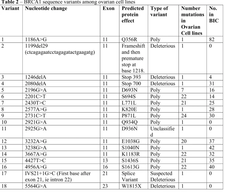
The clinical characteristics of the ovarian tumors from which the cell lines were established are presented in Table 1. Sequencing of BRCA1 revealed 18 different alterations in the gene sequence among 41 human ovarian cell lines (Table 2). The majority of alterations are nonpathogenic polymorphisms as described by the Breast Cancer Information Core (BIC) mutation database (http://research.nhgri.nih.gov/bic/). Three novel mutations in BRCA1 were identified in this study 1199del29, 2921 G>A, and 2925 G>A (Table 3). These variants have not been previously identified and are not present in the BIC or Myriad Genetics databases. 1199del29 is deleterious as it results in a frameshift and a premature stop codon; 2132 G>A is a polymorphism and 2136 G>A is a variant of unknown significance.
8 deleterious mutation at IVS21+1G>C, the first base after exon 21. However, this
mutation is also heterozygous.
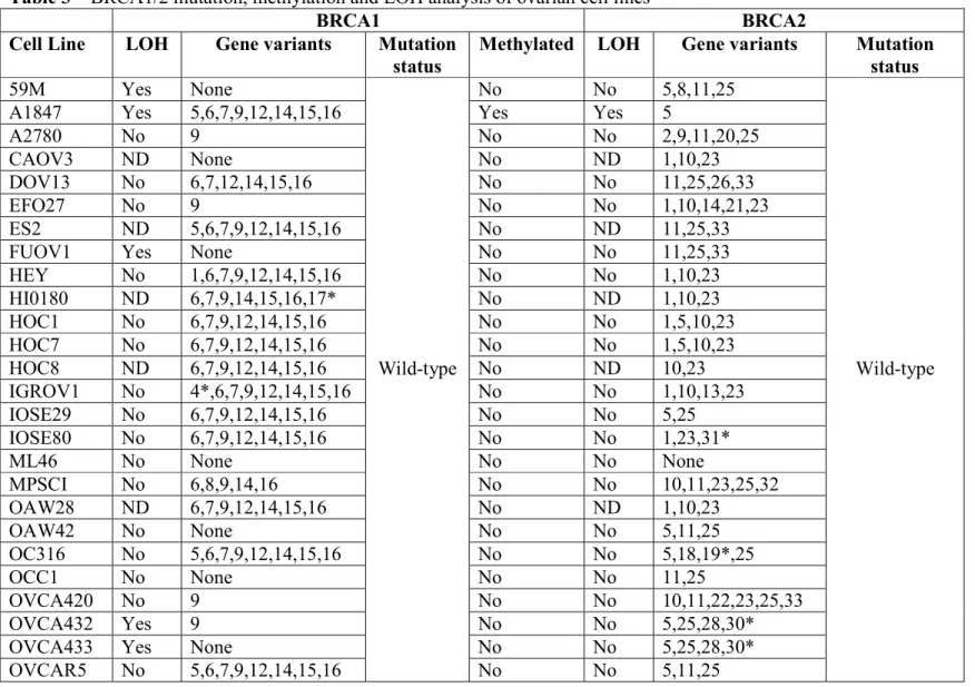
The 1199del29 mutation in BRCA1 is defined as deleterious as it results in a frameshift causing a premature stop codon. UPN-251 has the 1199del29 mutation however there is an additional mutation (1246delA), which restores the reading frame of the protein (Figure 1C). Therefore, despite having two mutations that would independently be deleterious, the UPN-251 cell line likely has a functional BRCA1 due to the combination of the two mutations. We believe this is an example of a deleterious mutation, followed by a reversion mutation.
SNU-251 has a homozygous deleterious mutation at 5564G>A which converts a tryptophan to a stop codon truncating the protein in exon 23. SNU-251 was developed from malignant ascites fluid from a Korean patient with endometrioid ovarian carcinoma (Yuan et al. 1997). The germline BRCA1 status of this patient is unknown.
BRCA2 Mutation Screening
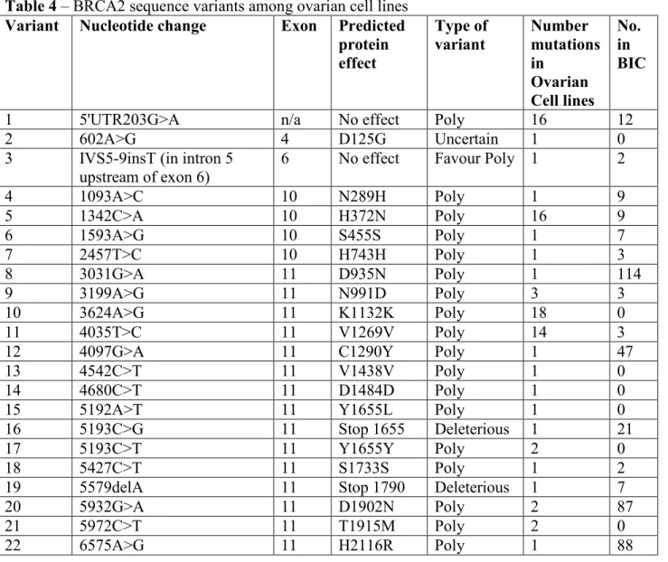
9 Deleterious BRCA2 mutations were identified in 5 ovarian cell lines IOSE80, OC316, OVCA432, OVCA433 and PEO1 (Tables 3 and 4). The deletion of an adenine at position 5579 in the OC316 cell line, leads to a premature stop of protein translation at amino acid 1790. However, OC316 is heterozygous, not homozygous for the 5579delA BRCA2 mutation. The germline genotype of BRCA2 in the patient who OC316 was derived from is unknown (Alama et al. 1996). The deleterious mutations in IOSE80, OVCA432, and OVCA433 are also all heterozygous.
The PEO1 cell line has a BRCA2 homozygous mutation 5193C>G, which would normally result in a stop codon at amino acid 1655. However, the PEO1 cell line has another mutation 5192A>T which prevents the stop codon. We believe that this is an example of a reversion mutation. The cell lines PEO4 and PEO6 were established from the ascites fluid of the same patient as PEO1 after further treatment with chemotherapy (Langdon et al. 1988). The PEO4 and PEO6 cells have a different mutation at 5193, it is now a thymine and the 5192A>T mutation seen in PEO1 is gone. This appears to be a different reversion mutation at the same site.
LOH, methylation and QPCR
10 Methylation was examined on the BRCA1 gene in all 41 cell lines and was found in 2 cell lines A1847 and OVCAR8 (Table 3). The methylated cell lines had a corresponding decrease in gene expression of BRCA1 (Figure 2). The deleterious mutated cell line SNU-251 had similar BRCA1 gene expression to the wild-type cell lines. The premature stop codon results from a single base change and this does not have an impact on the length of the mRNA produced.
Cytotoxicity and growth assays
11 panel in a 1 week assay. SNU-251 with a 2 week exposure is relatively sensitive to olaparib (Figure 3A & 3B).
The results for veliparib were similar (Figure 3E); however a higher dose of veliparib was needed than olaparib to achieve an IC50 across the cell line panel. The IC50s of olaparib and veliparib are also correlated in the panel, Pearsons-R 0.5393, p = 0.0311. The methylated cell lines OVCAR8 and A1847 were also on average more sensitive to veliparib than wild-type cells (Figure 3F). SNU-251 was also relatively resistant to veliparib in a 1 week cytotoxicity assay and more sensitive in a 2 week assay. Two cell lines were relatively sensitive to both olaparib and veliparib (A2780 and ES-2), and OVCAR420 was highly resistant to both agents. However, the next most resistant cell lines were different for olaparib (OVCAR432, HEY, IGROV-1) compared to veliparib (SKOV3, UPN-251).
12 Discussion
When BRCA1/2 mutations are detected in cell lines they represent either a germline mutation or a somatic mutation in the tumour, or a combination of both. Currently in clinical practice BRCA1/2 mutations are screened for in the germline with a blood test. This test is more economical, does not require tumor tissue and addresses the issue of familial risk of breast and ovarian cancer. However, with the advent of PARP inhibitors as a potential targeted therapy for BRCA1/2 mutated cancer additional tumour testing for somatic mutations may become a new standard as a significant number of somatic mutations are present in ovarian cancers (Hennessy et al. 2010).
The results of this study show that deleterious BRCA1/2 mutations are rare in ovarian cancer cell lines, only 1/41 cell lines studied (2.4%). The clinical characteristics of the patients the ovarian cell lines were derived from is presented in Table 1. Five cell lines in the panel represent later stages of cancer progression in patients cell lines were already derived from (HOC-7, PEA2, PEO4, PEO6, PEO14). Three cell lines are immortalised ovarian epithelial cells, HIO180, IOSE29 and IOSE80. Therefore, our panel of 41 cell lines represents 33 different ovarian cancer patients and the BRCA1/2 deleterious mutation rate per patient the cell lines were derived from is 1/33 (3.0%).
13 epithelial ovarian cancer (97.0%), which is similar to the reported clinical rates of
epithelial ovarian cancer (~90.0%) (Colombo et al. 2009). Out of the patients where the subtype of epithelial ovarian cancer is known (20), serous dominates over other epithelial subtypes in the cell line panel 14/20 (70.0%), slightly lower than what is seen in the clinic (~80-85%) (Colombo et al. 2010). This discrepancy is likely due to many cell lines being classified as adenocarcinoma, rather than into epithelial subgroups. Interestingly,
although the majority of BRCA1 mutations occur in serous ovarian cancer (Berchuck et al. 1998; Lakhani et al. 2004) the SNU-251 BRCA1 mutant is derived from an
endometrial ovarian tumour (Yuan et al. 1997). The PEO1, PEO4 and PEO6 cell lines which have reversions of a BRCA2 mutation are of also of serous origin from the same patient (Langdon et al. 1988)
Deleterious BRCA1 mutations have also been shown to be rare in breast cancer cell lines, 3/41 (7.3%) (Elstrodt et al. 2006). However, due to the higher reported rates of
deleterious BRCA1/2 mutations in unselected invasive ovarian versus unselected
14 Unusually, heterozygous mutations in BRCA1/2 were seen in 4 ovarian cancer cell lines studied (IGROV-1, OC316, OVCAR432 and OVCAR433), representing a frequency of 4/33 (12.12%). Heterozygous mutations in BRCA1/2 were also seen in two of the immortalised ovarian cell lines IOSE80 and HI0180. LOH was studied in all of the heterozygous mutated cancer cell lines and no LOH were observed (Table 3). One explanation for why heterozygous BRCA1/2 mutations may be observed in a cancer cell line is the cancer patient the cells were derived from was a BRCA1/2 mutation carrier but a second mutation in BRCA1/2 was not part of the carcinogenesis of their tumour. Breast cancers in patients who are heterozygous for BRCA1/2 are rarely ER+. However, there is a subset of patients that are both (Foulkes et al. 2004; Tung et al. 2010b), these patients may be ER+ because their cancer occurred due to hormonal carcinogenesis rather than loss of the second BRCA1/2 allele. One study examined 77 breast cancer patients women with BRCA1 germline mutations, 12 tumours remained heterozygous (15.5%), and 8 of these were ER+ (Tung et al. 2010a). It appears that heterozygous BRCA1/2 mutations may be more common in breast cancer where the lifetime risk of sporadic disease is higher than ovarian cancer, 1 in 8 vs. 1 in 72 (National Cancer Institute. 2010). In contrast to our observations with cell lines, of the BRCA1/2 mutant ovarian tumours identified to date by Myriad Genetics, only ~1/100 is heterozygous (Timms 2011).
When BRCA1/2 mutated cells are treated with chemotherapeutics, one mechanism of resistance that can occur is a reversion of the deleterious mutation. This has been shown to occur in cancer cell lines (Sakai et al. 2008) as well as in cancer patients after
15 that when the selective pressure of drug treatment is applied to BRCA1/2 mutated cells a common occurrence is a reversion of the mutation. The development of cell lines from tumour tissue is a process that involves multiple levels of selective pressure. Primary culture protocols involve physical disruption of the tissue, enzymatic digestion and selection of attached colonies in tissue culture. The more robust the cell, the more likely it is to survive the process. Cells without the full complement of DNA repair pathways, such as BRCA1/2 mutants may be at a disadvantage.
Loss of the second BRCA1/2 allele has also been shown to be heterogeneous within a tumour from BRCA1/2 mutation carriers (Martins et al. 2012). Mutations in BRCA1, PTEN and p53 were analysed at the single cell level in 55 BRCA1-associated breast tumours. Based on the frequency of each mutation loss of PTEN was the most common ‘first’ carcinogenic event associated with a basal-like subtype rather than BRCA1 (Martins et al. 2012). When a cell line is isolated from a heterogeneous tumour from a BRCA1/2 carrier cells with PTEN and p53 mutations could have been more robust in culture and BRCA1/2 mutated cells did not survive in the established cell line.
The heterozygous BRCA2 mutated cell line OC316 was derived from the malignant ascites of a cancer patient who had already received multiple rounds of chemotherapy (Alama et al. 1996) (Table 1). The metastasis and treatment with chemotherapy may have provided enough selective pressure for BRCA2 heterozygous cells to dominate the
16 is PTEN null (Cruet-Hennequart et al. 2003) but p53 wild-type (Casalini et al. 2001). It is possible that the primary tumour IGROV-1 was derived from was a mixture of PTEN and BRCA1 mutant cells and the PTEN mutation survived the selective pressure of cell culture.
17 The scientists who established SNU-251 commented on the difficulty developing cell lines from ovarian cancer patients, their success rate was only 17% (Yuan et al. 1997). Another BRCA1-mutated ovarian cell line (UWB1.289) was developed from a patient with a particularly aggressive cancer (DelloRusso et al. 2007). The authors of this study commented on the difficulty of establishing BRCA1 mutated cell lines in cell culture and speculate that the aggressiveness of this patient’s tumour may have facilitated the
propagation of this particular deleterious mutated cell line in vitro (DelloRusso et al. 2007).
An unselected breast cancer cell-line panel was skewed towards the triple-negative subgroup, 21/48 cell lines studied (43.75%) (Kao et al. 2009), higher than the incidence of triple negative breast cancer (TNBC) in unselected invasive breast cancer (12.4%) (Bauer et al. 2007). One would expect that BRCA1 mutations would be enriched in the panel as BRCA1 mutated tumours are common in TNBC (20.93%) (Merkel et al. 1989). However, BRCA1 mutations were relatively uncommon 2/14 (14.2%) of the TNBC cell lines examined (Kao et al. 2009). This suggests that the clinical aggressiveness of TNBC may make development of cell lines easier in the laboratory, but that the presence of BRCA1/2 mutations hinders the development of cell lines, resulting in a bias in the available cell lines.
18 mechanisms in addition to the BRCA1 mutation and more detailed study is needed to understand any DNA repair defects present. The deleterious mutation is also located near the end of the gene sequence and may have little impact on protein function. However, SNU-251 has a very slow growth rate compared to the other cell lines in the panel (Figure 3C). One of the causes of toxicity from PARP inhibitors is the generation of DSBs at collapsed replication forks (Rouleau et al. 2010). It is possible that the slow growth rate masks the true sensitivity of the cell line as the cells do not go through as many cell divisions as their wild-type counterparts. When SNU-251 was examined with a 2-week cytotoxicity assay their sensitivity to both olaparib and veliparib increases and they are more sensitive than wild-type cells.
The rate of BRCA1 methylation in the cell line panel 2/33 different patients (6.0%) is similar to the observed rate clinically in ovarian tumours 12/98 (12.2%) (Baldwin et al. 2000). BRCA1 methylation status has been shown to be stable in recurrent ovarian cancers (Baldwin et al. 2000). The BRCA1-methylated cell lines OVCAR8 and A1847 were more sensitive to PARP-inhibition than wild-type cell lines (Figure 3B, F). These cells also had a lower expression of BRCA1 mRNA (Figure 2). This data supports the synthetic lethality theory between PARP inhibition and BRCA1 dysfunction. This observation needs to be expanded upon but examining BRCA1 methylation may be relevant to predicting sensitivity to PARP inhibitors.
19 useful for the treatment of a broader spectrum of patients with ovarian cancer. Cells lines derived from metastases were significantly more resistant to veliparib than cells derived from primary tumours (Table 5). This is consistent with the concept that pathways that govern invasion and metastasis overlap with drug resistance (Liang et al. 2002). It is interesting that this observation is only the case for veliparib and not olaparib, suggesting a divergent mechanism of resistance between the two parp inhibitors. Some of the most resistant cell lines were different for olaparib (OVCAR432, HEY, IGROV-1) compared to veliparib (SKOV3, UPN-251). One possible explanation for this difference in
resistance mechanisms may be due to the expression of ABC transporters. There has been some evidence for olaparib being a P-glycoprotein substrate in an animal model
(Rottenberg et al. 2008) and for veliparib not being a P-glycoprotein substrate in a transfected cell line (Li et al. 2011).
Some studies have suggested that BRCA1/2 heterozygosity has a distinct phenotype and is associated with increased sensitivity to DNA damaging agents (Warren et al. 2003). Other studies have shown no difference in DNA repair capacity in response to radiation in BRCA1/2 heterozygotes (Nieuwenhuis et al. 2002). In this study we found no
20 Conclusions
We provide herein detailed BRCA1/2 status in a large panel of ovarian cancer cell lines to facilitate in vitro studies of parp inhibitors and other DNA damaging agents. The incidence of BRCA1/2 deleterious mutations in 1/41 cell lines, from 33 patients (3.0%) is much lower than the population incidence. The reversion mutations and high frequency of heterozygous mutations (14.6%) suggest that there is a selective pressure against BRCA1/2 mutations during adaption to cell culture similar to the selective pressure seen in the clinic after treatment with chemotherapy. PARP inhibitors may be useful in
21 Methods
Cell Culture
Cell lines HOC1, HOC7, HOC8, and IGROV1 were grown in DMEM (Invitrogen, Grand Island, NY, USA # 11995) 10% FBS (Hyclone, Logan, Utah, USA #sv30014.03); OAW42 and CAOV3 were grown in DMEM 10% FBS with the addition of sodium pyruvate (Sigma St. Louis, MO, USA, #S8636), 20ug/mL insulin (Invitrogen #12585-014), L-glutamine (Sigma #G7513) or NEAA (Sigma #M7145)/glucose respectively. FUOV1 was grown in DMEM:F12 (Invitrogen #11330) 10% FBS. OVCA420,
OVCA432, OVCA433 were grown in EMEM (ATTC, Manassas, VA, USA, #30-2003) 10% FBS; SW626 was grown in Leibovitz’s L15 (ATTC #30.2008) 10% FBS with no CO2. IOSE29 and IOSE80 were grown in M199:MCDB105 (Invitrogen #11150, Sigma
22 DNA Extraction
DNA extractions were performed using the Qiagen QIAamp DNA mini kit “Appendix B: Protocol for Cultured cells” spin column protocol adding 0.4 mg RNaseA to each sample prior to the AL buffer step.
RNA Extraction
All cell lines were grown in Advanced RPMI plus 3% FBS and 1%
penicillin/streptomycin 24-48 hours prior to harvesting cells. RNA was extracted using the Qiagen RNeasy mini kit “Animal cells spin” protocol using a QIAshredder for homogenisation and the optional on-column DNase digestion.
BRCA1/2 Analysis
BRCA1/2 mutation screening and qPCR was performed as previously published (Hennessy et al. 2010).
BRCA1 Promoter Methylation qPCR Assays
23 Cytotoxicity-Proliferation Assays
To determine the resistance to chemotherapy drugs, cells were plated into flat-bottomed, 96-well plates at the cell density shown of 1 x 10³cells/well and allowed to attach overnight. Slower growing cells (SNU-251) were plated at 2 x 10³cells/well. Olaparib (AZD2281) and veliparib (ABT888) were purchased from Selleck Chemicals (Boston, MA, USA) and made up in DMSO. Wells were treated in triplicate with serial dilutions of drug in a final volume of 200µL. Drug-free controls were included in each assay. DMSO controls were also performed for each cell line. Plates were incubated for a further 5 days at 37°C in a humidified atmosphere with 5% CO2 and cell viability was
determined using an acid phosphatase assay (Martin and Clynes 1993). For consistency, all cell lines for cytotoxicity and growth assays were growth in RPMI 10% FCS
containing L-glutamine and sodium bicarbonate (Sigma-R8758). Insulin (10µg/ml) was and Sodium pyruvate (1mM) was added to SNU-251. No antibiotics were used in the cell culture. The 2 week SNU-251 cytotoxicity assays followed the same procedure, the media containing drug was changed after 1 week of incubation.
Growth Assays
24 Conflict of Interest
Authors KT, DW, JP, TT, are all employees and shareholders of Myriad Genetics. The remaining authors have no conflict of interests to declare.
Acknowledgements
25 Figure Legends
Figure 1 – Sequence diagrams for BRCA1/2 deleterious mutations. A) Top: SNU-251 showing homozygous mutation W1815X (5564G>A) Bottom: BRCA1 wild-type sequence
B) Top: IGROV-1 showing heterozygous deletion - 2080delA Bottom: BRCA1 wild-type sequence
C) Top: UPN-251 showing homozygous deletions - 1199del29 + 1246delA reversion mutation. Bottom: BRCA1 wild-type sequence
Figure 2 – Quantitative PCR of BRCA1 in ovarian cell line panel. Wild-type cells are shown with blue circles, methylated cells with green circles and the deleterious mutation with a red circle.
Figure 3 – Cytotoxicity and doubling time in ovarian cell line panel. Wild-type cells are shown with blue bars, methylated cells with green bars and the deleterious mutation with a red bar. A) Cytotoxicity of olaparib in 17 ovarian cancer cell lines. B) Average
26 References
2012a. A2780 ECACC Datasheet. http://www.hpacultures.org.uk .
2012b. CRL-1978™ (ES-2) ATCC Datasheet. http://www.lgcstandards-atcc.org . 2012c. HTB-75™ (CAOV3) ATCC Datasheet. http://www.lgcstandards-atcc.org . 2012d. HTB-78™ (SW626) ATCC Datasheet. http://www.lgcstandards-atcc.org/ . 2000. Prevalence and penetrance of BRCA1 and BRCA2 mutations in a population-based series of breast cancer cases. Anglian Breast Cancer Study Group. British Journal of Cancer 83 1301-1308.
Aaronson,S.A. 2012. Personal Communication
Alama,A., Barbieri,F., Favre,A. et al. 1996. Establishment and Characterization of Three New Cell Lines Derived from the Ascites of Human Ovarian Carcinomas. Gynecologic Oncology 62 82-88.
Audeh,M.W., Carmichael,J., Penson,R.T. et al. 2010. Oral poly(ADP-ribose) polymerase inhibitor olaparib in patients with BRCA1 or BRCA2 mutations and recurrent ovarian cancer: a proof-of-concept trial. Lancet 376 245-251.
Baldwin,R.L., Nemeth,E., Tran,H. et al. 2000. BRCA1 Promoter Region
Hypermethylation in Ovarian Carcinoma: A Population-based Study. Cancer Research 60 5329-5333.
Bauer,K.R., Brown,M., Cress,R.D. et al. 2007. Descriptive analysis of estrogen receptor (ER)-negative, progesterone receptor (PR)-negative, and HER2-negative invasive breast cancer, the so-called triple-negative phenotype. Cancer 109 1721-1728.
Benard,J., Da Silva,J., De Blois,M.C. et al. 1985. Characterization of a Human Ovarian Adenocarcinoma Line, IGROV1, in Tissue Culture and in Nude Mice. Cancer Research 45 4970-4979.
Berchuck,A., Heron,K.A., Carney,M.E. et al. 1998. Frequency of germline and somatic BRCA1 mutations in ovarian cancer. Clinical Cancer Research 4 2433-2437.
Bryant,H.E., Schultz,N., Thomas,H.D. et al. 2005. Specific killing of BRCA2-deficient tumours with inhibitors of poly(ADP-ribose) polymerase. Nature 434 913-917.
27 Casalini,P., Botta,L., and Ménard,S. 2001. Role of p53 in HER2-induced Proliferation or Apoptosis. Journal of Biological Chemistry 276 12449-12453.
Choi,J.H., Choi,K.C., Auersperg,N. et al. 2005. Gonadotropins upregulate the epidermal growth factor receptor through activation of mitogen-activated protein kinases and phosphatidyl-inositol-3-kinase in human ovarian surface epithelial cells. Endocrine-Related Cancer 12 407-421.
Colombo,N., Peiretti,M., Castiglione,M. et al. 2009. Non-epithelial ovarian cancer: ESMO Clinical Recommendations for diagnosis, treatment and follow-up. Ann Oncol 20 iv24-iv26.
Colombo,N., Peiretti,M., Parma,G. et al. 2010. Newly diagnosed and relapsed epithelial ovarian carcinoma: ESMO Clinical Practice Guidelines for diagnosis, treatment and follow-up. Ann Oncol 21 v23-v30.
Cooke,S.L., Ng,C.K.Y., Melnyk,N. et al. 2010. Genomic analysis of genetic
heterogeneity and evolution in high-grade serous ovarian carcinoma. Oncogene 29 4905-4913.
Cruet-Hennequart,S., Maubant,S., Luis,J. et al. 2003. [alpha]v integrins regulate cell proliferation through integrin-linked kinase (ILK) in ovarian cancer cells. Oncogene 22 1688-1702.
DelloRusso,C., Welcsh,P.L., Wang,W. et al. 2007. Functional characterization of a novel BRCA1-null ovarian cancer cell line in response to ionizing radiation. Molecular Cancer Research: MCR. 5 35-45.
Elstrodt,F., Hollestelle,A., Nagel,J.H. et al. 2006. BRCA1 mutation analysis of 41 human breast cancer cell lines reveals three new deleterious mutants. Cancer Research 66 41-45. Emoto,M., Oshima,K., Ishiguro,M. et al. 1999. Establishment and Characterization of a Serous Papillary Adenocarcinoma Cell Line of the Human Ovary in a Serum-free Culture. Pathology - Research and Practice 195 238-243.
Fajac,A., Da Silva,J., Ahomadegbe,J.C. et al. 1996. Cisplatin-induced apoptosis and p53 gene status in a cisplatin-resistant human ovarian carcinoma cell line. International Journal of Cancer 68 67-74.
Farmer,H., McCabe,N., Lord,C.J. et al. 2005. Targeting the DNA repair defect in BRCA mutant cells as a therapeutic strategy. Nature 434 917-921.
28 Foray,N., Randrianarison,V., Marot,D. et al. 1999. Gamma-rays-induced death of human cells carrying mutations of BRCA1 or BRCA2. Oncogene 18 7334-7342.
Foulkes,W.D., Metcalfe,K., Sun,P. et al. 2004. Estrogen Receptor Status in BRCA1- and BRCA2-Related Breast Cancer. Clinical Cancer Research 10 2029-2034.
Furlong,M.T., Hough,C.D., Sherman-Baust,C.A. et al. 1999. Evidence for the Colonic Origin of Ovarian Cancer Cell Line SW626. J Natl Cancer Inst 91 1327-1328.
Hamilton,T.C. 2012. Personal Communication.
Hamilton,T.C., Young,R.C., McKoy,W.M. et al. 1983. Characterization of a Human Ovarian Carcinoma Cell Line (NIH:OVCAR-3) with Androgen and Estrogen Receptors. Cancer Research 43 5379-5389.
Helleday,T. 2011. The underlying mechanism for the PARP and BRCA synthetic lethality: Clearing up the misunderstandings. Molecular Oncology 5 387-393.
Hennessy,B.T., Timms,K.M., Carey,M.S. et al. 2010. Somatic mutations in BRCA1 and BRCA2 could expand the number of patients that benefit from poly (ADP ribose) polymerase inhibitors in ovarian cancer. Journal of Clinical Oncology 28 3570-3576. Hills,C.A., Kelland,L.R., Abel,G. et al. 1989. Biological properties of ten human ovarian carcinoma cell lines: calibration in vitro against four platinum complexes. British.Journal of Cancer 59 527-534.
Kao,J., Salari,K., Bocanegra,M. et al. 2009. Molecular Profiling of Breast Cancer Cell Lines Defines Relevant Tumor Models and Provides a Resource for Cancer Gene Discovery. PLoS One 4 e6146.
Kunzmann,R. and Hozel,F. 1987. Karyotype alterations in human ovarian carcinoma cells during long-term cultivation and nude mouse passage. Cancer Genetics and Cytogenetics 28 201-212.
Lakhani,S.R., Manek,S., Penault-Llorca,F. et al. 2004. Pathology of Ovarian Cancers in BRCA1 and BRCA2 Carriers. Clinical Cancer Research 10 2473-2481.
Langdon,S.P., Lawrie,S.S., Hay,F.G. et al. 1988. Characterization and Properties of Nine Human Ovarian Adenocarcinoma Cell Lines. Cancer Research 48 6166-6172.
Li,X., Delzer,J., Voorman,R. et al. 2011. Disposition and Drug-Drug Interaction Potential of Veliparib (ABT-888), a Novel and Potent Inhibitor of Poly(ADP-ribose) Polymerase. Drug Metabolism and Disposition 39 1161-1169.
29 Mackillop,W.J., Trent,J.M., Stewart,S.S. et al. 1983. Tumor Progression Studied by Analysis of Cellular Features of Serial Ascitic Ovarian Carcinoma Tumors. Cancer Research 43 874-878.
Martin,A. and Clynes,M. 1993. Comparison of 5 microplate colorimetric assays for in vitro cytotoxicity testing and cell proliferation assays. Cytotechnology 11 49-58. Martins,F.C., De,S., Almendro,V. et al. 2012. Evolutionary Pathways in BRCA1-Associated Breast Tumors. Cancer Discovery 2 503-511.
Merkel,D.E., Fuqua,S.A., Tandon,A.K. et al. 1989. Electrophoretic analysis of 248 clinical breast cancer specimens for P-glycoprotein overexpression or gene amplification. J Clin Oncol 7 1129-1136.
National Cancer Institute. 2010. EER Cancer Statistics Review 1975-2007. Lifetime Risk (Percent) of Dying from Cancer by Site and Race/Ethnicity: Males, Total US, 2005-2007 (Table 1.18) and Females, Total US, 2005-2007 (Table 1.19).
http://seer.cancer.gov/csr/1975_2007/results_merged/topic_lifetime_risk_death.pdf . Newman,B., Mu,H., Butler,L.M. et al. 1998. Frequency of Breast Cancer Attributable to BRCA1 in a Population-Based Series of American Women. JAMA 279 915-921.
Nieuwenhuis,B., Assen-Bolt,A.J.V., Van Waarde-Verhagen,W.H. et al. 2002. BRCA1 and BRCA2 heterozygosity and repair of X-ray-induced DNA damage. Int J Radiat Biol 78 285-295.
Pal,T., Permuth-Wey,J., Betts,J.A. et al. 2005. BRCA1 and BRCA2 mutations account for a large proportion of ovarian carcinoma cases. Cancer 104 2807-2816.
Papelard,H., De Bock,G.H., van Eijk,R. et al. 2000. Prevalence of BRCA1 in a hospital-based population of Dutch breast cancer patients. British Journal of Cancer 83 719-724. Pohl,G., Ho,C.L., Kurman,R.J. et al. 2005. Inactivation of the Mitogen-Activated Protein Kinase Pathway as a Potential Target-Based Therapy in Ovarian Serous Tumors with KRAS or BRAF Mutations. Cancer Research 65 1994-2000.
Risch,H.A., McLaughlin,J.R., Cole,D.E. et al. 2001. Prevalence and penetrance of germline BRCA1 and BRCA2 mutations in a population series of 649 women with ovarian cancer. American Journal of Human Genetics 68 700-710.
Rottenberg,S., Jaspers,J.E., Kersbergen,A. et al. 2008. High sensitivity of BRCA1-deficient mammary tumors to the PARP inhibitor AZD2281 alone and in combination with platinum drugs. Proc Natl Acad Sci U S A 105 17079-17084.
30 Rubin,S.C., Blackwood,M.A., Bandera,C. et al. 1998. BRCA1, BRCA2, and hereditary nonpolyposis colorectal cancer gene mutations in an unselected ovarian cancer
population: relationship to family history and implications for genetic testing. American Journal of Obstetrics.& Gynecology. 178 670-677.
Russell,P.A., Pharoah,P.D., De Foy,K. et al. 2000. Frequent loss of BRCA1 mRNA and protein expression in sporadic ovarian cancers. International Journal of Cancer 87 317-321.
Sakai,W., Swisher,E.M., Karlan,B.Y. et al. 2008. Secondary mutations as a mechanism of cisplatin resistance in BRCA2-mutated cancers.[see comment]. Nature 451 1116-1120. Sakai,W., Swisher,E.M., Jacquemont,C. et al. 2009. Functional Restoration of BRCA2 Protein by Secondary BRCA2 Mutations in BRCA2-Mutated Ovarian Carcinoma. Cancer Research 69 6381-6386.
Schilder,R.J., Hall,L., Monks,A. et al. 1990. Metallothionein gene expression and resistance to cisplatin in human ovarian cancer. International Journal of Cancer 45 416-422.
Sikic,B.I. 2012. Personal Communication.
Sood,A.K., Fletcher,M.S., Gruman,L.M. et al. 2002. The Paradoxical Expression of Maspin in Ovarian Carcinoma. Clinical Cancer Research 8 2924-2932.
Stronach,E.A., Alfraidi,A., Rama,N. et al. 2011. HDAC4-Regulated STAT1 Activation Mediates Platinum Resistance in Ovarian Cancer. Cancer Research 71 4412-4422. Swisher,E.M., Sakai,W., Karlan,B.Y. et al. 2008. Secondary BRCA1 mutations in BRCA1-mutated ovarian carcinomas with platinum resistance. Cancer Research 68 2581-2586.
Syed,V., Ulinski,G., Mok,S.C. et al. 2001. Expression of Gonadotropin Receptor and Growth Responses to Key Reproductive Hormones in Normal and Malignant Human Ovarian Surface Epithelial Cells. Cancer Research 61 6768-6776.
Timms,K.M. 2011. Personal Communication.
Tung,N., Miron,A., Schnitt,S. et al. 2010a. Prevalence and predictors of loss of wild type BRCA1 in estrogen receptor positive and negative BRCA1-associated breast cancers. Breast Cancer Research 12 R95.
31 Tutt,A., Robson,M., Garber,J.E. et al. 2010. Oral poly(ADP-ribose) polymerase inhibitor olaparib in patients with BRCA1 or BRCA2 mutations and advanced breast cancer: a proof-of-concept trial. The Lancet 376 235-244.
Warren,M., Lord,C.J., Masabanda,J. et al. 2003. Phenotypic effects of heterozygosity for a BRCA2 mutation. Human Molecular Genetics 12 2645-2656.
Wong,A.S.T., Roskelley,C.D., Pelech,S. et al. 2004. Progressive changes in Met-dependent signaling in a human ovarian surface epithelial model of malignant transformation. Experimental Cell Research 299 248-256.
Wong,W.S.F., Wong,Y.F., Ng,Y.T.A. et al. 1990. Establishment and characterization of a new human cell line derived from ovarian clear cell carcinoma. Gynecologic Oncology 38 37-45.
Young,T.N., Pizzo,S.V., and Stack,M.S. 1995. A Plasma Membrane-associated Component of Ovarian Adenocarcinoma Cells Enhances the Catalytic Efficiency of Matrix Metalloproteinase-2. Journal of Biological Chemistry 270 999-1002.
Yuan,Y., Kim,W.H., Han,H.S. et al. 1997. Establishment and characterization of human ovarian carcinoma cell lines. Gynecologic.Oncology 66 378-387.
Table 1 – Clinical characteristics of ovarian tumors from which cell lines were established.
Pre-Isolation Post-Isolation
Cell Line Original Tumor Histology Isolated From Treatment Received Response Treatment Received
Response References
59M Endometriod/Clear Cell Ascites None N/A IFO, MEL PR, Died (Hills et al. 1989) A1847 Undifferentiated carcinoma Metastasis Unknown Unknown Unknown Unknown (Aaronson 2012)
A2780 Unknown Primary Tumour None N/A Unknown Unknown ( 2012a)
CAOV3 Adenocarcinoma Unknown Unknown Unknown Unknown Unknown ( 2012c)
DOV13 Adenocarcinoma Unknown Unknown Unknown Unknown Unknown (Young et al. 1995) EFO27
Mucinous Solid Metastasis None N/A Unknown Unknown (Kunzmann and
Hozel 1987)
ES2 Serous/Clear Cell Primary Tumour None N/A Chemotherapy Refractory ( 2012b) (Sikic 2012) FUOV1 Serous Primary Tumour None N/A CIS, ASR, CYC Responded (Emoto et al. 1999) HEY
Serous Peritoneal Deposit and Xenograft
Radiotherapy, Radium CR CIS, ADR, CHL Died (Buick et al. 1985; Hills et al. 1989)
HI0180 Normal OSE Normal OSE N/A N/A N/A N/A (Sood et al. 2002)
HOC1 Serous Ascites MEL, CIS, ADR, CYC PR, PR None N/A (Buick et al. 1985; Mackillop et al. 1983)
HOC7~ Serous Ascites MEL, CIS, ADR, CYC PR, PR Unknown Unknown (Buick et al. 1985; Mackillop et al. 1983)
HOC8 Serous Ascites MEL PR (Filmus et al. 1986;
Filmus and Buick 1985)
IGROV1 Endometriod/Clear Cell Primary Tumour None N/A Unknown Unknown (Benard et al. 1985)
IOSE29 Normal OSE Normal OSE + SV40 N/A N/A N/A N/A (Wong et al. 2004)
IOSE80 Normal OSE Normal OSE + SV40 N/A N/A N/A N/A (Choi et al. 2005)
ML46
Serous Primary Tumour + SV40
Unknown Unknown Unknown Unknown (Yu et al. 2007)
MPSCI Serous Unknown Unknown Unknown Unknown Unknown (Pohl et al. 2005)
OAW28 Adenocarcinoma Ascites CIS, MEL NR, NR Unknown Died (Hills et al. 1989)
OAW42 Serous Ascites CIS CR Unknown PD, Died (Hills et al. 1989)
OVCA432 Serous Unknown Unknown Unknown Unknown Unknown (Syed et al. 2001) OVCA433 Serous Unknown Unknown Unknown Unknown Unknown (Syed et al. 2001) OVCAR5 Adenocarcinoma Unknown None N/A Unknown Unknown (Schilder et al. 1990)
OVCAR3
Serous Ascites CYC, CIS, DOX Unknown Unknown Unknown (Hamilton et al. 1983; Hills et al. 1989; Schilder et al. 1990) OVCAR8 Adenocarcinoma Unknown CAR PD Unknown Unknown (Schilder et al. 1990) OVCAR1
0
Adenocarcinoma Unknown CIS, CAR PD Unknown Unknown (Schilder et al. 1990) PA1 Germ Cell Tumor Ascites Chemotherapy NR Unknown Died (Hills et al. 1989) PEA1 Adenocarcinoma Pleural Effusion None N/A CIS, PDM Relapsed (Langdon et al. 1988) PEA2* Adenocarcinoma Ascites CIS, PDM Relapsed Unknown Unknown (Langdon et al. 1988) PEO1 Serous Ascites CIS, CHL, 5-FU CR CIS, CHL, 5-FU CR (Langdon et al. 1988;
Sakai et al. 2009) PEO4# Serous Ascites CIS, CHL, 5-FU CR Higher dose CIS PD (Langdon et al. 1988;
Sakai et al. 2009) PEO6# Serous Ascites CIS, CHL, 5-FU, CIS CR, PD None Died (Langdon et al. 1988;
Sakai et al. 2009)
PEO14 Serous Ascites None N/A CIS, CHL Relapsed (Langdon et al. 1988)
PEO23@ Serous Ascites CIS, CHL Relapsed Unknown Unknown (Langdon et al. 1988) SKOV3 Adenocarcinoma Ascites THI Unknown Unknown Unknown (Hills et al. 1989) SNU251 Endometriod Ascites CYC, ADR, CIS Unknown Unknown Unknown (Yuan et al. 1997) SW626 Adenocarcinoma Primary Tumor or
Ovarian Metastasis
Unknown Unknown Unknown Unknown ( 2012d; Furlong et al. 1999)
UPN251 Unknown Unknown Pt/TAX, TAX NR Unknown Unknown (Hamilton 2012)
Table 2 – BRCA1 sequence variants among ovarian cell lines Variant Nucleotide change Exon Predicted
protein effect Type of variant Number mutations in Ovarian Cell lines No. in BIC
1 1186A>G 11 Q356R Poly 1 82
2 1199del29
(ctcagagaatcctagagatactgaagatg)
11 Frameshift
and then premature stop at base 1218.
Deleterious 1 0
3 1246delA 11 Stop 393 Deleterious 1 4
4 2080delA 11 Stop 700 Deleterious 1 31
5 2196G>A 11 D693N Poly 7 16
6 2201C>T 11 S694S Poly 22 14
7 2430T>C 11 L771L Poly 21 25
8 2577A>G 11 K820E Poly 1 28
9 2731C>T 11 P871L Poly 24 30
10 2921G>A 11 Q934Q Poly 1 0
11 2925G>A 11 D936N Unclassifie
d
1 0
12 3232A>G 11 E1038G Poly 20 37
13 3238G>A 11 S1040N Poly 1 42
14 3667A>G 11 K1183R Poly 22 32
15 4427T>C 13 S1436S Poly 21 35
16 4956A>G 16 S1613G Poly 22 40
17 IVS21+1G>C (First base after
exon 21, ie intron 22)
21 Splice
Variant
Suspected Deleterious
1 0
Table 3 – BRCA1/2 mutation, methylation and LOH analysis of ovarian cell lines
BRCA1 BRCA2
Cell Line LOH Gene variants Mutation status
Methylated LOH Gene variants Mutation status
59M Yes None
Wild-type
No No 5,8,11,25
Wild-type
A1847 Yes 5,6,7,9,12,14,15,16 Yes Yes 5
A2780 No 9 No No 2,9,11,20,25
CAOV3 ND None No ND 1,10,23
DOV13 No 6,7,12,14,15,16 No No 11,25,26,33
EFO27 No 9 No No 1,10,14,21,23
ES2 ND 5,6,7,9,12,14,15,16 No ND 11,25,33
FUOV1 Yes None No No 11,25,33
HEY No 1,6,7,9,12,14,15,16 No No 1,10,23
HI0180 ND 6,7,9,14,15,16,17* No ND 1,10,23
HOC1 No 6,7,9,12,14,15,16 No No 1,5,10,23
HOC7 No 6,7,9,12,14,15,16 No No 1,5,10,23
HOC8 ND 6,7,9,12,14,15,16 No ND 10,23
IGROV1 No 4*,6,7,9,12,14,15,16 No No 1,10,13,23
IOSE29 No 6,7,9,12,14,15,16 No No 5,25
IOSE80 No 6,7,9,12,14,15,16 No No 1,23,31*
ML46 No None No No None
MPSCI No 6,8,9,14,16 No No 10,11,23,25,32
OAW28 ND 6,7,9,12,14,15,16 No ND 1,10,23
OAW42 No None No No 5,11,25
OC316 No 5,6,7,9,12,14,15,16 No No 5,18,19*,25
OCC1 No None No No 11,25
OVCA420 No 9 No No 10,11,22,23,25,33
OVCA432 Yes 9 No No 5,25,28,30*
OVCA433 Yes None No No 5,25,28,30*
OVCAR3 Yes None No Yes 1,10,23
OVCAR8 Yes 5,6,7,12,14,15,16, Yes Yes None
OVCAR10 ND 9,10,11 No ND 3,9,11,20,24,25,27
PA1 No 13 No No 5,12,25
PEA1 Yes None No Yes 1,10,23
PEA2 Yes None No Yes 1,10,23
PEO1 Yes 6,7,9,12,14,15,16 No Yes 11,15@,16#,25
PEO4 Yes 6,7,9,12,14,15,16 No Yes 11,17@,25
PEO6 Yes 6,7,9,12,14,15,16 No Yes 11,17@,25
PEO14 ND 5,6,7,9,12,14,15,16 No ND 1,5,10,23,25
PEO23 Yes 5,6,7,9,12,14,15,16 No No 1,5,10,23,25
SKOV3 No None No No 1,10,21,23
SNU251 ND 18 Mutant No ND 5,25,29
SW626 Yes None Wild-type No Yes 4,6,7,9
UPN251 Yes 2#,3# No Yes 5,25
Table 4 – BRCA2 sequence variants among ovarian cell lines
Variant Nucleotide change Exon Predicted
protein effect
Type of variant
Number mutations in
Ovarian Cell lines
No. in BIC
1 5'UTR203G>A n/a No effect Poly 16 12
2 602A>G 4 D125G Uncertain 1 0
3 IVS5-9insT (in intron 5
upstream of exon 6)
6 No effect Favour Poly 1 2
4 1093A>C 10 N289H Poly 1 9
5 1342C>A 10 H372N Poly 16 9
6 1593A>G 10 S455S Poly 1 7
7 2457T>C 10 H743H Poly 1 3
8 3031G>A 11 D935N Poly 1 114
9 3199A>G 11 N991D Poly 3 3
10 3624A>G 11 K1132K Poly 18 0
11 4035T>C 11 V1269V Poly 14 3
12 4097G>A 11 C1290Y Poly 1 47
13 4542C>T 11 V1438V Poly 1 0
14 4680C>T 11 D1484D Poly 1 0
15 5192A>T 11 Y1655L Poly 1 0
16 5193C>G 11 Stop 1655 Deleterious 1 21
17 5193C>T 11 Y1655Y Poly 2 0
18 5427C>T 11 S1733S Poly 1 2
19 5579delA 11 Stop 1790 Deleterious 1 7
20 5932G>A 11 D1902N Poly 2 87
21 5972C>T 11 T1915M Poly 2 0
23 7470A>G 14 S2414S Poly 18 10
24 7829C>T 15 A2534V Favour Poly 1 5
25 IVS16-14C>T (in intron 16
upstream of exon 17)
17 None Poly 25 15
26 8688A>C 19 V2820V Poly 1 0
27 9145C>T 22 R2973C Favour Poly 1 14
28 IVS24-16T>C (intron 24
upstream of exon 25)
25 None Poly 3 6
29 IVS26-19G>A (intron 26
upstream of exon 27)
27 None Poly 1 0
30 10204A>T 27 Stop 3326 Poly 3 293
31 10323delCins11 27 Stop 3369 Poly 1 11
32 10349C>T 27 T3374I Poly 1 8
Table 5 – Response of BRCA1/2 wild-type, unmethylated cells to parp inhibitors based on patient characteristics of original tumors.
Number of Cell Lines
Mean Olaparib IC50
Fold T-test Mean Veliparib IC50
Fold T-test
Primary Tumour 3 2.44 ± 3.14 24.82 ± 29.06
Metastatic Disease 6 2.92 ± 1.49 1.19 0.87 59.76 ± 11.12 2.00 0.03 Chemotherapy Naïve * 4 2.96 ± 2.76 31.69 ± 27.42
Chemotherapy Pre-treated* 7 2.91 ± 1.40 0.99 0.98 58.15 ± 17.71 1.83 0.08 Responder to Chemotherapy 4 2.92 ± 1.79 59.00 ± 12.67
Non-Responder to Chemotherapy 4 2.74 ± 1.60 0.93 0.89 45.53 ± 30.48 0.77 0.45 Serous Histology 6 3.50 ± 3.59 34.91 ± 32.84
Non-Serous Histology 2 3.03 ± 1.71 0.86 0.80 44.90 ± 20.04 1.28 0.61
BRCA1/2 +/+ 15 2.29 ± 1.75 42.00 ± 25.99
A
B
C
0 1 2 3 4 5 Wildtype
Deleterious Mutation Methylated
BRCA1 DeltaCt
Mut ated
2 W eeks Met hyla ted Wild type Mut ated
1 W eek 0 5 10 15 A v e ra g e O la p a ri b I C 5 0 ( u M ) A27 80 CA OV 3 ES-2
SN U-2
51 2 wee ks OV CA R8 HO C1 PEO
1 OV CA R43 3 SK OV 3 OV CA R10 DO V13 OC 316 OA
W42A1847 OV
CA R5
UPN -251
OV CA
R43 2
HEY IGR
OV -1
SN U-2
51 1 wee k OV CA R42 0 0 10 20 30 O la p a ri b I C 5 0 ( u M )
A
B
C
D
E
F
A27 80ES-2
OV CA R43 3 OV CA R8 CA OV 3 OV CA 10 SN U-2
51 2 Wee ks OV CA R43 2 DO V13 HO C1 OV CA R5 A18 47 IGR OV -1 PE O1
SNU -251
1 w eekHEY
OC 316 OA W42 SK OV 3 UP N-2 51 OV CA 420 0 20 40 60 80 100 120 V e li p a ri b I C 5 0 ( u M ) Mut ated
2 W eeks Met hyla ted Wild type Mut ated
1 W eek 0 20 40 60 80 100 120 A v e ra g e V e li p a ri b I C 5 0 ( u M ) HE Y A27 80 SK OV 3 OV CA R43 3 OC 316 OV CA 10
ES2 IGR
OV -1
OA W42HOC
1
UPN -251 DO V13 OV CA 420 OV CA R-8 OV CA R43 2 CA OV 3 A18 47