http://www.scirp.org/journal/ojim ISSN Online: 2162-5980
ISSN Print: 2162-5972
DOI: 10.4236/ojim.2018.82014 Apr. 28, 2018 131 Open Journal of Internal Medicine
Expression of Circulating Plasma Long
Non-Coding RNA uc063iod.1 and Its
Relationship with Coronary Artery Disease
Vaidya Rajan1, Genshan Ma2*, Hong Jin2
1School of Medicine, Southeast University, Nanjing, China
2Department of Cardiology, Zhongda Hospital, Southeast University, Nanjing, China
Abstract
Objective: To investigate the expression of LncRNA uc063iod.1 in patients with different diseases and to explore the correlation between expression level of LncRNA uc063iod.1 and Coronary Artery Disease (CAD). Method: Plasma samples were prepared using the blood extracted from total 101 patients un-dergoing coronary artery angiogram (63 CAD patients and 38 non CAD pa-tients). The expression level of LncRNA uc063iod.1 was detected using quan-titative real time polymerase chain reaction (qRT-PCR) and the relationship between expression level of LncRNA uc063iod.1 and CAD was explored. The clinicopathological features of patients with CAD were also discussed in our study. Results: The expression of LncRNA uc063iod.1 was significantly in-creased in the plasma of patients with CAD (p < 0.05). AUC was 0.701 (95%CI: 0.598 - 0.804), and the sensitivity and specificity were 77.8% and 57.9% respectively. Conclusion: Upregulation of LncRNA uc063iod.1 in plasma can be used as a biomarker for the diagnosis of CAD.
Keywords
LncRNA uc063iod.1, LncRNA E330013P06, Coronary Artery Disease, Plasma, qRT-PCR
1. Introduction
Cardiovascular diseases (CVD) are the leading cause of morbidity and mortality in the world. An estimated 17.7 million people died from CVDs in 2015, repre-senting 31% of all global deaths. Of these deaths, an estimated 7.4 million were How to cite this paper: Rajan, V., Ma, G.S.
and Jin, H. (2018) Expression of Circu-lating Plasma Long Non-Coding RNA uc063iod.1 and Its Relationship with Coro-nary Artery Disease. Open Journal of In-ternal Medicine, 8, 131-145.
https://doi.org/10.4236/ojim.2018.82014
Received: February 24, 2018 Accepted: April 25, 2018 Published: April 28, 2018
Copyright © 2018 by authors and Scientific Research Publishing Inc. This work is licensed under the Creative Commons Attribution International License (CC BY 4.0).
DOI: 10.4236/ojim.2018.82014 132 Open Journal of Internal Medicine due to Coronary artery Disease (CAD). Atherosclerotic disease and its throm-botic complication are the common manifestation of CVDs that leads to the de-velopment of CAD; if untreated, it progresses into Myocardial Infarction and Heart Failure [1]. Reducing lipids, antiplatelet agents, anticoagulants, Percuta-neous Coronary Intervention (PCI), and Coronary Artery Bypass Graft (CABG) surgery are available effective treatment measures for CAD. However, those treatments are challenged by the adverse effects of drugs, drug tolerance and high recurrence rate, resulting no change in mortality. Therefore, the develop-ment of a new non-invasive method that can diagnose CAD early in its onset is critical in improving its prognosis.
Modern advancement in genome-wide analyses has identified large number of long non-coding RNAs (LncRNAs), which are transcribed in human genome. LncRNAs are non-protein coding RNA, which are 200 to 10,000 nucleotides in length [2]. Even though LncRNAs are new to the research world, they have al-ready been used as promising biomarkers for diagnosis of several diseases. LncRNA PCA3 is already a well-established biomarker for prostate cancer [3]. Likewise, LncRNA ATB can serve as biomarker to detect Coal Worker Pneu-moconiosis [4]. And LncRNA LIPCAR is shown to have potential to serve as a biomarker for heart failure [5]. Recent studies have already claimed several LncRNAs are associated with initiation and progression of coronary heart dis-eases [6].
LncRNA uc063iod.1 is human equivalent of LncRNAE330013P06 (E33), which is also named as Cardiac Mesoderm Enhancer-associated Noncoding RNA (CARMN) or MIR143HG [7]. In an animal model study, the LncRNAE33 was found to induce foam cell formation promoting atherosclerosis [8]. And in the same study, LncRNA uc063iod.1 was upregulated in monocytes derived from type II diabetic patients. Study has proven that monocytes play essential roles in initiation, progression and development of atherosclerosis and CAD [9]. E33 is associated with M2 macrophage; and M2 macrophage accelerates the cholesterol uptake and induces foam cell formation promoting atherosclerosis [10]. Intra-plaquehemorrhage accelerates atherosclerosis via oxidant stress and contributes to lesion development and destabilization. M2 macrophage is also found in intra plaque hemorrhage in coronary atheroma [11]. LncRNA uc063iod.1 is host gene of miR-143 and miR-145. Several studies suggested that mir-143 and mir-145 is associated with progression development of atherosclerosis and CAD [12] [13] [14] [15]. Therefore, it will be reasonable to speculate that the LncRNA uc063iod.1 may be involved in the progression of CAD.
DOI: 10.4236/ojim.2018.82014 133 Open Journal of Internal Medicine
2. Materials and Methods
2.1. Study Population
Plasma samples were prepared using the blood extracted from total 101 patients undergoing coronary artery angiogram (63 CAD patients and 38 Non-CAD pa-tients) in Zhongda Hospital, Southeast University, Nanjing PR China, between May 2017 and October 2017. The study was approved by ethics committee of medical faculty of the Zhongda Hospital, and written informed consent was taken from all included patients or their families in accordance with the Declara-tion of Helsinki [16].
2.1.1. Inclusion Criteria
1) Case: Patients with >50% stenosis of at least one of the major coronary ar-teries (left main coronary trunk, anterior descending branch, circumflex artery and right coronary artery) confirmed by two independent intervention cardiolo-gist during percutaneous coronary artery angiogram.
2) Control: No stenosis in any coronary arteries confirmed by two independ-ent intervindepend-entional cardiologists.
3) Undergone percutaneous coronary artery angiogram for the first time. 4) Age >50 yrs to <75 yrs.
2.1.2. Exclusion Criteria
1) Patients with severe valvular disease, malignant tumors, other systemic disease like renal failure and hepatic disease.
2) Patients who rejects to take part in the study.
3) Patients with serious acute infections in recent 4 weeks periods.
2.2. Blood Sample Collection
10 ml of arterial blood were collected in EDTA tube from radial artery, after the insertion of the arterial catheter before the administration of anticoagulants. Plasma were collected after centrifugation at 2000 rpm at 4˚C for 10 mins and stored at −80˚C before use.
2.3. RNA Extraction and qRT-PCR
Total RNA was extracted from plasma using TRIzol reagent (Invitrogen, USA) and cDNA synthesis was carried out by 1 μl of RNA, using 5xPrimeScriptRT Master Mix (Takara Biomed Co. Ltd., Beijing) 4 μl, in total volume of 20 μl reac-tion mixture. SYBR Green Real-Time PCR Master Mixes (Nanjing VazymeBio-Tech Co. Ltd.) was used to prepare PCR reaction system. The following primers were used in PCR reactions: 5’-TCTTTCTCACAGGCCGCATT-3’ (forward) and 5’-TGATTTCTCCACGGTCAGGC-3’ (reverse) for LncRNA uc063iod.1; 5’- GCACCGTCAAGGCTGAGAAC-3’ (forward) and
de-DOI: 10.4236/ojim.2018.82014 134 Open Journal of Internal Medicine naturation at 95˚C for 1 minute, followed by 40 cycles of denaturation 95˚C for 5 seconds annealing at 60˚C for 31 seconds. Data were analysed using 2−∆∆CT
methods, and relative expression level of LncRNA uc063iod.1 was normalized to endogenous control GAPDH. Each sample was independently tested for three times.
2.4. Statistical Analysis
SPSS 20.0 software was used to analyse all data. The test of normality of data was checked by Shapiro-Wilk test. Independent t-test or Mann-Whitney U-test were used to assess the differences in the expression levels of genes between CAD and Non-CAD groups. Continuous variables were tested with student t test and 2 × 2 contingency table analysis of χ2 was used for categorical variables. Abnormally
distributed data were processed using non-parametric Mann-Whitney U test. Kruskal-Wallis test was used to test the significance of expression of the gene with respect to the degree of lesion. The correlation between the expression lev-els of genes was performed by Spearman correlation coefficient. Finally, the Re-ceiver Operating Characteristic (ROC) curve analysis was performed to analyse the sensitivity and specificity of the expression level of LncRNA uc063iod.1. The area under the ROC curve (AUC) was used to summarise the diagnostic accu-racy of the genes. A value of p < 0.05 was considered statistically significant.
3. Results
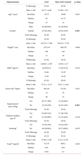
3.1. Clinicopathological Characteristics of the Study Population Case control study was designed; blood samples were collected from total 101 patients, which were divided into two groups, 63 patients with CADas Case and 38 Non-CAD as Control. Then we conducted a comparison analysis of clinico-pathological characteristics of the study population. In this study we found that CAD patients were aged 64.17 ± 6.60 years, comparable with 61.88 ± 3.93 years in Non-CAD group. There was significance differences between CAD and Non-CAD in status of gender (p = 0.001), hypertension (p = 0.001), HDL (p = 0.042), WBC (p = 0.048), neutrophil (p = 0.027), monocyte (p = 0.027) and HbA1c (p = 0.028), whereas parameters like age, height, BMI, heart rate, diabe-tes mellitus II, smoking, trop I, LVEF, lymphocyte, TC, TG and LDL could not show any significance difference (Table 1).
3.2. LncRNA uc063iod.1 Expression in the Plasma of CAD and Non-CAD Patients
DOI: 10.4236/ojim.2018.82014 135 Open Journal of Internal Medicine
Table 1. Distribution of clinical characteristics in CAD patients and control group.
Characteristics CAD Non-CAD Control p value
AgeΩ (year)
N (Missing) 63 (0) 38 (0)
0.055 Mean ± Std. 64.17 ± 6.60 61.88 ± 3.93
Min/Max 52/75 54/68
Median 65 61.75
Range 23 14
Gender#
Male 44 (69.84%) 14 (36.84%)
0.001 Female 19 (30.16%) 24 (63.16%)
Total (Missing) 63 (0) 38 (0)
HeightΩ (cm)
N (Missing) 63 (0) 38 (0)
0.060 Mean ± Std. 167.25 ± 7.296 164.42 ± 7.176
Min/Max 147/178 148/178
Median 168 164
Range 31 30
BMIΩ (kg/m2)
N(Missing) 63 (0) 38 (0)
0.974 Mean ± Std. 24.004 ± 2.587 23.98 ± 3.17
Min/Max 18.29/30.11 17.85/32.34
Median 23.66 23.29
Range 11.82 14.49
Heart rateΩ (bpm)
N (Missing) 63 (0) 38 (0)
0.415 Mean ± Std. 77.21 ± 12.75 79.47 ± 14.64
Min/Max 60/120 51/126
Median 74 79
Range 60 75
Hypertension# (mm of Hg)
Yes 49 (77.78%) 14 (36.84%)
0.001 No 14 (22.22%) 24 (63.16%)
Total (Missing) 63 (0) 38 (0)
Diabetes mellitus type II#
Yes 38 (60.31%) 17 (40.74%)
0.128 No 25 (39.69%) 21 (55.26%)
Total (Missing) 63 (0) 38 (0)
Smoking#
Yes 19 (30.16%) 10 (26.32%)
0.679 No 44 (69.84%) 28 (73.68%)
Total (Missing) 63 (0) 38 (0)
TropIØ (ng/ml)
N (Missing) 61 (2) 36 (2)
0.070 Mean ± Std. 0.706 ± 3.09 0.14 ± 0.178
Min/Max 0.1/19 00/0.1
Median 0.01 0.01
DOI: 10.4236/ojim.2018.82014 136 Open Journal of Internal Medicine
Continued
HbA1CØ (%)
N (Missing) 49 (14) 24 (14)
0.028 Mean ± Std. 0.706 ± 3.09 0.672 ± 3.83
Min/Max 5.20/14.30 5.20/10.50
Median 7.10 6.15
Range 9.10 5.30
LVEFØ (%)
N (Missing) 52 (11) 33 (5)
0.481 Mean ± Std. 66.25 ± 7.17 67.57 ± 6.65
Min/Max 52/80 55/80
Median 67 68
Range 28 25
WBCØ (109/L)
N (Missing) 61 (2) 37 (1)
0.048 Mean ± Std. 6.818 ± 2.268 5.9611 ± 1.812
Min/Max 3.05/15.56 3.36/13.01
Median 6.51 5.6200
Range 12.51 9.65
NeutrophilØ (109/L)
N (Missing) 61 (2) 37 (1)
0.027 Mean ± Std. 4.624 ± 2.051 3.856 ± 1.638
Min/Max 1.63/13.26 1.61/11.22
Median 4.22 3.53
Range 11.63 9.61
LymphocyteΩ (109/L)
N (Missing) 61 (2) 37 (1)
0.986 Mean ± Std. 1.633 ± 0.739 1.635 ± 0.561
Min/Max 0.34/5.69 0.53/2.71
Median 1.47 1.62
Range 5.35 2.18
MonocyteØ (109/L)
N (Missing) 61 (2) 37 (1)
0.027 Mean ± Std. 0.411 ± 0.171 0.339 ± 0.126
Min/Max 0.19/0.89 0.16/0.74
Median 0.40 0.31
Range 0.70 0.58
TCΩ (mmol/L)
N (Missing) 58 (5) 31 (7)
0.646 Mean ± Std. 4.355 ± 1.225 4.4729 ± 0.972
Min/Max 2.16/8.86 2.89/6.94
Median 4.25 4.57
Range 6.70 4.05
TGØ (mmol/L)
N (Missing) 58 (5) 31 (7)
0.288 Mean ± Std. 1.768 ± 0.0677 1.554 ± 1.0732
Min/Max 0.7/4.9 0.5/4.9
Median 1.355 1.230
DOI: 10.4236/ojim.2018.82014 137 Open Journal of Internal Medicine
Continued
HDLΩ (mmol/L)
N (Missing) 58 (5) 31 (7)
0.042 Mean ± Std. 1.088 ± 0.242 1.200 ± 0.25
Min/Max 0.75/1.88 0.81/1.60
Median 1.055 1.20
Range 1.13 0.79
LDLΩ (mmol/L)
N (Missing) 58 (5) 31 (7)
0.616 Mean ± Std. 3.093 ± 1.259 2.58 ± 0.738
Min/Max 1.10/6.52 1.41
Median 2.81 4.24
Range 5.42 2.83
[image:7.595.237.513.340.541.2]BMI = Body mass index, bpm = beats per minute, TG = triglyceride, TC = total cholesterol, HDL = high density lipoprotein, LDL = low density lipoprotein, WBC = white blood cell, LVEF = left ventricular ejec-tion fracejec-tion, TropI = troponin I, HbA1c = glycated haemoglobin. Data are presented as mean ± standard deviation (std.), Minimum/Maximum (Min/Max), Median and Range. N = number of patients. P < 0.05 was considered significant. ΩStudent T-test. #2 × 2 contingency table analysis of χ2. ØMann-Whitney U-test.
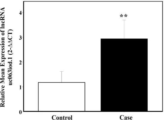
Figure 1. The relative mean expression of LncRNA uc063iod.1 in the plasma of CAD pa-tients and Non-CAD control. **compared with control group, p < 0.01.
3.3. Expression of LncRNA uc063iod.1 in Patients with Respect to the Coronary Artery Lesion
DOI: 10.4236/ojim.2018.82014 138 Open Journal of Internal Medicine
Figure 2. The mean expression of LncRNA uc063iod.1 in plasma of CAD patients with respect to diseased number of coronary arteries and the control with no lesion. **p < 0.01 and ***p < 0.001.
3.4. Expression of LncRNA uc063iod.1 in CAD Patients with Stable Angina Pectoris, Unstable Angina Pectoris and Acute
Myocardial Infarction
Plasma levels of LncRNA uc063iod.1 in patients with Stable Angina Pectoris, unstable Angina Pectoris and Acute Myocardial Infarction found to have sig-nificant difference with the control (p < 0.01). As shown in Figure 3, patients presented with SAP and AMI appeared to have significantly higher levels of LncRNA uc063iod.1, although there was no significant difference observed among these three sub-groups (Figure 3).
3.5. Correlation Ship between LncRNA uc063iod.1 and Coromarker
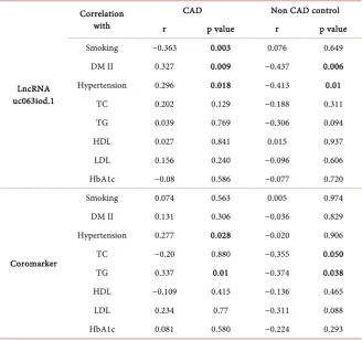
Coromarker is already an established biomarker of CAD [17]. A case control study done by Yue et al. has claimed Coromarker to be highly sensitive and spe-cific for the diagnosis of CAD. They also validated it for its diagnostic accuracy in a prospective way. They found that patients with overly expressed plasma Coromarker were later diagnosed with CAD during coronary artery angiogram. In this study we also evaluate the expression of Coromaker, and the result was consistent with their findings, i.e. Coromarker is overly expressed in plasma of CAD patients, p < 0.05. We also had bivariate spearman correlation analysis between LncRNA uc063iod.1 and Coromarker which showed the expression of LncRNA uc063iod.1 in plasma was positively correlated with the expression of Coromarker the correlation coefficient r = 0.224 and p = 0.024, shown in Table 2.
3.6. Correlation Ship of genes with Some Clinical Features of CAD and Non CAD Patients
DOI: 10.4236/ojim.2018.82014 139 Open Journal of Internal Medicine
Figure 3. The relative expression of LncRNA uc063iod.1 in plasma of CAD patients showed significant difference with control group, **p < 0.01 and ***p < 0.001. SAP = Sta-ble Angina Pectoris, UAP = UnstaSta-ble Angina Pectoris, AMI = Acute Myocardial Infarc-tion.
Table 2. Correlation ship between LncRNA uc063iod.1 and Coromarker.
LncRNA uc063iod.1
Coromarker
r p value
0.225 0.024
patients were calculated by bivariate spearman correlation analysis, which showed positive correlation ship between the expression levels of LncRNA uc063iod.1 with DM type II (r = 0.327, p = 0.009), hypertension (r = 0.296, p = 0.018) and negative correlation ship with Smoking (r = −0.363, p = 0.003) in CAD patients. There was negative correlation ship between the expression levels of LncRNA uc063iod.1 with diabetes mellitus II (r = −0.437, p = 0.006), hyper-tension (r = 0.413, p = 0.01) among Non CAD patients, shown in Table 3. There were no significant correlation ship establish on status of TC, TG, HDL, LDL, HbA1c.
3.7. Plasma LncRNA uc063iod.1 Is Sensitive for CAD
[image:9.595.214.538.358.408.2]DOI: 10.4236/ojim.2018.82014 140 Open Journal of Internal Medicine (a)
(b)
[image:10.595.278.468.75.654.2](c)
DOI: 10.4236/ojim.2018.82014 141 Open Journal of Internal Medicine
Table 3. Correlation ship of genes with some clinical features of CAD and Non CAD pa-tients.
LncRNA uc063iod.1
Correlation with
CAD Non CAD control
r p value r p value
Smoking −0.363 0.003 0.076 0.649
DM II 0.327 0.009 −0.437 0.006
Hypertension 0.296 0.018 −0.413 0.01
TC 0.202 0.129 −0.188 0.311
TG 0.039 0.769 −0.306 0.094
HDL 0.027 0.841 0.015 0.937
LDL 0.156 0.240 −0.096 0.606
HbA1c −0.08 0.586 −0.077 0.720
Coromarker
Smoking 0.074 0.563 0.005 0.974
DM II 0.131 0.306 −0.036 0.829
Hypertension 0.277 0.028 −0.020 0.906
TC −0.20 0.880 −0.355 0.050
TG 0.337 0.01 −0.374 0.038
HDL −0.109 0.415 −0.136 0.465
LDL 0.234 0.77 −0.311 0.088
HbA1c 0.081 0.580 −0.224 0.293
DM II = Diabetes Mellitus type II, TG = triglyceride, TC = total cholesterol, HDL = high density lipoprote-in, LDL = low density lipoprotelipoprote-in, HbA1c = glycatedhemoglobin.
4. Discussion
DOI: 10.4236/ojim.2018.82014 142 Open Journal of Internal Medicine showed significant differences in their circulating level between Acute Myocar-dial Infarction (AMI) patients and control subjects and they found that the ex-pression of LncRNAZFAS1 is reduced and exex-pression of LncRNACDR1AS is increased in AMI [25]. In another study by R. Kumaraswamy et al., found that LncRNA uc022bqs.1 was overly expressed in heart failure patients following AMI incident. And they discovered the LncRNA uc022bqs.1 can predict the sur-vival in patients with heart failure [5]. Therefore, it can be suggested that LncRNAs are closely associated with the development of Coronary Heart Dis-eases; from atherosclerotic cardiovascular diseases (ASCVD) to chronic heart fail-ure. Therefore the LncRNAs have potential to be a great breakthrough in diag-nosis and management of cardiovascular diseases.
LncRNA uc063iod.1 being host gene of mir-143 and mir-145, is a crucial regulator of cardiac cell differentiation and homeostasis [26]. In our study we found that the expression of plasma LncRNA uc063iod.1 in CAD patients is upregulated than the Non CAD patients. A ROC curve was analyzed for distin-guishing CAD patients from the Non CAD patients, and the results demon-strated that the AUC was 0.701, (95%CI: 0.598 - 0.808), and the sensitivity and specificity were 77.8% and 57.9% respectively. The result strongly suggests to-wards the feasibility of the LncRNA uc063iod.1 as biomarker for CAD. We found significance differences between CAD and Non-CAD in status of gender, hypertension, HDL, WBC, neutrophil, monocyte and HbA1c. We further stud-ied the association of the expression level with the number of coronary arteries involved and we found significant association between expression of LncRNA uc063iod.1 and degree of lesions among CAD and Non CAD group. But we could not find any significant relationship with the degree of lesions among CAD patients. We also found significant higher level of expression of LncRNA uc063iod.1 instable angina pectoris, unstable angina pectoris and acute myocar-dial infarction patients than Non CAD patients. This result suggests that there is no varying in levels of the lncRNA uc063iod.1 and the severity of Coronary Ar-tery Disease.
DOI: 10.4236/ojim.2018.82014 143 Open Journal of Internal Medicine diabetes in Non CAD patients. The LncRNA uc063iod.1 is host gene of mir-143 and mir-145. And miR-143/-145 in human peripheral blood mononuclear cells have been found to be upregulated in patients with essential hypertension [27]. The positive correlation ship between the level of LncRNA uc063iod.1 and hy-pertension in CAD patients may be explain by linking up with this study, but the negative correlation between the level of LncRNA uc063iod.1 and hypertension in Non CAD patients cannot be explained on the basis of this study. So the exact mechanism underlying for these correlations cannot be explained and need fur-ther study. We were not able to establish any statistical significant correlation ship between plasma expression level of LncRNA uc063iod.1 with HbA1c and lipids among CAD patients. And it should be noted that all these findings are very preliminary in view of low sample size.
5. Limitation and Conclusion
The study was conducted in a Chinese Han Population who underwent coronary artery angiogram in Zhongda Hospital, Nanjing, China. Further studies ex-panded to different population groups and regions may ensure the true signifi-cance of the association between LncRNA uc063iod.1 and CAD. Selection bias might have affected our results. The sample size is relatively very small. There-fore, multicentre large number of studies are needed to be conducted for valida-tion and consideravalida-tion of LncRNA uc063iod.1 as biomarker of CAD. In conclu-sion, the expression for LncRNA uc063iod.1 was upregulated in the plasma of patients with CAD. However, the mechanism and pathway of the LncRNA uc063iod.1 in progression of CAD need further exploration.
Conflict of Interest
The authors declare that there is no conflict of interest.
References
[1] (2017) WHO Fact Sheet, Cardiovascular Disease.
[2] Ponting, C.P., Oliver, P.L. and Reik, W. (2009) Evolution and Functions of Long Noncoding RNAs. Cell, 136, 629-641. https://doi.org/10.1016/j.cell.2009.02.006
[3] Verhaegh, G.W., et al. (2003) DD3, a Very Sensitive and Specific Marker to Detect Prostate Tumors. Cancer Research, 62, 2695-2698.
[4] Ma, J.X., Cui, X.Q., Rong, Y., Zhou, Y., Guo, Y., Zhou, M., Xiao, L. and Chen, W.H. (2016) Plasma, LncRNA-ATB, a Potential Biomarker for Diagnosis of Patients with Coal Workers’ Pneumoconiosis: A Case-Control Study. International Journal of
Molecular Sciences, 17, 1367. https://doi.org/10.3390/ijms17081367
[5] Kumarswamy, R., Bauters, C., Volkmann, I., et al. (2014) Circulating Long Non-coding RNA, LIPCAR, Predicts Survival in Patients with Heart Failure. Circulation
Research, 114, 1569-1575. https://doi.org/10.1161/CIRCRESAHA.114.303915
[6] Vaidya Rajan, M.G.S., Sabina, S., Naresh, K., Jasmine, S. and Farhan, K. (2018) Long Non-Coding RNAs and Coronary Artery Disease. International Journal of Science
DOI: 10.4236/ojim.2018.82014 144 Open Journal of Internal Medicine
[7] HUGO Gene Nomeclature Committe.
[8] Reddy, M., et al. (2014) Regulation of Inflammatory Phenotype in Macrophages by a Diabetes-Induced Long Noncoding RNA. 63.
[9] Ghattas, A., et al. (2013) Monocytes in Coronary Artery Disease and Atherosclero-sis: Where Are We Now? Journal of the American College of Cardiology, 62, 1541-1551. https://doi.org/10.1016/j.jacc.2013.07.043
[10] Oh, J., et al. (2012) Endoplasmic Reticulum Stress Controls M2 Macrophage Dif-ferentiation and Foam Cell Formation. 287, 11629-11641.
[11] Boyle, J.J., et al. (2009) Coronary Intraplaque Hemorrhage Evokes a Novel Athero-protective Macrophage Phenotype. American Journal of Pathology, 174, 1097-1108. [12] Gao, H., Guddeti, R.R., Matsuzawa, Y., Liu, L.-P., Su, L.-X., Guo, D., et al. (2015) Plasma Levels of microRNA-145 Are Associated with Severity of Coronary Artery Disease. PLoS ONE, 10, e0123477.https://doi.org/10.1371/journal.pone.0130780
[13] Shrestha, S., Ren, L. and Vaidya, R. (2018) miRNAs as Biomarkers for Diagnosis and Assessment of Prognosis of Coronary Artery Disease. Open Journal of Internal
Medicine, 8, 10.
[14] Sala, F., et al. (2014) MiR-143/145 Deficiency Attenuates the Progression of Atherosclerosis in Ldlr-/-Mice. Thrombosis and Haemostasis, 112, 796-802. [15] D’alessandra, Y., et al. (2013) Diagnostic Potential of Plasmatic MicroRNA
Signa-tures in Stable and Unstable Angina. PLoS ONE, 8, e80345.
[16] World Medical Association (2013) World Medical Association Declaration of Hel-sinki: Ethical Principles for Medical Research Involving Human Subjects. JAMA, 310, 2191-2194.https://doi.org/10.1001/jama.2013.281053
[17] Cai, Y., et al. (2016) Circulating “lncRNA OTTHUMT00000387022” from Mono-cytes as a Novel Biomarker for Coronary Artery Disease. Cardiovascular Research, 112, 714-724.
[18] Shemirani, H. and Nayeri-Torshizi, E. (2015) Electrocardiographic Characteristics of Posterior Myocardial Infarction in Comparison to Angiographic Findings. ARYA
Atherosclerosis, 11, 30-35.
[19] McPherson, R., et al. (2007) A Common Allele on Chromosome 9 Associated with Coronary Heart Disease. Science, 316, 1488-1491.
https://doi.org/10.1126/science.1142447
[20] Helgadottir, A., et al. (2007) A Common Variant on Chromosome 9p21 Affects the Risk of Myocardial Infarction. Science, 316, 1491-1493.
https://doi.org/10.1126/science.1142842
[21] Zhuang, J., et al. (2012) Methylation of p15INK4b and Expression of ANRIL on Chromosome 9p21 Are Associated with Coronary Artery Disease. PLoS ONE, 7, e47193.https://doi.org/10.1371/journal.pone.0047193
[22] Zhou, X., et al. (2016) Long Non-Coding RNA ANRIL Regulates Inflammatory Re-sponses as a Novel Component of NF-κB Pathway. RNA Biology, 13, 98-108.
https://doi.org/10.1080/15476286.2015.1122164
[23] Lee, K., et al. (2016) Inhibition of VCAM-1 Expression on Mouse Vascular Smooth Muscle Cells by Lobastin via Downregulation of p38, ERK 1/2 and NF-κB Signaling Pathways. Archives of Pharmacal Research, 39, 83-93.
https://doi.org/10.1007/s12272-015-0687-3
[24] Pan, J.-X. (2017) LncRNA H19 Promotes Atherosclerosis by Regulating MAPK and NF-κB Signaling Pathway. European Review for Medical and Pharmacological
DOI: 10.4236/ojim.2018.82014 145 Open Journal of Internal Medicine
[25] Zhang, Y., et al. (2016) Reciprocal Changes of Circulating Long Non-Coding RNAs ZFAS1 and CDR1AS Predict Acute Myocardial Infarction. Scientific Reports, 6, Ar-ticle No. 22384.
[26] Ounzain, S., et al. (2015) CARMEN, a Human Super Enhancer-Associated Long Noncoding RNA Controlling Cardiac Specification, Differentiation and Homeosta-sis. Journal of Molecular and Cellular Cardiology, 89, 98-112.