Electroencephalographic Changes after a Marathon at
4300 M of Altitude
Ivana Gritti1, Marco Martignoni1, Renato Calcaterra1, Giulio Sergio Roi2 1
Luigi Sacco Clinical Department, Milan State University, Milan, Italy 2
Education & Research Department, Isokinetic Medical Group, Bologna, Italy Email: [email protected]
Received April 5, 2012; revised April 30, 2012; accepted May 10,2012
ABSTRACT
Running at altitude is gaining greater popularity but it may expose participants to the risk of acute mountain sickness (AMS). The study investigated electroencephalographic (EEG) changes and eventual symptoms suggestive of AMS in 5 well-trained lowland native male runners (average age, 38.2 ± 4.6 years; VO2 peak 61.4 ± 2.7 mL·kg–1·min–1 at sea
level; best marathon performance at sea level under 3 hours), who completed a marathon at 4300 m altitude. EEG, per-centage of peripheral arterial oxygen saturation (% SpaO2) and heart rate (HR) were recorded during wakefulness at rest
(supine position) and in comfort: 1) at sea level; 2) at 3600 m after 32 - 38 hours of acute acclimatization; 3) at 4300 m after 145 - 153 hours of chronic acclimatization; and 4) at 4300 m immediately after a marathon race. Symptoms of AMS were evaluated with the Lake Louise questionnaire before any ECG recording. There was a significant decrease in low-voltage high-frequency activities at rest after acute hypoxic-hypobaric exposure at 3600 m as compared to sea level. After six days of acclimatization at 4300 m there was a significant increase in the power of low-voltage high-frequency activities, particularly beta and gamma, indicating an aroused waking state and an integrated activity across widely dis-tributed cortical regions. An increase in the power of low-voltage high-frequency activities over the entire cortex was observed, particularly after completion of the marathon at 4300 m. The increase in the high-frequency activities was probably due to direct and indirect reflex activation of the forebrain and reticular activating system involved in behav-ioral and metabolic integration of autonomic control and arousal and due to residual activation of the somatomotor and parietal cortex after the end of the marathon. Lake Louise score always resulted lower than 3, indicating no signs of AMS in all the runners. The results of this study indicate that in well-trained and acclimatized athletes, arousal has a protective role in preventing excessive oxygen deprivation also after an endurance exercise performed at high altitude. The absence of AMS fond in our study bear out that well trained and acclimatized runners, can safely participate in a marathon at high altitude that gives rise to temporary EEG changes without inducing paroxysmal phenomena.
Keywords: High Altitude; EEG Spectral Analysis; Reticular Activating System; Endurance Exercise
1. Introduction
With the increasing popularity of long-distance running, marathon races are now organized also at high altitude, where decreased barometric pressure and oxygen tension in the ambient air are the primary environmental changes affecting individual maximal aerobic power and conse-quently performances [1].
The natural hypobaric hypoxic environment gives rise to immediate and delayed physiological responses to com- pensate tissue hypoxia [2-4]. However, Acute Mountain Sickness (AMS) usually develops after rapid ascent to high altitude and/or soon after exercise performed before acclimatization to altitude [5].
Relative hypoxia can have also a direct influence on the Central Nervous System (CNS) and some typical consequences of exposure to high altitude (e.g.
dehy-dration, lack of appetite, and AMS) are thought to be responsible for paroxysmal EEG phenomena [6,7], which resolve after adequate acclimatization and hydra-tion [7,8].
It was demonstrated that exposure to high altitude leads to symptoms indicating impaired neuronal func-tions [2-4]. Electrocephalographic (EEG) abnormalities and sleep disruption are the major neurological symp-toms following ascent to high altitude, and acute hy-poxia is one of the main systemic factors that produces a widespread slowing down of the EEG and depression of its power [8]. Furthermore, Ozaki et al. [9] have suggested a link between AMS and EEG changes and that the EEG can be useful to predict the risk of devel-oping AMS.
ctrical activity and the eventual onset of AMS symptoms in low-land native runners during acclimatization and after a marathon race performed at 4300 m altitude.
2. Material and Methods
2.1. Subjects
The study population was 5 male runners (mean age, 34.8 ± 4.9 years; body mass, 67.0 ± 4.8 kg; height, 176 ± 4 cm) who ran a marathon at high altitude. All were ex-perienced high-altitude marathon runners whose best per- formance at sea level was under 2:42:04 ± 0:11:58 h: mm:ss.
Assessment included assessed administration of a medical and fitness history questionnaire and physical examination prior to EEG recording at sea level. Exclu-sion criteria were clinical evidence of hematochemical, cardiac, pulmonary, gastrointestinal, endocrine and psy-chological abnormalities.
The study protocol was approved by the Scientific Board of the International Federation for Sport at Alti-tude. The runners were recruited by advertisement as volunteers to participate in the study; they gave their written consent after having been fully informed of any risks or discomfort associated with the study.
2.2. Characteristics of the Marathon at Altitude
The marathon (42,195 m long) took place on the Tingri Plateau (Tibet, China) at 4300 m altitude. The marathon race route was flat along most of its course, with some areas of rough ground. The race began at 9:30 a.m. and ended before 3:30 p.m. (local time). There were restora-tion points every 6000 m along the marathon, and the runners were allowed to drink water and/or carbohydrate beverages ad libitum. The mean time taken to complete the race was 4:25:54 ± 0:54:30 h:mm:ss.
2.3. Journey to the Marathon Site
After about 13 hours flight time, the runners arrived in Lhasa (Tibet, China) at 3800 m altitude. The time differ-ence between the sites of the start and the end of the fight was 7 hours.
The runners stayed for 3 days in Lhasa (2 nights) and then travelled by bus to Tingri (Tibet, China; 4300 m altitude) in 5 days (about 600 km/day), riding between 6 and 8 hours every day on the bus, and sleeping 4 nights in comfortable hotels. Before the marathon race, the runners stayed for 3 days (two nights) in Tingri. During the journey all the runners ran daily for 1 - 2 hours at intensity below the aerobic threshold.
2.4. Experimental Protocols
The aerobic characteristics of the subjects were
deter-mined at sea level, 4 to 9 weeks prior to the departure for the marathon. Measurements were performed by means of the standard open circuit spirometry breath-by-breath method (Sensorimedics 4000, TC, Yorba Linda, CA). For each athlete VO2peak was measured at the end of an
in-cremental running test on a treadmill, starting at 8 km·h–1, with increments of 2 km·h–1 every 3 minutes, until vol-untary exhaustion.
2.5. Polygraphic Recording Procedures
EEG activity was recorded with six Ag/AgCl EEG scalp electrodes placed according to the international 10-20 system over both hemispheres on the frontal (F3, F4),
parietal (P3, P4), and occipital (O1, O2) cortices. A
refer-ential electrode was clipped onto the right ear (A2). The
electrode assembly was fixed to the scalp with an elastic plastic cap and filled before and after fixing with a saline solution mixed with conductive Grass EEG betonies paste using a syringe with a smoothed needle.Resistance was checked before and after the end of EEG acquisition and was often found lower than 5 - 32 kΩ for a couple of electrodes. All the electrodes were colored with a saline solution before each recording session. The electrodes were positioned after having cleaned the skin with a so-lution composed of alcohol, ether and acetone at ap-proximately 95% - 100% degree.
Using a high-impedance differential amplifier module, the EEG signals were balanced with different amplifica-tions (×10, ×100 and ×1000) and then forwarded for ac-quisition. The module was composed of a DBK13 IOtech amplifier module and a multiplexer DaqBook/100 (IO-tech, Cleveland, OH) accommodating 16 differential analogical amplifier input channels with an A/D con-verter and independent gain levels (1×, 10×, 100×, 1000×). The DaqBook was developed for the acquisition of analogical data (Data Acquisition System) for portable application. The DaqBook was matched with the parallel connector door to a laptop for data storage. The Daq- Book/100 has 12/16 bit input channels with 1×, 2×, 4× and 8× gains with three amplifier stages with a wide gain range (from 10× up to 8,000,000×). The acquisition set was powered by a set of rechargeable lead batteries (Dry- fit A500, Sonnenschein, Ft. Lauderdale, FL). The power supplies of all the instruments were battery assemblies that were recharged by internal, external or solar elec-trogenic groups.On-line signal visualization on the mo- nitor enabled us to control the quality and the balance of the acquired signals.
Amplified EEG signals were acquired from F3-F4;
F3-P3; P3-A2; F4-P4; P4-A2; P3-O1; P4-O2; O1-O2
sent to the computer equipped with software for on-line digitalization, signal recording and display (Extensa 355, Texas Instruments, Dallas, TX; DaqBook-100, IOtech) and for subsequent off-line spectral analysis (LabView, National Instruments, Austin, TX). The signals were analyzed using the rapid fast Fourier transform, by which the compressed prospected spectral arrays were devel-oped. Each map represented 8 seconds of the EEG signal with a definition of 0.25 Hz. The maps were recorded on the hard disk as a numerical tabulation and visualized with Excel software. The relative and absolute power activity was determined in artefact-free 8-second spectra in the following bands: Theta (4.5 - 8.0 Hz); Alpha (8.5 - 11.5);
Beta (15.5 - 30.0 Hz); and Gamma (30.5 - 47.0 Hz). All signals were recorded in darkened room, or near the end of the finish line in a camp tent after the mara-thon. Efforts were made to reduce the noise in and out-side of the rooms or around the camp tent.
During the EEG recording both ears were covered with 4 cm thick compressed gauze to reduce the discom-fort of the plastic cap on the ears. All recordings were performed with the subject in a resting supine position and mentally relaxed, with both eyes closed and covered with a black mask. The medical staff informed the sub-jects to keep their eyes in a fixed position during the re-cording. Recordings were performed in the absence of muscle contractions of the face, the frontal-occipital skull and the neck. The neck was fixed with a plastic adaptable frame.
After completing the marathon and before starting the recordings, the runners towel-dried their bodies to avoid the effect of perspiration on the EEG. During data acqui-sition, they were instructed to keep their bodies as re-laxed as possible.
2.6. Arterial Oxygen Saturation and Heart Rate
Peripheral arterial oxygen saturation (% SpaO2) and heart
rate (HR, beat·minute–1) were measured during EEG re-cording. The % SpaO2 was recorded with a pulse
oxyme-tre (8500 Nonin Medical, Plymouth, MN) with a sensor placed on the middle finger of the left hand. Heart rate was recorded with cardiac monitors (Polar Vantage, Po-lar Electro, Kempele Finland).
2.7. Lake Louise Questionnaire
The Lake Louise scoring system was selected to diag-nose and quantify AMS symptoms because it is short, easy to complete in difficult situations, and sensitive enough to detect AMS [5]. The diagnosis of AMS is based on a recent rise in altitude, the presence of head-ache and at least one of the following symptoms: gastro-intestinal symptoms; fatigue; dizziness; and difficulty sleeping. The minimum total score is 3 (scoring range, from 0 to 15). The questionnaires were self-reported by each runner just before the EEG recordings.
The EEG recordings were done:
1) at sea level indoors for 30 minutes ([S1] sea level;
Brescia, Italy, Pb: 745 mm Hg);
2) at 3600 m altitude indoors for 20 minutes ([A1]
acute acclimatization) 32 - 38 hours after reaching Lhasa, (Tibet, China, Pb: 485 mm Hg);
3) at 4300 m altitude indoors for 20 minutes ([A2]
chronic acclimatization) 145 - 153 hours after reaching Tingri, Tibet, China, Pb: 444 mm Hg);
4) after the end of the marathon ([M1] at Tingri Plateau
(Tibet, China, Pb: 444 mm Hg) in a camp tent (22˚C -
24˚C) for 17 ± 4 minutes.
2.8. Statistical Analysis
Differences between values measured under the four con- ditions were assessed by means of the Student’s paired
t-test (P < 0.05).
3. Results
3.1. Maximal Aerobic Power
The maximal aerobic power (VO2peak) at sea level was
4.1 ± 0.4 L·min–1 (i.e. 61.4 ± 2.7 mLO2·kg–1·min–1).
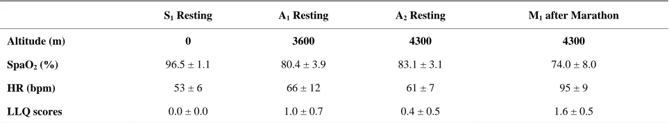
3.2. Peripheral Arterial Oxygen Saturation and Heart Rate
The % SpaO2 was higher at rest at S1 than that recorded
at A1 (P < 0.0005) and A2 (P < 0.0005); it was
signifi-cantly lower (P < 0.05) after the marathon than that ob-served at A2 (Table 1).
[image:3.595.57.539.645.734.2]The heart rate recorded at S1 was significantly lower
Table 1. Mean ± SEM values of peripheral arterial oxygen saturation (SpaO2; %), heart rate (HR; bpm) and Lake Louise Questionnaire (LLQ) scores.
S1 Resting A1 Resting A2 Resting M1 after Marathon
Altitude (m) 0 3600 4300 4300
SpaO2 (%) 96.5 ± 1.1 80.4 ± 3.9 83.1 ± 3.1 74.0 ± 8.0
HR (bpm) 53 ± 6 66 ± 12 61 ± 7 95 ± 9
LLQ scores 0.0 ± 0.0 1.0 ± 0.7 0.4 ± 0.5 1.6 ± 0.5
compared to that recorded at A1 (P < 0.05) and A2 (P <
0.05); it was significantly higher after the marathon than that observed at rest before the race (P < 0.0025) (Table 1).
3.3. EEG after Acute Acclimatization (A1) versus Sea Level (S1)
There was a significant decrease in Theta (F3-P3, A2-P3; P <
0.0001), Alpha (F3-P3, A2-P3, O1-O2; P<0.001), Beta
(F3-P3, A2-P3, O1-O2; P < 0.001), and Gamma (F3-P3,
A2-P3, O1-O2; P < 0.005) power activities over the entire
scalp at A1 at rest compared to S1 (Table 2).
3.4. EEG after Chronic Acclimatization (A2) versus Sea Level (S1)
A decrease in Theta (F3-P3, A2-P3; P < 0.0001) and Alpha
powers (F3-P3; P < 0.05) at A2 compared to S1. The Beta
power decreased in the frontal (F3-P3; P < 0.01), parietal
(A2-P3; P < 0.001) and occipital cortex (O1-O2; P <
0.001). The Gamma power was higher than at S1 (F3-P3;
P < 0.05) (Table 2).
3.5. EEG after Chronic Acclimatization (A2) versus Acute Acclimatization (A1)
There was a significant decrease in Theta power in the occipital cortex (P < 0.01) and an increase in Alpha
(F3-P3; P < 0.001; A2-P3, P < 0.001; O1-O2; P < 0.001), Beta (F3-P3; P < 0.001; A2-P3; P < 0.001; O1-O2; P <
0.001) and Gamma (F3-P3; P < 0.025; A2-P3, P < 0.001;
O1-O2; P < 0.005) powers at A2 compared to A1 (Table
2).
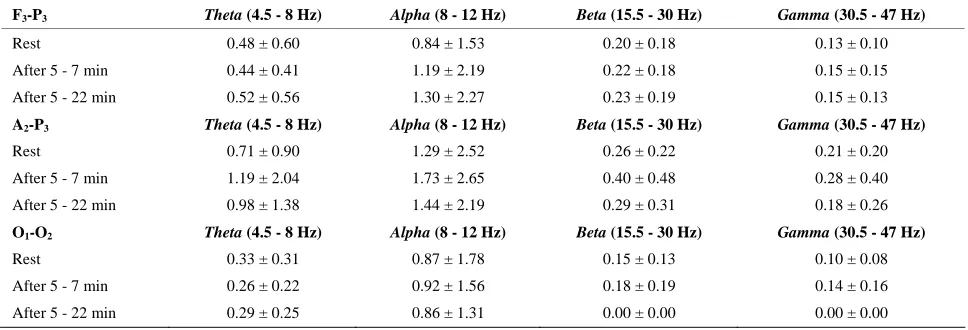
3.6. EEG after the Marathon (M1) versus Chronic Acclimatization (A2)
After the marathon (M1), a decrease was noted in Theta
power in the occipital cortex (P < 0.01) and an increase in the frontoparietal cortex (P < 0.0005). Alpha power increased (P < 0.0005) in the rostral cortex. Beta power increased over the parietal cortex (P < 0.05). The Beta
(F3-P3; P < 0.0005) and Gamma (F3-P3; P < 0.0005)
[image:4.595.56.539.377.533.2]power activities increased significantly in the frontopa-rietal cortex, but the Gamma power decreased significantly (P < 0.05) over the parietal cortex (A2-P3) (Table 3).
Table 2. EEG activity (mV2; mean and SEM) at rest at three different altitudes. Statistical differences are shown in the text.
F3-P3 Theta (4.5 - 8 Hz) Alpha (8 - 12 Hz) Beta (15.5 - 30 Hz) Gamma (30.5 - 47 Hz)
Sea level (S1) 0.87 ± 1.52 0.95 ± 1.51 0.24 ± 0.26 0.13 ± 0.10
3600 m (A1) 0.45 ± 0.65 0.64 ± 1.35 0.14 ± 0.15 0.06 ± 0.05
4300 m (A2) 0.48 ± 0.60 0.84 ± 1.53 0.20 ± 0.18 0.13 ± 0.10
A2-P3 Theta (4.5 - 8 Hz) Alpha (8 - 12 Hz) Beta (15.5 - 30 Hz) Gamma (30.5 - 47 Hz)
Sea level (S1) 1.02 ± 1.40 1.34 ± 2.00 0.33 ± 0.41 0.21 ± 0.27
3600 m (A1) 0.69 ± 0.88 0.85 ± 1.37 0.15 ± 0.14 0.07 ± 0.07
4300 m (A2) 0.71 ± 0.90 1.29 ± 2.52 0.26 ± 0.22 0.21 ± 0.20
O1-O2 Theta (4.5 - 8 Hz) Alpha (8 - 12 Hz) Beta (15.5 - 30 Hz) Gamma (30.5 - 47 Hz)
Sea level (S1) 0.39 ± 0.45 0.94 ± 1.63 0.18 ± 0.19 0.10 ± 0.07
3600 m (A1) 0.38 ± 0.47 0.58 ± 1.02 0.09 ± 0.08 0.05 ± 0.04
4300 m (A2) 0.33 ± 0.31 0.87 ± 1.78 0.15 ± 0.13 0.10 ± 0.08
Table 3. EEG activity (mV2; mean and SEM) before at rest (A2) and after the marathon (M1) at 4300 m. Statistical differ- ences are shown in the text.
F3-P3 Theta (4.5 - 8 Hz) Alpha (8 - 12 Hz) Beta (15.5 - 30 Hz) Gamma (30.5 - 47 Hz)
Rest 0.48 ± 0.60 0.84 ± 1.53 0.20 ± 0.18 0.13 ± 0.10
After 5 - 7 min 0.44 ± 0.41 1.19 ± 2.19 0.22 ± 0.18 0.15 ± 0.15
After 5 - 22 min 0.52 ± 0.56 1.30 ± 2.27 0.23 ± 0.19 0.15 ± 0.13
A2-P3 Theta (4.5 - 8 Hz) Alpha (8 - 12 Hz) Beta (15.5 - 30 Hz) Gamma (30.5 - 47 Hz)
Rest 0.71 ± 0.90 1.29 ± 2.52 0.26 ± 0.22 0.21 ± 0.20
After 5 - 7 min 1.19 ± 2.04 1.73 ± 2.65 0.40 ± 0.48 0.28 ± 0.40
After 5 - 22 min 0.98 ± 1.38 1.44 ± 2.19 0.29 ± 0.31 0.18 ± 0.26
O1-O2 Theta (4.5 - 8 Hz) Alpha (8 - 12 Hz) Beta (15.5 - 30 Hz) Gamma (30.5 - 47 Hz)
Rest 0.33 ± 0.31 0.87 ± 1.78 0.15 ± 0.13 0.10 ± 0.08
After 5 - 7 min 0.26 ± 0.22 0.92 ± 1.56 0.18 ± 0.19 0.14 ± 0.16
After 5 - 22 min 0.29 ± 0.25 0.86 ± 1.31 0.00 ± 0.00 0.00 ± 0.00
[image:4.595.56.540.571.736.2]4. Lake Louise Questionnaire
The Lake Louis Questionnaire scores prior to EEG re-cordings were <3 for all the runners.
5. Discussion
5.1. Acute Acclimatization at 3800 m Altitude (A1)
A significant decrease in EEG power has been described during acute acclimatisation to altitude in low-land tives, in acclimatised subjects, and in high-altitude na-tives [10-12]. In agreement with these observations, we found a significant topographical decrease in Theta, Al-pha, Beta and Gamma activities at A1 and a slowing
down of the high frequency component of Alpha activity and its anteriorization. These finding are suggestive of a deterioration in vigilance [9,12-16].
Moreover, acute hypoxia leads to hyperventilation and thus to arteriolar hypocapnia; this last may produce re-gional cerebral hypocapnic vasoconstriction, with changes in cerebral blood flow, alkalosis and increased pH, EEG synchronization and lethargy. The present data are in agreement with other observations showing that the slowing down of the EEG activity is one of the most constant features of cerebral hypoxia [3].
5.2. Chronic Acclimatisation at 4300 m Altitude (A2)
There were significant increases in Theta, Alpha, Beta
and Gamma activities over the entire scalp at A2 as
com-pared to A1. These activities were consistently lower than
those recorded at sea level (S1). Also, there were some
topographical variations in the power of the so-called “altitude sensitive” Theta and Alpha powers, but still indicating hypoxia-induced deterioration in vigilance.
We found also a positive correlation between the The- ta activity in the frontal areas and hypoxemia (y = 0.022x – 1.397, r2 = 1:F3P3). Moreover, a study with psychometric
tests performed by the same subjects and published elsewhere showed that some brain functions of the left frontotemporal lobe were temporarily impaired under hypobaric-hypoxic conditions [17].
We suggest that the EEG increase in low-voltage high frequency observed after acclimatisation at 4300 m alti-tude was due to activation of the forebrain and reticular activating system involved in behavioural and metabolic integration of autonomic control and arousal. This is re-lated to reflex activation of the ascending reticular acti-vating system in the ventral medullary reticular and the central pontin-midbrain reticular formation, either di-rectly via the carotid baroreceptors and/or indidi-rectly on carotid and aortic chemoreceptors and/or hypocapnic- central chemoreceptors [2,4]. Indeed, it would not be
surprising if exposure to hypobaric hypoxia were to elicit, in parallel with increased ventilation and cardiac output, stimulation of the catecholaminergic systems which ac-tivate the reticular activating system and the pituitary- adrenocortical system, cortical activation and arousal, together with slowing down of frontal EEG [18-24].
5.3. Marathon at Altitude (M1)
After the marathon at altitude we found an increase in the
Theta, Alpha, Beta and Gamma activities. Since our ex-periments involved well-trained and acclimatised runners, we suggest that the marathon race performed at altitude might have been responsible for the cortical arousal and, as at low altitude, is due to the stimulation of the reticular activating system involved in the behavioral and meta-bolic integration of autonomic control and arousal [25- 29].
The increase in high-frequency rhythms recorded just after the end of the marathon document the persistence of motor tasks in the neocortical somatomotor cortices. The parallel increase in the so-called rhythmic slow activity on the parietal region just after the end of the race was probably due to residual activation of the chemical af-ferents from the subcortical nuclei of the limbic system reaching the frontal and parietal cortices through the cingulated cortex. As previously observed by Hobson [22], it is also possible that the increase in body tem-perature due to the long distance run might have influ-enced the EEG cortical activity as a result of an alteration in hypothalamic function. Above all, the results suggest that short-term arousal may have a protective role in preventing excessive oxygen deprivation at altitude [2, 11].
All EEG changes recorded after the marathon at alti-tude tended to return to A2 values within 20 minutes
(Table 2), indicating that the marathon-induced EEG changes were transitory and that the main changes re-corded after acclimatization (i.e., at A2) were due to the
hypobaric hypoxic conditions typical of a high-altitude environment.
Furthermore, during acclimatization and after the race (A2 and M1) the runners showed no signs of AMS, as
revealed by the Lake Louise Questionnaire, and we were unable to detect any pathological paroxysmal phenomena during the EEG recordings.
Exercise without EEG paroxysmal phenomena was reported to be possible at altitude only in subjects with an aerobic power of 60 - 65 mL·kg–1·min–1, a value very similar to that recorded at sea level in our subjects, and the most important characteristic for “extreme” physical performances at high altitude is claimed to be an excel-lent cardiorespiratory function [7,9,30].
mara-thon at around 4300 m altitude is safe for well-trained mountain runners, as confirmed by other studies [8,31, 32].
6. Conclusions
After completion of a marathon at an altitude of 4300 m, the increase in the power of low-voltage high-frequency activities over the entire cortex was probably due to the direct and indirect reflex activation of the forebrain and the reticular activating system which has a protective role in preventing excessive cerebral oxygen deprivation.
Although exercise at high altitude, without oxygen supply, has been held to be unsafe [1,33], the results of this study indicate that arousal plays a protective role in preventing excessive oxygen deprivation. The absence of AMS fond in our study bear out that well trained and acclimatized runners can safely participate in a marathon at high altitude that gives rise to temporary EEG changes without inducing paroxysmal phenomena.
6. Acknowledgements
This study was supported by the Federation for Sport at Altitude (FSA), Biella, Italy, and MURST and FIRST grants. We thank Marco Rosa, M.D., and the staff of the Marathon Sports Medical Centre, Brescia, Italy, for their technical assistance during the measurements at sea level.
REFERENCES
[1] G. S. Roi, M. Giacometti and S. P. Von Duvillard, “Ma- rathons in Altitude,”Medicine & Science in Sports & Ex-ercise, Vol. 31, No. 5, 1999, pp. 723-728.
doi:10.1097/00005768-199905000-00016
[2] J. H.Coote, “Medicine and Mechanisms in Altitude Sick-ness. Recommendations,” Sports Medicine,Vol. 20, No. 3, 1995, pp. 148-159.
doi:10.2165/00007256-199520030-00003
[3] J.V.Weil, “Sleep at High Altitude,” High Altitude Medi-cine and Biology, Vol. 5, No. 2,2004, pp. 180-189. doi:10.1089/1527029041352162
[4] J.V. Weil,“Respiratory Physiology: Sleep at High Alti-tude,” In: M. H. Kryger, T. Roth and W. C. Dement, Eds.,
Principle and Practice of Sleep Medicine, W. B. Sauders Company, Toronto, 2004, pp. 242-254.
[5] R. C. Roach, P. Bärtsch, O. Oelz and P. H.Hackett, “The Lake Louise Acute Mountain Sickness Scoring System,” In: J. R. Sutton, C. S. Houston and G. Coates, Eds., Hy-poxia and Molecular Medicine, Queen City Press, Bur-lington, 1993, pp. 272-274.
[6] P. Abraham, “Electroencephalography and Everest Climb-ers,” Journal of the Royal Army Medical Corps, Vol. 124, 1978, pp. 84-85.
[7] Z. Ryn, “Problem of Treating Mental Disorders at High
Altitudes,” Psychiatria Polska,Vol. 4, No. 5, 1970, pp. 589-595.
[8] T. P. Finnegan and P. Abraham, “High-Altitude Hypoxia and the Brain,” The Lancet, Vol. 20, 1986, p. 695. [9] H. Ozaki, S. Watanabe and H. Suzuki,“Topographic EEG
Changes Due to Hypobaric Hypoxia at Simulated High Altitude,” Electroencephalography & Clinical Neurophy- siology, Vol. 94, No. 5, 1995, pp. 349-356.
doi:10.1016/0013-4694(94)00311-8
[10] H. V. Forster, R. J. Soto, J. A. Dempsey and M. J. Hosko, “Effect of Sojourn at 4300 m Altitude on Electroence- phalogram and Visual Evoked Response,” Journal of Ap-plied Physiology, Vol. 39,No. 1, 1975, pp. 109-113. [11] W.Selvamurthy, R. K. Saxena, N. Krishnamurthy, M. L.
Suri and M. S. Malhotra, “Changes in EEG Pattern during Acclimatization to High Altitude (3500 m) in Man,”
Aviation Space & Environmental Medicine, Vol. 49, No. 8, 1978, pp. 968-971.
[12] V. Kraaier, A. C. Van Huffelen and G. H. Wieneke,“Quan- titative EEG Changes Due to Hypobaric Hypoxia in Nor- mal Subjects,” Electroencephaography and Clinical Neuro- physiology, Vol. 69, No. 4, 1987, pp. 303-312.
[13] J. S. Meyer and A. G.Waltz,“Arterial Oxygen Saturation and Alveolar Carbon Dioxide during Electroencephalo-graphy. I. Comparison of Hyperventilation and Induced Hypoxia in Subjects without Brain Disease,” Archives of Neurology, Vol. 2, No. 6, 1960, pp. 631-643.
doi:10.1001/archneur.1960.03840120037005
[14] J. S. Meyer and F. Gotoh, “Metabolic and Electroen-cephalographic Effects of Hyperventilation. Experimental Studies of Brain Oxygen and Carbon Dioxide Tension, pH, EEG and Blood Flow during Hyperventilation,” Ar-chives of Neurology, Vol. 3, No. 5, 1960, pp. 539-552. doi:10.1001/archneur.1960.00450050059007
[15] N. A. M. Schellart, “Transient and Maintained Changes of the Spontaneous Occipital EEG during Acute Systemic Hypoxia,” Aviation, Space, and Environmental Medicine, Vol. 72, No. 5, 2001, pp. 462-470.
[16] H. B. Van der Worp, “Quantitative EEG Changes during Progressive Hypercapia and Hypoxia. Hyperventilation Induced EEG Changes Reconsidered,” Electroencepha-lography and Clinical Neurophysiology, Vol. 79, 1991, pp. 1335-1341.
[17] G. Pelamatti, M. Pascotto and C.Semenza, “Verbal Free Recall in High Altitude: Proper Names vs Common Names,” Cortex, Vol. 39, No. 1, 2003, pp. 97-103. doi:10.1016/S0010-9452(08)70077-7
[18] G.Banfi, M. Marinelli, G. S. Roi, A. Colombini, M. Pon-tillo, M. Giacometti and S.Wade, “Growth Hormone and Insulin-Like Growth Factor I in Athletes Performing a Marathon at 4000 m of Altitude,” Growth Regulation, Vol. 4, No. 2, 1991, pp. 82-86.
[19] G. Banfi, M. Marinelli, P. Bonini, I. Gritti and G. S. Roi, “Pepsinogens and Gastrointestinal Symptoms in Moun-tain Marathon Runners,” International Journal of Sports Medicine, Vol. 17, No. 8, 1996, pp. 554-558.
doi:10.1055/s-2007-972894
Roi, “Thyrotropin and Free Thyroid Hormones in Ath-letes during and after Ultra Endurance Sport Perform-ances,”Journal of Clinical Ligand Assay, Vol. 21, No. 3, 1998, pp. 331-334.
[21] I. Gritti, G. Banfi and G. S.Roi, “Pepsinogens: Physiol-ogy, PharmacolPhysiol-ogy, Pathophysiology and Exercise,” Phar- macological Research, Vol. 41, No. 3, 2000, pp. 265-281. doi:10.1006/phrs.1999.0586
[22] J. A. Hobson, “Sleep after Exercise,” Science,Vol.162, No. 3861, 1968, pp. 1503-1505.
doi:10.1126/science.162.3861.1503
[23] K. J. Maloney, E. G. Cape, J. Gotman and B. E. Jones, “High Frequency γ Electroencephalogram Activity in Association with Sleep-Wake States and Spontaneous Behaviours in the Rat,” Neurosciences, Vol.76, No. 2, 1997, pp. 541-555. doi:10.1016/S0306-4522(96)00298-9 [24] F. H. Lopes da Silva, “The Generation of Electric and
Magnetic Signals of the Brain by Local Networks,” In: R. Greger and U. Windhorst, Eds., Comprehensive Human Physiology from Cellular Mechanisms to Integration, Springer, New York, Vol. 2, 1996, pp. 509-532.
[25] R. Llinás and U. Ribary, “Coherent 40-Hz Oscillation Characterizes Dream State in Humans,” Proceedings of the National Academy of Sciences, Vol. 90, No. 5, 1993, pp. 2078-2081.doi:10.1073/pnas.90.5.2078
[26] V. N. Murthy and E. E. Fetz, “Coherent 25- to 35-Hz Oscillations in the Sensorimotor Cortex of Awake Be-having Monkeys,” Proceedings of the National Academy of Sciencesof the United States of America, Vol. 89, No. 12, 1992, pp. 5670-5674. doi:10.1073/pnas.89.12.5670
[27] D. Pinault and M. Deschenes, “Control of 40-Hz Firing of Reticular Thalamic Cells by Neurotransmitters,” Neuro-science, Vol.51,No.2, 1992, pp. 259-268.
doi:10.1016/0306-4522(92)90313-Q
[28] M. Steriade, R. C. Dossi, D. Paré and G.Oakson,“Fast Oscillations (20 - 40 Hz) in Thalamocortical Systems and Their Potentation by Mesopontine Cholinergic Nuclei in the Cat,” Proceedings of the National Academy of Sci-encesof the United States of America, Vol. 88,No.10, 1991, pp. 4396-4400.doi:10.1073/pnas.88.10.4396 [29] C. B.Saper, “Central Autonomic System. The Rat
Nerv-ous System,” 1995, pp. 107-128.
[30] J. R. Sutton, “Effect of Acute Hypoxia on the Hormonal Response to Exercise,” Journal of Applied Physiology:
Respiratory, Environmental & Exercise Physiology,Vol. 42, No. 4, 1977, pp. 587-592.
[31] T. P. Finnegan, P. Abraham and T. B. Docherty, “Ambu-latory Monitoring of the Electroencephalogram in High Altitude Mountaineers,” Electroencephalography & Cli- nical Neurophysiology, Vol. 60, No. 3, 1985, pp. 220- 224.doi:10.1016/0013-4694(85)90034-3
[32] G. S. Roi, M. Giacometti, G. Banfi, M. Zaccaria, I. Gritti and S. P. Von Duvillard, “Competitive Running at High Altitude, Is it Safe?” Medicine & Science in Sports & Ex-ercise, No. 5, 1999, p. S191.