RESEARCH ARTICLE
STUDY OF CYTOLOGICAL SPECTRUM OF MALIGNANT LESIONS IN LYMPH NODE
*Dr. Lakshmibai B. Mallappa
Department of Pathology, Bangalore Medical College and Research Institute, Fort Bangalore- 560002,
Karnataka, India
ARTICLE INFO ABSTRACT
Background: Lymphadenopathy is a common presenting symptom in various diseases.¹ Lymphadenopathy, which is defined as an abnormality in the size or character of lymph-nodes, is caused by the invasion or propagation of either inflammatory cells or neoplastic cells into the node.²
Objective: The present study was conducted to evaluate the usefulness of Fine Needle Aspiration Cytology as a diagnostic tool in cases of Malignant Lymphadenopathy.
Study design: This prospective study was conducted in the Department of Pathology; Victoria Hospital; Bangalore Medical College Bangalore over a period of 6 months i.e from 1.1.11 to 30.6.11.
Material and Methods: No of Fine Needle Aspirations done in this period were 1349. Out of which 74 cases were that of Malignant Lymphadenopathy (involving cervical, axillary, inguinal and generalised group of
lymph-nodes). Malignant Lymphadenopathy comprises of both primary and secondary (metastatic) disease. Out of 441
cases of lymphnode fnac, 74 cases were diagnosed as malignant cytologically and clinically.
Results: Out of 1349, 74 cases were diagnosed to have Malignant Lymphadenopathy. Of the 74 cases, 5 cases were suspicious of malignancy. The age group involved was between 5-95 yrs. The male to female ratio was 2:1. Among them, there were 9 cases (12.16%) of primary lymphoid malignancy and 60 cases (81.08%) of metastatic malignancy and about 5 cases (6.75%) were suspicious of malignancy. The most common metastatic deposit was of squamous cell carcinoma (58.10%) followed by breast carcinoma (8.10%) and malignant melanoma (4.05%). Conclusion: FNAC remains a useful investigation in diagnosing Malignant Lymphadenopathy with good certainty. It is an economical and convenient alternative to open biopsy.
Copyright, IJCR, 2013, Academic Journals. All rights reserved.
INTRODUCTION
Lymphadenopathy is a common presenting symptom in various diseases¹. Key risk factors for malignancy include older age, firm, fixed nodal character, duration of greater than two weeks and supra-clavicular location². Knowledge of these risk factors is critical in determining the management of unexplained lymphadenopathy². The rate of malignant etiologies of lymphadenopathy is very low in childhood but increases with age². Environmental exposures such as tobacco, alcohol and ultraviolet radiation may raise suspicion for metastatic carcinoma of the internal organs, cancers of the head and neck and skin malignancies respectively². Lymphadenopathy may be the first sign of malignancy in a patient. Fine needle aspiration cytology (FNAC) not only confirms the presence of metastatic disease, but also gives clues regarding the nature and origin of the primary tumour, in patients with enlarged lymph-nodes and previously documented malignancy³. The cytomorphological features obtained in Needle Aspiration, frequently correlate very well with histologic appearance of the same lesion and in some situations has qualities of a microbiopsy4. The value of FNAC, besides making a diagnosis also lies in early direction of appropriate investigations5.
The metastasis from Squamous Cell Carcinoma, may lead to necrosis, abscess formation or cystic change leading to a false negative diagnosis. Moreover, the primary sites sometimes remain occult contributing to the delay in treatment6. It can be carried out at bed side or in the out-patient clinic, requiring neither local nor general anaesthesia7. FNAC is particularly helpful in the work-up of nodules because surgical biopsy of lymphadenopathy is a more invasive procedure8.
*Corresponding author: [email protected]
The aim of this study is
To evaluate the role of FNAC in various lymphadenopathies. To study the cytomorphological patterns of both primary and
metastatic lesions in Malignant Lymphadenopathy.
MATERIALS AND METHODS
A prospective study was conducted in the Department of Pathology, at our institution, over a period of six months i.e. from 1.1.11 to 30.6.11. A total of 1349 cases were aspirated. Out of which 74 cases were of Malignant lymphadenopathy involving cervical, axillary and inguinal Lymph-Nodes. In each case a brief history, physical examination and relevant investigations were carried out. The technique of the procedure was explained to the patient and the formal consent was obtained. FNAC was performed using a 22-23G needles with 5ml or 10ml syringes. Multiple sites were aspirated. Smears were made and stained with Giemsa and Haematoxylin & Eosin. These slides were later on interpreted to come to a probable diagnosis. They were either reported as suspicious or positive for malignancy.
RESULTS
During this period a total of 1349 FNACs from all sites were performed. Out of which 74 cases were that of Malignant Lymphadenopathy (involving cervical, axillary, inguinal and generalized lymphnode groups). The age group was between 5-95yrs.The maximum number of cases were seen in the 5th decade (41-50yrs) and 6th decade (51-60yrs). There were 50 males and 24 females. Male to female sex ratio was 2:1. Out of the 74 cases
ISSN: 0975-833X
International Journal of Current Research
Vol. 5, Issue, 04, pp.961-965, April,2013
INTERNATIONAL JOURNAL
OF CURRENT RESEARCH
Article History:
Received 15th January, 2012
Received in revised form
26th February, 2013
Accepted 24th March, 2013
Published online 13th April, 2013
9 cases (12.16%) were primary lesions. 60 cases (81.08%) were of metastatic deposits and about 5 cases (6.75%) were suspicious of malignancy [Table 1, 2]. The commonest lymph-node group with primary malignancy was the Cervical Group (81.08%) followed by Inguinal Group(9.45%), Axillary Group (8.108%) and Generalised Group (1.35%) [Table 3a]. The metastatic lymphnode-groups involving the primary sites are cervical, axillary, inguinal groups. About 28 cases were from unknown primary sites. The primary sites identified were from tongue, thyroid, buccal mucosa, vocal cord, lower lip, oropharynx, kidney, tonsil, stomach/oesophagus, maxilla, breast, salivary gland, blood (in the form of leukemic infiltrates),
cervix, lower extremities, prostate and penis [Table 3b].
[image:2.595.83.518.66.164.2]The most common type of malignancy was squamous cell carcinoma (58.11%), breast carcinoma (8.10%), lymphoproliferative disease (6.75%), non-hodgkin’s disease (5.40%), malignant melanoma (4.05%), 1 case each of renal cell carcinoma (sarcomatoid variant), adenocarcinoma, plasmacytoma, malignant epithelial lesion from salivary gland, leukemic infiltrates. 5 cases (6.57%) were suspicious of malignancy [Table 4].
Table 2. Lymph-node groups involved in Malignant Lymphadenopathy along with Sex Distribution
Lymph-Node Group Sex No.of
Cases
Percentage (%)
Cervical Group
Male 46
81.08
Female 14
Inguinal Group Male 03
9.45
Female 04
Axillary Group Male 00 8.108
Female 06
Generalised Group Male 01 1.35
Female 00
Table 3a . Distribution of Primary Malignancy in Lymphadenopathy
Lymph-Node Group No. of
Cases
Primary Malignancy
Cervical
9
Lymphoproliferative Disease,
Hodgkin’s Disease,
Non-Hodgkin’s Lymphoma
Axillary _ _
Inguinal _ _
Generalised 1 Non-Hodgkin’s Lymphoma
Table 3b. Distribution of Metastatic Group of Lymph-Nodes with Primary Sites
Lymph-node Group
No.of cases Primary Sites
Cervical 25
Tongue, Thyroid, Buccal Mucosa, Right Vocal Cord, Lower Lip,
Oropharynx, Kidney, Tonsil,
Stomach, Oesophagus, Maxilla, oral cavity, Breast,
Salivary Gland,
Supraglottis, hypopharynx
Axillary 6
Breast,
Leukemic Infiltrates.
Inguinal 6 Cervix, Lower Extremities, Prostate,
Penis Generalised
___ Unknown
Sites
[image:2.595.38.288.210.315.2]28 ?
Table 4. Distribution of type of Malignancy in Lymphadenopathy
Type of Malignancy No. of Cases Percentage
Squamous Cell Carcinoma 43 58.108
Lymphoproliferative disease & anaplastic large cell lymphoma
09 +01 13.51
Breast carcinoma 06 8.10
Malignant melanoma 03 4.05
Papillary carcinoma 02 2.7
RCC (Sarcomatoid variant) 01 1.35
Adenocarcinoma 01 1.35
Malignant epithelial lesion from salivary gland
01 1.35
Plasmacytoma 01 1.35
Leukemic infiltrates 01 1.35
[image:2.595.347.526.378.540.2]Suspicious malignancy 05 6.75
Figure 1. Cytology of Non-Hodgkin’s Lymphoma (H and E, 10Xmagnification)
Figure showing non-hodgkin’s lymphoma smears with monomorphic cell pattern consisting of lymphoblasts or lymphocytes
Figure 2a. Cytology of Squamous Cell Carcinoma, keratinizing type (H and E 10Xmagnification)
Figure showing keratinising type of sqamous cell carcinoma with predominant single cell keratinisation, keratinized malignant cells, refractile cytoplasm.
Table 1. Age and Sex Distribution chart of Malignant Lymphadenopathy
Age in Years No.of Cases Primary Disease
M F
Metastatic Disease
M F Suspicious of Malignancy
0-10 1 0 1 0 0 0
11-20 0 0 0 0 0 0
21-30 3 3 0 0 0 0
31-40 5 0 1 0 4 0
41-50 19 2 1 6 6 ³
51-60 19 0 0 14 5 0
61-70 15 1 0 11 3 0
>70 12 0 0 9 2 2
[image:2.595.43.283.531.766.2] [image:2.595.342.530.571.710.2]DISCUSSION
Lymphadenopathy is one of the common clinical presentation of various ongoing disease process inside the body. The lesions arising in the Lymph-Node can be found in patients ranging from early to advanced age1. Family history may raise suspicion for certain neoplastic causes of lympadenopathy such as carcinomas of the breast or familial dysplastic nevus syndrome and Melanoma2. More specific review questions such as whether pain occurs in the area of lymphadenopathy after even limited alcohol ingestion, may bring out a rare but fairly specific finding of a neoplasm such as Hodgkin’s Lymphoma2. In our study the age of the patients ranged between 5-95yrs. This is almost similar to the work done by Hirachand S.et al9 with the age of patients ranged from 3 to 85yrs. In our study, the age group which had maximum number of cases was above 40 yrs. These findings were similar to the study of S.Shamshad Ahmad et al4, where malignant lesions were encountered in the 6th decade. Ruchi Khajuria et al5 showed that metastatic carcinoma were prevalent over 40 yrs of age whereas cases of lymphomas were distributed in all age groups. Also, a study conducted by M.Bezabih
et al8 also showed adults more than 40yrs of age were more commonly affected in malignant nodal enlargements. Male to female ratio was 2:1. This was similar to Ruchi Khajuria et al 5 also showing male preponderance with same ratio. The commonest group of lymph-nodes involved in our study were that of cervical
Figure 2b. Cytology of Squamous Cell Carcinoma (non-keratinising type) ) H and E 40Xmagnification
Figure showing non keratinizing type of tumors usually more cohesive and present as multilayered fragments. Their nuclei are usually spindle shaped or elongated with dense irregular distributed chromatin.
[image:3.595.59.271.53.219.2]Figure 3. Cytology of Papillary Carcinoma-Thyroid Gland Giemsa 40x
Figure showing cellular smears with, syncytial aggregates and sheets of cells with a distinct anatomical border, focally nuclear crowding and overlapping, papillary tissue fragments with or without a fibrovascular
core. Enlarged, ovoid, strikingly pale, finely granular, powdery
[image:3.595.323.530.54.234.2]chromatin. Multiple distinct nucleoli; intranuclear cytoplasmic inclusions ;nuclear grooves;
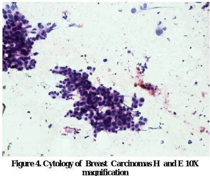
Figure 4. Cytology of Breast Carcinomas H and E 10X magnification
Figure showing breast carcinomas usually displays poor glandular differentiation, while cell balls and single files are common. Some tumors form a monolayered of dispersed cells with intact cytoplasm
Figure 5. Cytology of Malignant Melanoma H and E 10X magnification
Smears of Malignant Melanoma may show total dissociation of cells, well defined cytoplasm, eccentric nuclei, Prominent anisokaryosis, a uniformly dense chromatin which doesnot vary much from nucleus to nucleus, often large nucleoli, binucleate cells, intranuclear vacuoles.
[image:3.595.57.273.289.452.2]Some cells with intracytoplasmic melanin pigment11.
Figure 6. Cytology of Adenocarcinoma H and E 10X magnification
Glandular cells, moderately pleomorphic, arranged in a gland-in-gland
[image:3.595.326.542.297.479.2] [image:3.595.56.271.548.729.2]group (81.08%) followed by inguinal group (9.45%), axillary group (8.108%) and generalised lymphadenopathy (1.35%).The lymph-node group involvement was similar to that of S.Shamshad Ahmad et al4 where in the cervical group(73.6%) was the major group involved followed by axillary group(10.6%) & inguinal group of lymph-nodes (6.0%). In their study, Ruchi Khajuria et al5
showed the involvement of cervical neck nodes ,inguinal group followed by axillary group5. A study done by Hirachand S. et al9
found that the commonest site of lymphadenopathy was in the neck constituting 66 cases. Among the remaining cases, 20 were axillary, 18 submandibular, 14 supraclavicular and 12 inguinal lymph-nodes. In our study, primary lymphoid malignancies constituted 9 cases (12.16%), lymphoproliferative diseases 5 cases and non-hodgkin’s lymphoma 4 cases. Out of the 4, 1case of anaplastic large cell lymphoma was diagnosed.
In a study conducted by Ruchi Khajuria et al5 reported 13 cases of Malignant Lymphoma,8 Non-Hodgkin’s Lymphoma and 5 Hodgkin’s Disease. Muhammad Javaid et al7 reported 9.52% cases of lymphoproliferative disorder and 4.76% cases of lymphomas. Out of 8 cases(6%) of Lymphoma, 6 were Non-Hodgkin’s Lymphoma and 2 were Hodgkin’s Disease, in the study by Hirachand S. et al9. 15.3% of cases were primary tumours i.e Lymphoma reported by Kiran Alam et al10. In our study 1 case suspicious of Hodgkin’s Lymphoma was noted. Among the metastatic malignancies, the most common lymph-node group involved was the cervical group followed by axillary and inguinal lymph-node group. About 28 cases showed deposits from unknown sites. Two studies conducted by Ruchi Khajuria.K.et al5 and M.Bezabih et al 8 correlated with the same findings where in the cervical group was largely involved followed by axillary and inguinal group of lymph-nodes with regards to metastatic malignancies. In the category of distribution of metastatic group of lymph-nodes showing the origin of primary sites are as follows-in the cervical group; tongue, thyroid, buccal mucosa, right vocal cord, lower lip, oropharynx, kidney, tonsil ,ca stomach/oesophagus, maxilla, salivary gland, oral cavity, supraglottis and hypopharynx. Squamous cell carcinoma was the commonest primary tumour in our study. 2 cases showed areas of necrosis and inflammation, another 2 cases showed cystic change. Papillary carcinoma-thyroid, renal cell carcinoma-sarcomatoid variant, breast carcinoma were the other primary tumors.
In the Axillary chain of lymph-nodes metastases from Breast Carcinomas & Leukemic Infiltration were noted. The inguinal group of lymph nodes were involved by squamous cell carcinoma metastases from the cervix & penis. While from the lower-extremities deposits of malignant melanoma was seen in this group of lymph nodes. deposits of adenocarcinoma from the prostate were also seen.
A study conducted by Deepti Agarwal et al1 also showed that the major cause of lymphadenopathy was metastatic carcinoma involving the Cervical Group .The type of tumour deposits were SCC(53.12%) arising commonly in the tongue, alveolus, buccal mucosa and palate. Similar, findings were noted by I.N.Bagwan et al3 with (36.81%). The aspiration of the Metastatic Nodes with cystic change often becomes hypocellular due to presence of fluid. A centrifuged deposit should be carefully examined for any malignant cells. A repeat aspiration from any palpable mass left after aspiration of fluid may yield cellular material. An image- guided FNAC from the solid area of the swelling can also be helpful. Smears with large amounts of inflammatory cell infiltration and abscess formation should be carefully searched for malignant squamous cell. An important clue to the diagnosis of metastatic SCC in the presence of necrosis and keratinisation, which is better appreciated on Pap Stain than on H&E Stain.The presence of keratinous debris and foreign body giant cell formation indicates the possibility of kertinising Squamous Cell Carcinoma6
A repeat FNAC may be helpful in the smears that are hypocellular, inadequate or doubtful6 A study conducted by I.N.Bagwan et al3 also showed metastatic deposits into the Neck Nodes by Papillary Carcinoma-Thyroid, Salivary Gland Tumour. Other primary sites
include breast, Lung, Kidney, Prostate and Gonads etc. Melanomas arising in the eyeball, scalp or head and neck region may metastasize to neck nodes, but occasionally it presents as a metastases from an occult primary3 In the axillary group of Lymph-Nodes the commonest metastatic tumour was from the Breast Carcinomas which was reported by M.Bezabih et al8.The same author also reported 4 cases of Metastatic Melanomas and 1 unspecified carcinoma in the inguinal region.
1 case of AML and CML infiltration into the Lymph-node was reported by S. Shamshad Ahmad et al4 Lastly, the commonest type of malignancy in Lymphadenopathy in our study were as follows; Squamous cell Carcinomas 43(58.108%) cases, Breast Carcinomas(8.10%),Malignant Melanoma 3(4.05%) cases; Papillary Carcinoma Thyroid 2(2.7%) cases.1 case each of Renal Cell Carcinoma(sarcomatoid variant), Adenocarcinoma, Plasmacytoma, Malignant Epithelial Lesion from Salivary Gland & Leukemic infiltrates. All the above constitutes(1.35%) . Lymphoproliferative disease 5 cases (6.75%),Non-Hodgkin’s Lymphoma 4 cases(5.40) and 5 suspicious cases. Out of which 1 case of Hodgkin’s Lymphoma and 4 cases of Squamous Cell Carcinoma. About 28 cases (37.83%) were from unknown sites. Metastatic Malignancy was the most frequent causative agent of Lymphadenopathy with Squamous Cell variety being predominant1,3,4,9.
Conclusion
Our study has proved to be a useful tool in diagnosing malignancy with good certainity.A thorough study of the morphological details of the individual tumour cells helps in suggesting the most likely primary sites of tumour along with clinicocytological correlation. Our preliminary aim was to help the clinician in arriving at an early diagnosis in cases of malignant Lymphadenopathy. Once a diagnosis of Malignancy was made the cases were referred to higher cancer Institutes for further treatment. Thus FNAC offers a very important screening procedure for Lymph-Node Malignancies. Squamous Cell Carcinoma still remains as the metastatic Malignant tumour of the Head &Neck region.
REFERENCES
1. D. Agarwal, P. Bansal, B. Rani, .Sharma, S. Chawla, V. Bharat, S. Sharma-Evaluation of etiology of lymphadenopathy in
different age groups using Fine Needle Aspiration Cytology:A retrospective study.The Internet Journal of Pathology.2010 Volume 10 Number2
2. Andrew. W. Bazemore and Douglas R. Smucker-Lymphadenopathy and Malignancy:American Family Physician 2002 Dec1;66(11):2103-2111.
3. I. N. Bagwan, S. V. Kane & R. F. Chinoy: Cytologic Evaluation of the Enlarged Neck Node:FNAC Utility in Metastatic Neck Disease.The Internet Journal of Pathology.2007Volume 6 Number2.
4. S.Shamshad Ahmad,Shakeel Akhtar,KafilAkhtar,Shano Naseem,Tariq Mansoor-Study of Fine Needle Aspiration Cytology in Lymphadenopathy with special Reference to Acid-fast Staining in Cases of Tuberculosis.JK Science Vol. 7 No. 1, January-March 2005.
5. Ruchi Khajuria,K.C.Goswami,K.Singh,V.K.Dubey-Pattern of Lymphadenopathy on Fine Needle Aspiration Cytology in Jammu.JK Science Vol.8 No.3, July-September 2006
6. Karabi Konar,Sulekha Ghosh,Tapan Ghosh,Subodh Bhattacharya, Saurabh Sanyal-Pitfalls in the cytodiagnosis of metastatic squamous cell carcinoma in the head and neck:A retrospective study.Year2008,Volume:25 Issue:4 Page:119-122 7. Muhammad Javiad, Niamatullah, Khurshid Anwar, Muhammad
8. M. Bezabih and D. W. Mariam-Determination of Aetiology of Superficial enlarged Lymph-Nodes using Fine Aspiration Cytology.East African Medical Journal Vol.80 No.11 November 2003.
9. Hirachand. S, Lakhey M, Akhter J, Thapa B-Evaluation of fine needle aspiration cytology of lymph nodes in Kathmandu Medical College,Teaching hospital.Kathmandu University Medical Journal (2009),Vol.7,Issue 26,139-142
10. K. Alam, V. Maheshwari, N. Haider, F. Siddique, A. Jain & A.Khan:Fine needle aspiration cytology (FNAC),a handy tool for metastatic lymphadenopathy. The Internet Journal of Pathology.2010 Volume 10 Number 2.
11. Svante. R. Orell, Gregory F.Sterrett and Darrel Whiterker:-Fine Needle Aspiration Cytology,4th Edition.