ISSN Print: 2160-8792
DOI: 10.4236/ojog.2018.810092 Aug. 27, 2018 891 Open Journal of Obstetrics and Gynecology
Retrospective Descriptive Analysis of the
Combined First Trimester of Pregnancy
Screening in the Period Included from
February 2016 to March 2017, Maternal Fetal
Medicine Unit, San Juan de Dios Hospital
Carina Breuning Velásquez, Oscar Durán Soto, Joaquin Bustillos Villavicencio
*,
Jorge Mora Sandí, Eugenio Calderon, Veronica Saborio, Leonardo Jimenez, Pablo Parra
Hospital San Juan de Dios, San José, Costa Rica
Abstract
A retrospective descriptive study is conducted at the San Juan de Dios Hos-pital, San José, Costa Rica, during the period from February 2016 to March 2017, with a total of 37 patients from which a combined screening during the first trimester of pregnancy was conducted, evaluating maternal age, bio-chemical and sonographic methods that together can predict the risk of fetal chromosomal alterations during pregnancy. The purpose of using combined screening as a noninvasive method is to identify high risk gestations and to minimize the number of invasive procedures to detect the highest number of cases. Four patients with higher risk of aneuploidy during pregnancy were identified through this screening.
Keywords
Protocol, Combined Screening, First Trimester, Aneuploidy, Trisomy
1. Introduction
The following paper is based on the results obtained from the application of a first trimester of pregnancy screening tests that suggest fetal aneuploidy, focused in 21, 13, 18 trisomies, Down, Patau and Edwards syndrome respectively with the final goal of educating parents and foresee the causes of associated perinatal morbidity and mortality.
How to cite this paper: Velásquez, C.B., Soto, O.D., Villavicencio, J.B., Sandí, J.M., Calderon, E., Saborio, V., Jimenez, L. and Parra, P. (2018) Retrospective Descriptive Analysis of the Combined First Trimester of Pregnancy Screening in the Period In-cluded from February 2016 to March 2017, Maternal Fetal Medicine Unit, San Juan de Dios Hospital. Open Journal of Obstetrics and Gynecology, 8, 891-899.
https://doi.org/10.4236/ojog.2018.810092
Received: March 15, 2018 Accepted: August 24, 2018 Published: August 27, 2018
Copyright © 2018 by authors and Scientific Research Publishing Inc. This work is licensed under the Creative Commons Attribution International License (CC BY 4.0).
DOI: 10.4236/ojog.2018.810092 892 Open Journal of Obstetrics and Gynecology The first trimester of pregnancy echography and structural chromosomic mutation screening protocol is stablished at the unit of obstetrics of Hospital San Juan de Dios and the latter is applied during the period between February 2016 and March 2017. The objective of the screening pretends to reduce the number of invasive procedures during pregnancy that might carry a risk of miscarriage as well as to reduce the monetary costs that this measure generates [1].
Given that the screening based on maternal age represents a method with a low sensibility and specificity, the combined method that includes variables such as maternal age, biochemical tests and echography altogether has a superior ac-curacy, achieving index of detection for Trisomy 21 as high as 90% and a 5% of false positives [1] [2].
The exclusion criteria used to apply the screening method are: • Younger than 15 years old and older than 35 years old.
• History of aneuploidy as well as congenital malformations or genetic syn-drome.
• Pregestational diabetes. • Epilepsy.
• History of 3 or more abortions.
• Pregnancies achieved through in vitro fertilization.
• Patients exposed to radiation during the first trimester of pregnancy. All of these exclusion criteria are related to high risk of fetal chromosomic al-teration, high risk of metabolic or genetic disease as well as a higher risk of peri-natal infection.
The protocol contemplates two types of exploration: the ultrasound between the 11 - 13.6 weeks of gestation and biochemical analysis of the beta fraction of the chorionic gonadotropic hormone and a protein associated to pregnancy from maternal blood samples. Both results are expressed as multiples of the me-dian (MoM) for each gestational age. In case of trisomy 21 the β-HCG values are elevated while PAPP-A is reduced. In case of trisomy 13 and 18 both values are reduced.
The most important echography markers of aneuploidy are the nuchal trans-lucency and the nasal bone. An increase in nuchal transtrans-lucency is due to liquid accumulation behind the fetal neck which is observed during ultrasonographic evaluation during the first trimester of pregnancy and is associated with trisomy 21. 75% of the fetuses with T21 are reported to have an increased nuchal trans-lucency. The absence of the nasal bone not necessarily increases the index of de-tection of down syndrome, but it can be used as a marker to decrease false posi-tives.
DOI: 10.4236/ojog.2018.810092 893 Open Journal of Obstetrics and Gynecology other abnormalities, they don’t indicate aneuploidy or perinatal pathology, there is need for highly trained personal to carry both explorations and so they are re-served for population of an intermediate to high risk.
In the protocol there is also the possibility to perform a biochemical evalua-tion during the second trimester of pregnancy in those cases that were not screened during the first trimester of pregnancy. Given such a case, ACOG (American College of Obstetrics and Gynecology) recommends a quadruple scrutiny to be performed. The latter includes: alfa fetal protein, β-HCG, estriol and inhibin A; as well as the evaluation with ultrasound between the week 18 and 20 of pregnancy. The ultrasonographic markers of the second trimester in-clude: choroid plexus cyst, intracardiac echogenic foci, increased nuchal fold, echogenic bladder, one unique umbilical artery, ventriculomegaly, abscense or hypoplasia of nasal bone, right aberrant subclavian artery, hydronephrosis, fe-moral and/or humeral shrinkage. All the above-mentioned markers have elevated incidence in fetuses with aneuploidy compared to normal ones [1] [2] [3] [4].
The software used for the screening makes a basic calculation where all three fundamental variables (age, nuchal translucency, biochemical analysis report) plus gestational age are integrated and expressed in terms of a fraction. The used cut point for positive screening for risk of aneuploidy is 1/270 at 35 years [1].
In case of a positive screening test, it is recommended to perform a fetal ka-ryotype. The elected method is the biopsy of chorionic villi preferably between weeks 11 and 13.6. After week 15 of gestation the gold standard is amniocentesis.
Chorionic villi biopsy must be performed after the 10th week of gestation ei-ther by transabdominal route or by transcervical means depending on the expe-rience and preferences of the physician or the location of the placenta. The com-plications related to this procedure include the risk of miscarriage (reported be-tween 0.2% and 2% of cases) and vaginal bleeding reported in 10% of cases [1] [5] [6].
Factors associated with increased risk of miscarriage after chorionic villi bi-opsy reported in previous retrospective trials include: African Americans, at least two needle aspirations or insertions, abundant bleeding during procedure, ma-ternal age less than 25 years or gestational age less than 10 weeks.
Low levels of PAPP-A are related with placentation-preeclampsia issues, rea-son why it is also related to higher miscarriage risk [6].
Amniocentesis must be done after the 15th week of gestation by transabdo-minal means and guided by ultrasound. The complications reported after this procedure include the risk of fetal loss that varies between 0.1% and 1%, in-creased risk of amniotic fluid loss up to week 24 of gestation. There has been a reduced risk of chorioamnioitis and uterine infection after an amniocentisis, the risk below 0.7%.
amni-DOI: 10.4236/ojog.2018.810092 894 Open Journal of Obstetrics and Gynecology ocentesis.
Another test performed in the United States is the analysis of fetal blood sam-ple in search of chromosomal mosaicism, which is obtained by transabdominal means after the 18th week of gestation.
Eligibility criteria for the procedure are: • High risk for chromosomic fetal alterations. • High risk of metabolic or genetic diseases. • High risk of perinatal infection.
Before any invasive procedure, detailed family counsel as well as awareness of associated risks should be done.
2. Methods
A retrospective descriptive trial was performed using data from San Juan de Dios Hospital’s maternal fetal unit. 37 patients total were included in the period of time from February 2016 to March 2017. These patients all received the first trimester screening test during their pregnancy.
Data were drawn from the archive of screening results for Down Syndrome of the unit. Both maternal and fetal variables were analyzed. Patients did not sign an informed consent since the screening is already part of the services offered to the pregnant patient in our center. A thorough file search was performed and factors such as maternal age, number of fetuses, gestational age, ethnicity, histo-ry of trisomy 21, and the values of the screening tests were recorded. Afterwards a frequency and rate analysis was performed to obtain the main results. All of the data was recorded in Microsoft Office Excel, graphs were also performed us-ing this software [1] [2] [5] [7] [8] [9] [10].
Using the exclusion criteria, patients followed at San Juan de Dios Hospital from February 2016 to March 2017 were included in our study. A recording data table was created and 41 patients were gathered. From this main group, 4 pa-tients lost their control in our center so at the end 37 papa-tients were finally in-cluded.
3. Objectives
Perform first trimester screening test to identify asymptomatic individuals at in-creased risk of having a pregnancy with chromosomic fetal alterations.
Diminish the amount of invasive procedures either by chorionic villi biopsy or amniocentesis that could result in fetal loss.
4. Results
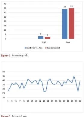
During the period of February 2016 to March 2017 patients were screened with the combined first trimester screening test during their first trimester of preg-nancy. From the 37 included patients three presented risk for trisomy 21 and two presented high risk in the combined test (see Table 1).
DOI: 10.4236/ojog.2018.810092 895 Open Journal of Obstetrics and Gynecology age (see Figure 1 and Figure 2). 23 of the patients included for screening were Caucasian and 14 had no ethnicity recorded.
The screening was performed in patients with pregnancies with gestational ages between 10.4 weeks and 14.2 weeks in average the gestational age was of 12.4 weeks.
The values of the BHCG are located between 0.2 and 2.51 MOM with an av-erage of 0.80 MoM.
The values of PAPP-A were found to be at ranges between 0.27- 3.54 MOM with an average of 1.02.
For the sonographic evaluation the nuchal translucency and the cranium rump length (CRL) were recorded and the cases were the nasal bone could be observed were reported (Table 2 and Table 3).
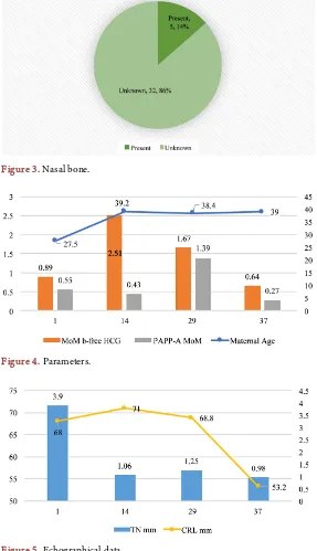
The range of the CRL was 38 - 82.34 mm with an average of 62.30 mm. For the nuchal translucency a range between 0.4 and 3.9 with an average of 1.41 mm. The nasal bone was present in 5 of the 37 evaluated fetuses (Figure 3).
In Figure 4 and Figure 5 the results of the serum analysis from high risk
[image:5.595.233.512.331.717.2]pa-tients are summarized.
Figure 1. Screening risk.
DOI: 10.4236/ojog.2018.810092 896 Open Journal of Obstetrics and Gynecology
Figure 3. Nasal bone.
[image:6.595.230.517.81.580.2]Figure 4. Parameters.
Figure 5. Echographical data.
Table 1. Individuals at risk in the analysis of the combined screening.
Subject 1 14 29 37
Maternal age 27.5 39.2 38.4 39
T21 combined risk >1:50 1:62 1:118 1:318
Double test risk >1:50 >1:50
[image:6.595.208.540.616.730.2]DOI: 10.4236/ojog.2018.810092 897 Open Journal of Obstetrics and Gynecology
Table 2. Analyzed data in the combined screening.
Free β-HCG MoM PAPP-A MoM CRL mm TN mm .. MoM
Average 0.80 1.02 62.30 1.41 0.88
Minimum 0.2 0.27 38 0.4 0.32
Maximum 2.51 3.54 82.84 3.9 2.27
Range 0.2 - 2.51 0.27 - 3.54 38 - 82.84 0.4 - 3.9 0.32 - 2.27
Table 3. Laboratory analyzed data.
Estriol PAPP-A lab HCG libre protein UI/ml Alpha fetal mUI/ml HCG
Average 0.13 5.61 28.78 15.89 87,714.7
Minimum 0.07 1.22 7 6.36 18,394
Maximum 0.6 10 90 33.3 78,248
5. Discussion
The analysis of the results from the combined screening in the first trimester of pregnancy done during one year at Hospital San Juan de Dios, reflects a total of four patients of high risk detected by this protocol. In individuals at risk the av-erage age was of 36 years, which matches with the universal statistics that place increased maternal age as the principal risk correlated to fetal aneuploidy.
It is worth noting that nuchal translucency of one fetus was reported at 3.9, superior to the cut associated with trisomy 21. Nevertheless when following up those cases identified as high risk, after an appropriate counseling to the patients and their family, only one patient was reported and amniocentesis is still pend-ing. Two of the remaining patients rejected the procedure and one of them never came back to prenatal control at the unit, reason why there is a bias of informa-tion in this case.
This study concluded 10% of affected cases and, as suggested by foreign sources, in our environment amniocentesis will be performed on the patient who accepted the procedure, corresponding respectively to 1.85% of the screened pa-tients.
Since we practice in social care and our country has limited economical re-sources, the screening is made on selected patients with inclusion risk criteria that might implicate an increased risk for the gestation, reason why the sample is not significant.
[image:7.595.210.540.221.304.2]DOI: 10.4236/ojog.2018.810092 898 Open Journal of Obstetrics and Gynecology nuchal translucency tests. The accuracy of noninvasive prenatal screening with Fetal cell-free DNA reduces the number of invasive tests needed for a definitive diagnosis. In general obstetric population, Fetal cell-free DNA have lower false-positive rates and higher positive predictive values than standard screening
[11].
This method has the ability to detect small elevations in the amount of one respective chromosome in maternal plasma in a pregnancy due to a trisomy. Studies have demonstrated that this method might detect up to 99% of the cases of trisomy 21, 97% of trisomy 18, 92% of the cases of trisomy 13. The false posi-tive rate is of 0.1% for T21, 0.2% for T18 and 0.2% for T13 respecposi-tively. The findings in the screening of free cell DNA in maternal blood in the general pop-ulation resemble those of previous studies in high-risk pregnancies.
It has been shown that this method is feasible for routine screening and allows a reliable diagnosis of aneuploidy, with false-positive rates significantly lower than those of combined screening [12] [13].
Although this screening method is not publicly available, it is ideal for all pregnant patients to have at least one first-trimester ultrasound that includes the measurement of the nuchal translucency, which is a marker not only of fetal chromosomal alterations but also of cardiac defects and other genetic syn-dromes.
6. Conclusions
The combined screening of the first trimester of pregnancy represents an ade-quate screening strategy in this population to fundamentally reduce invasive di-agnostic procedures and their complications in the case of suspected aneuploidy in the fetus.
In our study, despite being a sample of high-risk pregnant women, 4 patients with a risk of chromosomopathies were detected, which equal 10% of the study population, concordant with published data and demonstrating that counseling for just as screening is essential for the final follow-up of these patients.
This confirms that ultrasound aimed at screening patients with some risk in the first trimester of pregnancy should be a fundamental and mandatory study as a measure even of initial uptake of the patient during a pregnancy and, when combined with other epidemiological variables and biophysical in this popula-tion, we stratify the individual risk early on in each patient and thus we can offer a higher quality care in this very determinant stage of being a human being.
Acknowledgements
To the radioimmune analysis laboratory and to the Obstetrics Department of Hospital San Juan de Dios for all the support and collaboration in this project.
DOI: 10.4236/ojog.2018.810092 899 Open Journal of Obstetrics and Gynecology
References
[1] Bustillos Villavicencio, J. (2014) Protocolo: Ecografía de primer trimestre y screen-ing de alteraciones cromosómicas y estructurales en el primer trimestre del emba-razo. Servicio de Obstetricia, Hospital San Juan de Dios.
[2] Rao, R. and Platt, L.D. (2016) Ultrasound Screening: Status of Markers and Efficacy of Screening for Structural Abnormalities. Seminars in Perinatology. WB Saunders, 67-78. https://doi.org/10.1053/j.semperi.2015.11.009
[3] Chi, T., Huggon, I., Zosmer, N. and Nicolaides, K.H. (2001) Incidence of Major Structural Cardiac Defects Associated with Increased Nuchal Translucency But Normal Karyotype. Ultrasound in Obstetrics & Gynecology, 18, 610-614.
https://doi.org/10.1046/j.0960-7692.2001.00584.x
[4] Agathokleous, M., Chaveeva, P., Poon, L.C.Y., Kosinski, P. and Nicolaides, K.H. (2013) Meta-Analysis of Second-Trimester Markers for Trisomy 21. Ultrasound in Obstetrics & Gynecology, 41, 247-261. https://doi.org/10.1002/uog.12364
[5] Howard, C. (2000) Biochemical Screening for Down Syndrome. European Journal of Obstetrics & Gynecology and Reproductive Biology, 92, 97-101.
https://doi.org/10.1016/S0301-2115(00)00431-0
[6] Ghi, T., et al. (2016) ISUOG Practice Guidelines: Invasive Procedures for Prenatal Diagnosis. Ultrasound in Obstetrics & Gynecology, 48, 256-268.
https://doi.org/10.1002/uog.15945
[7] Nicolaides, K.H., Heath, V. and Cicero, S. (2002) Increased Nuchal Translucency at 11 - 14 Weeks.Prenatal Diagnosis, 22, 308-315. https://doi.org/10.1002/pd.308 [8] Nicolaides, K.H., Sebire, N. and Snijders, R. (1999) Nuchal Translucency and
Chromosomal Defects. In: Nicolaides, K.H., Ed., The 11 - 14 Week Scan. The Di-agnosis of Fetal Abnormalities. Diploma in Fetal Medicine Series, The Parthenon Publishing Group, London, 3-65.
[9] Cicero, S., Curcio, P., Papageorghiou, A., Sonek, J. and Nicolaides, K.H. (2001) Ab-sence of Nasal Bone in Fetuses with Trisomy 21 at 11-14 Weeks of Gestation: An Observational Study. Lancet, 358, 1665-1667.
https://doi.org/10.1016/S0140-6736(01)06709-5
[10] Alfirevic, Z. and Brigham, S.K. (2003) Amniocentesis and Chorionic Villus Sam-pling for Prenatal Diagnosis. Cochrane Database of Systematic Reviews, 3, CD003252. https://doi.org/10.1002/14651858.CD003252
[11] Wagner, A., Mitchell, M. and Tomita-Mitchell, A. (2014) Use of Cell-Free Fetal DNA in Maternal Plasma for Noninvasive Prenatal Screening. Clinics in Perinatol-ogy, 41, 957-966. https://doi.org/10.1016/j.clp.2014.08.013
[12] Quezada, M.S., Gil, M.M., Francisco, C., Oròsz, G. and Nicolaides, K.H. (2015) Screening for Trisomies 21, 18 and 13 by Cell-Free DNA Analysis of Maternal Blood at 10 - 11 Weeks Gestation and the Combined Test at 11 - 13 Weeks. Ultra-sound in Obstetrics & Gynecology, 45, 36-41. https://doi.org/10.1002/uog.14664 [13] Gil, M.M., Quezada, M.S., Revello, R., Akolekar, R. and Nicolaides, K.H. (2015)