CLINICAL AND HISTOPATHOLOGICAL STUDY OF ORBITAL TUMORS
*Dr. Tanushree, V., Dr.
Department of Ophthalmology,
ARTICLE INFO ABSTRACT
Web
presenting at a tertiary care centre. Methods
hospital from May 2010 to May 2014.Details on patient’s history, clinical examination, radiological investigations, histopathological profiles were retrieved from
Results (range 2 pseudotumors. Conclusion:
and lacrimal gland tumors to be the common
Copyright © 2015 Tanushree et al. This is an open access article distributed under the Creative Commons Att distribution, and reproduction in any medium, provided the original work is properly cited.
INTRODUCTION
The eye is not only an organ of vision but also an index of beauty in the human race (Saha Somnath
volume of the orbit is small and confined by bony walls on all sides except anteriorly. Within this space is a juxtaposition of numerous structures that subserve visual as well as extraorbital functions. Therefore, it is not surprising that ophthalmic symptoms are common presenting findings with most orbital diseases (Yanoff and Duker, ?). The majority of orbital tumors originate between the bony orbital wall and the extraocular muscle cone and 90% of the orbital tumors present with proptosis (Rootman, 1988). Orbital tumors constitute a heterogeneous array of lesions and, as such, pose numerous challenges in terms of diagnosis, imaging and management. Given the variety of structures within the relatively confined orbit, a systematic approach is necessary to understand the classification and clinical features of orbital tumors
et al., 2000). Close co-operation between an oculo surgeon, otorhinolaryngologist, neurosurgeon, radiologist, oncologist and histopathologist is required for the management of orbital tumors (Rizvi et al., 2010). The aim of this study is to report the clinical profile and histopathology of orbital tumors in patients presenting at a tertiary eye care centre.
*Corresponding author: Dr. Tanushree, V.
Department of Ophthalmology, Mysore Medical College and Research Institute, Mysore.
ISSN: 0975-833X
Article History:
Received 17th December, 2014
Received in revised form
10th January, 2015
Accepted 25th February, 2015
Published online 31st March,2015
Key words: Orbit, Proptosis, Tumors, Orbitotomy.
RESEARCH ARTICLE
CLINICAL AND HISTOPATHOLOGICAL STUDY OF ORBITAL TUMORS
Dr. H.T. Venkate Gowda, Dr. Uma Balakrishnan and
Dr. Amar Kulkarni
Ophthalmology, Mysore Medical College and Research Institute, Mysore
ABSTRACT
Web Purpose: To report the clinical profile and histopathology of orbital presenting at a tertiary care centre.
Methods: This is a retrospective case series of patients with orbital
hospital from May 2010 to May 2014.Details on patient’s history, clinical examination, radiological investigations, histopathological profiles were retrieved from medical records.
Results: 48 patients were studied (22 males & 26 females). Mean age range 2 – 75 yrs). 31 of the tumors were benign, 5 were pseudotumors.
Conclusion: This study demonstrates the prevalence of dermoid cysts,
lacrimal gland tumors to be the common space occupying lesions in the orbit.
is an open access article distributed under the Creative Commons Attribution License, which distribution, and reproduction in any medium, provided the original work is properly cited.
The eye is not only an organ of vision but also an index of
et al., 2002). The
volume of the orbit is small and confined by bony walls on all sides except anteriorly. Within this space is a juxtaposition of numerous structures that subserve visual as well as extraorbital functions. Therefore, it is not surprising that ophthalmic symptoms are common presenting findings with most orbital
The majority of orbital tumors originate between the bony orbital wall and the extraocular muscle cone and 90% of the orbital tumors present with
Orbital tumors constitute a heterogeneous array of lesions and, as such, pose numerous challenges in terms of diagnosis, imaging and management. Given the variety of structures within the relatively confined ry to understand the
classification and clinical features of orbital tumors (Dutton operation between an oculo-plastic
surgeon, otorhinolaryngologist, neurosurgeon, radiologist, uired for the management . The aim of this study is to report the clinical profile and histopathology of orbital tumors in patients presenting at a tertiary eye care centre.
Department of Ophthalmology, Mysore Medical College and
MATERIALS AND METHODS
A total of 48 patients with orbital tumors who presented to our institute were taken up in this study. The duration of the study was from May 2010 to May 2014. All the patients in the present study underwent detailed history
examination that included best
(BCVA), exophthalmometry, extraocular muscle movements, retropulsion test, palpation of orbital margins and eyelids, IOP measurement in different gazes, orbital palpation for thrills and auscultation for bruits, visual field examination and ophthalmoscopy. A detailed general physical examination including the patient’s skin and oropharynx, regional and distant lymph nodes and cranial nerves examination was also done.
All the patients in the present series
investigation. CT scan was done in all cases to evaluate the extent of tumor and localize the lesion. Complete haemogram was done for all patients. Chest X ray and USG abdomen was done to rule out distant metastasis. The lesions were approached as per their anatomical location, size, extent and suspected pathology. Primary orbital tumor was approached by different orbitotomy routes. Anterior orbitotomy either by the trans-conjunctival or trans- cutaneous route was performed for lesions anterior to the equator of the globe and in the anterior intraconal space. All deep-seated lesions posterior to equator and in the intraconal space and lesions lateral to optic nerve were approached by the lateral orbitotomy route. Excision was
Available online at http://www.journalcra.com
International Journal of Current Research
Vol. 7, Issue, 03, pp.13954-13958, March, 2015
INTERNATIONAL
z
CLINICAL AND HISTOPATHOLOGICAL STUDY OF ORBITAL TUMORS
Dr. Uma Balakrishnan and
and Research Institute, Mysore
To report the clinical profile and histopathology of orbital tumors in patients
This is a retrospective case series of patients with orbital tumors who presented to the hospital from May 2010 to May 2014.Details on patient’s history, clinical examination, radiological
medical records.
. Mean age of presentation was 38.5 benign, 5 were malignant and the rest 12 being
This study demonstrates the prevalence of dermoid cysts, pseudotumors, hemangiomas space occupying lesions in the orbit.
ribution License, which permits unrestricted use,
MATERIALS AND METHODS
A total of 48 patients with orbital tumors who presented to our institute were taken up in this study. The duration of the study was from May 2010 to May 2014. All the patients in the present study underwent detailed history-taking and meticulous on that included best-corrected visual acuity , exophthalmometry, extraocular muscle movements, retropulsion test, palpation of orbital margins and eyelids, IOP measurement in different gazes, orbital palpation for thrills and , visual field examination and ophthalmoscopy. A detailed general physical examination including the patient’s skin and oropharynx, regional and distant lymph nodes and cranial nerves examination was also
done for superficial tumors. Few patients were referred to higher center for further management if required. All the lesions of the present series were biopsied after excision for their histopathological diagnosis and were treated accordingly.
RESULTS
All the patients with orbital tumor in this study were analyzed with regard to their age, symptoms, surgical approaches employed and histopathological diagnosis.
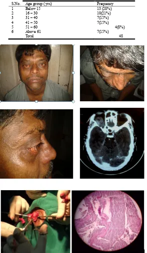
[image:2.595.326.539.166.220.2]The age of the patients ranged from 2 years to 75 years. Maximum patients were in 2nd – 3rd decade. Common lesions in this group were dermoids followed by cavernous hemangioma. Malignant lesions were seen in older age group which is consistent with most of the literatures. Common lesions were in this age group were pleomorphic adenoma of lacrimal gland and lymphoma.
Table 2. Common Symptoms of presentation
S. No. Symptom Overall percentage
1 Protrusion of eyeball 32(63 %)
Axial 21(32%)
Eccentric 11(11%)
[image:2.595.163.457.252.759.2]2 Swelling or Mass 16(27 %)
Table 1. Age distribution
S.No. Age group ( yrs) Frequency
1 Below 15 13 (28%)
2 16 – 30 10(21%)
3 31 – 40 7(15%)
4 41 – 50 7(15%)
5 51 – 60 4(8%)
6 Above 61 7(15%)
Total 48
About two third (63%) of the patients presented with the complaint of protrusion of eyeball. (32%) of them had axial proptosis (cavernous hemangioma, meningioma,
schwanomma) and the rest (27%) had eccentric proptosis (lacrimal gland tumor, lymphoma). Cases of dermoids
presented only with a swelling round about the orbit. The other associated symptoms were pain in and around the eye (15 cases), diplopia (14 cases), diminution of vision (11 cases), watering (10 cases)
[image:3.595.34.561.194.606.2]Dermoids were excised completely. A lateral orbitotomy, with temporary removal of the lateral orbit wall was done for lesions in the lacrimal fossa and lateral orbit, including the areas above and below the optic nerve. Ten lateral orbitotomies were done. Most of them were for cavernous hemangioma followed by pleomorphic adenoma of lacrimal gland. Seven anterior orbitotomies were done. They were performed for Cavernous hemangioma, meningioma, pleomorphic adenoma of lacrimal gland, diffuse large B cell lymphoma. An upper lid splitting technique was used for superior lesions.
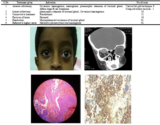
Table 3. Management
S.No. Treatment given Indication No of cases
1 Anterior orbitotomy Cavernous hemangioma, meningioma, pleomorphic adenoma of lacrimal gland,
diffuse large B cell lymphoma
Vertical lid split technique: 6 Using subcilliary incision : 1
2 Lateral orbitotomy pleomorphic adenoma of lacrimal gland , Cavernous hemangioma, 10
3 Conservative treatment Pseudotumor 12
4 Excision of lesion Dermoid 16
5 Exentration Mucoepidermoid carcinoma of lacrimal gland 01
6 Referred to higher center Metastatic plasmacytoma and meningioma 02
[image:3.595.138.471.657.791.2]
Figure 2. Primary peripheral primitive nuerectodermal tumor of orbit
Table 3. Diagnosis
S.No. Histopathological diagnosis Number
1 Dermoid 16(33%)
2 Pseudotumor 12(25%)
3 Cavernous Hemangioma 5(10%)
4 Pleomorphic adenoma of Lacrimal Gland 4(8%)
5 Meningioma 3(6%)
6 Schwannoma 2(4%)
7 Nasopharyngeal Carcinoma 1(2%)
8 Metastatic Plasmacytoma 1(2%)
9 Non Hodgkin’s Lymphoma 1(2%)
10 Large B cell Lymphoma 1(2%)
11 Mucoepidermoid carcinoma of Lacrimal Gland 1(2%)
12 P-NET 1(2%)
TOTAL 48
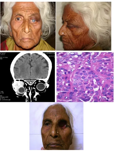
Orbital exenteration which included excision of the eyelids and the removal of the conjunctiva, globe, optic nerve, extra- ocular muscles, the lacrimal gland, and all the soft tissues of the orbit was done for one case. Pseudotumor cases were diagnosed as a diagnosis of exclusion after detailed clinical, histopalthological and radiological investigations and were treated conservatively. One case each of metastatic plasmacytoma and meningioma were referred to higher center for management. Overall, dermoids were the most common tumors in our study (33%) followed by pseudotumor (25%) and hemangiomas (10%). Malignant tumors accounted for 10% of cases among which lymphoma was the most common (4%) and benign lesions for 90% among which dermoids were most common (33%).
DISCUSSION
Orbital tumors can occur at various age groups, and because of their myriad presentation, their diagnosis pose a great challenge. Orbital tumors encompass a heterogeneous variety of lesions. Careful evaluation of a patient’s history and examination along with modern high-resolution imaging studies provide invaluable information regarding the possible origin of an orbital lesion.
[image:4.595.105.504.53.587.2]Although imaging studies graphically illustrate tissue definition, pathological conditions can be assessed definitely only by obtaining tissue specimen surgically (Radha and Ani
Sreedhar, 2005). In our study of 48 patients, dermoids were found to be most common accounting for 33% of cases. Pseudotumors accounted for 25% of all orbital lesions. Orbital pseudo tumor is one distinct disease albeit with many clinical and histopathological guises. It can occur in any age and sex
(Radha and Ani Sreedhar, 2005). Hence it is a diagnosis of exclusion. Lacrimal gland tumors accounted for 10% of all orbital tumors. Of the 5 cases of lacrimal gland tumors, all were of epithelial origin four being pleomorphic adenoma and one mucoepidermoid carcinoma. Benign epithelial tumors are generally common. Although pleomorphic adenomas are histologically benign, incomplete excision will lead to relentless recurrences and even malignant transformation. Hence when suspected, lateral orbitotomy is mandatory and entire tumor must be excised enbloc (Halli et al., 2011)
Majority of malignant tumors are adenoid cyst carcinoma- Major cause of death is intracranial spread hastened by perineural invasion. Treatment is exenteration and post op radiotherapy (Radha and Ani Sreedhar, 2005).
Optic nerve tumors included 3 meningiomas (6%) and 2 schwannomas (4%). All the patients had axial proptosis with other findings such as defective pupillary reactions, dimunition of vision and optic atrophy. Lymphomas accounted for 4 % of all orbital tumors. Orbital lymphomas have worse prognosis. In our study, there was 1 case of Non Hodgkins lymphoma and one case of Large B cell lymphoma. Demirci et al. have showed that this neoplasm has a predilection for older patients and it is the most common neoplasm in older adult group
(Demirci et al., 2002). The best strategy for managing orbital tumors is largely determined by the nature of the lesion. Orbital approaches can be made more extensive when the lesion is posterior to the equator of the eye (Tim et al., 2001). Definitive surgical treatment remains the mainstay of therapy for the majority of symptomatic orbital tumors. The type of anesthesia and the most appropriate surgical approach to the lesion are dependent on its anatomical location, proximity to vital structures, and extent of involvement as visualized on CT scanning and MR imaging. Orbital and nasal approaches can be warranted, especially when the tumor does not involve the apex or extend beyond the confines of the orbit.
To sum up, this study was undertaken to determine the number and percentage of various orbital tumors that presented to our hospital. Based on the results of our study, most common orbital tumors include dermoids (33%), pseudotumors (25%), hemangiomas (10%) and lacrimal gland tumors (10%). The relative incidence of malignant tumors is more in the elderly. These figures correlated with the previous studies conducted by Shield’s et al. (2004).
Conclusion
Orbital tumors are a great challenge for the ophthalmologist since they present with a variety of signs and symptoms and they are often difficult to diagnose at the initial stages. The patient’s age should be kept in mind while evaluating a patient with possible orbital malignancy. Availability of CT arid MRI however should not neglect necessity of careful history, detailed systemic and ocular examination.
REFERENCES
Demirci H, Shields CL, Shields JA, Honavar SG, Mercado GJ, Tovilla JC. Orbital tumors in the older adult population. Ophthalmology, 2002;109:1288-9.
Dutton JJ, Byrne SF, Proia AD: Diagnostic Atlas of Orbital Diseases. Philadelphia: WB Saunders, 2000
Halli RC, Mishra S, Kini YK, et al. Modified lateral orbitotomy approach: a novel technique in the management of lacrimal gland tumors. J Craniofac Surg., 2011; 22(3):1035–38.
Hassler W, Unsöld R,et al Orbital Tumors: Diagnosis and Surgical Treatment Dtsch Arztebl 2007; 104(8): A 496– 501
Radha. J., Ani Sreedhar. Orbital Tumors - A Clinico Pathological Study. KSOS September 2005 Vol.27 Issue 3
Rizvi SAR, Gupta Y, Gupta M Surgical treatment and histopathological analysis of proptosis Nep J Oph
2010;2(3):31-34
Rootman J. Frequency and Differential Diagnosis. 2nd ed. In: Diseases of the orbit. Philadelphia: JB Lippincott; 1988.
Saha Somnath, Saha Vedula Padmini et al. orbital and paraorbital tumors- clinicopathological profile and surgical management. Indian Journal of Otolaryngology
and Head and Neck Surgery, Vol. 54 No. 2, April – June
2002.
Shields JA, Shields CL, Scartozzi R. Survey of 1264 patients with orbital tumors and simulating lesions: The 2002 Montgomery Lecture, part 1. Ophthalmology,
2004;111(5):997–1008.
Tim E. Darsaut, Giuseppe Lanzino, et al. An introductory overview of orbital tumors. Neurosurg Focus 10 (5):Article 1, 2001
Yanoff and Duker: Ophthalmology, 3rd ed.Section3 – Orbit and Lacrimal gland Chapter 12.12 – Orbital Diseases