J. exp. Biol. 139, 1-30 (1988)
printed in Great Britain © The Company of Biologists Limited 1988
STIMULUS-SECRETION COUPLING: A PERSPECTIVE
HIGHLIGHTING THE CONTRIBUTIONS OF PETER BAKER
BY T. J. RINK
Smith Kline & French Research Ltd, The Fry the, Welwyn, Herts AL6 9AR
AND D. E. KNIGHT
MRC Secretory Mechanism Group, Department of Physiology, King's College London, Strand, London WC2R 2LS
Summary
Many investigators are using numerous preparations for contributing to our present understanding of stimulus-secretion coupling, by which we mean stimu-lus-dependent exocytosis, sometimes known as the regulated pathway. However, a few model systems have been particularly illuminating and several of these were exploited by Peter Baker and his close associates: namely, the motor nerve terminal, the adrenal chromaffin cell, the sea urchin egg and the blood platelet. In fact, Peter's first real contribution in this area came from his seminal studies on calcium transport in his favourite preparation, the squid giant axon, where he investigated Ca2 +/Na+ exchange, Ca2+ distribution and voltage-gated Ca2+ entry. More direct investigations into stimulus-secretion coupling came from work on neurone transmitter release in collaboration with Andrew Crawford, and on catecholamine secretion from the adrenal medulla in collaboration (with TJR). His most important generic contribution to this field was in the development (with DEK), of the electropermeabilized cell, which allows control of the low molecular weight components of the cytosol while leaving the exocytotic apparatus and process intact. In the initial experiments on the cells it was finally proved that Ca2+-dependent secretion of catecholamines is indeed from the granules and not from the cytosol. The quantification of the Ca2+ requirement of secretory exocytosis was an important step, as was the investigation of many factors purported to be important in the coupling mechanism or in the exocytotic process itself. Work with the human platelet, using this technique, has proved to be especially valuable in unravelling the complex interactions between different second messengers and has been neatly complemented by work in intact cells containing Ca2+-indicator fluorescent dyes. Peter was also intrigued by post-secretory events both in the early seventies, and at the end of his career when he embarked on analysis of the membrane retrieval process and the associated uptake of extracellular medium.
2 T. J. RINK AND D. E. KNIGHT
Introduction
We have two main objectives for this article: to outline some of the main steps on the road to our present understanding of stimulus-secretion coupling and the 'regulated' pathway of secretory exocytosis; and to highlight the main contri-butions made by our mentor and colleague, the late Peter Baker, and by some of the scientists he introduced to this field.
The term secretion is used to describe several rather different biological processes, ranging from neurotransmitter release to the production of milk. Here, we will be considering stimulus-dependent discharge of the contents of specific intracellular granules or vesicles by exocytosis. This is an exceedingly neat biological trick which we now recognize as having at least two major functions. It provides a way for the rapid and specific release of substantial amounts of carefully selected substances which can be of high molecular weight, very hydrophilic, and unlikely to pass readily through the cell membrane. It is also a way of rapidly inserting new membrane components into the surface membrane and increasing the surface area of specific parts of the membrane. Two important examples of this lesser known function of exocytosis are the insertion of water-permeable channels into the luminal membrane of distal nephron cells under the influence of anti-diuretic hormone, and the insertion of proton-pumping vesicles into the apical membrane of the parietal cells of the gastric mucosa.
We can also note the diversity and versatility of this mechanism throughout an enormous range of biological function and evolutionary scale, and this was one of the aspects of secretion that appealed to Peter Baker. Secretory exocytosis underlies the millisecond events of neurotransmitter release, many forms of hormone secretion, fundamental processes of fertilization, the acrosome reaction of sperm and the cortical reaction of the egg and, more primitively yet, the discharge of the contractile vacuole of free-living protozoans such as amoeba.
It can be helpful for both conceptual purposes and to gain a historical perspective, to subdivide stimulus-secretion coupling into a number of steps or processes. First we will consider the storage of secreted products into specific granules or vesicles and the demonstration of their stimulated release by exocytosis. Next comes a consideration of the role of cytosolic calcium. More recently has come an investigation into other factors that can trigger and regulate secretory exocytosis. Then we will consider various theories and models for the mechanism and processes of the exocytotic event itself and, finally, briefly consider membrane retrieval.
Stimulus—secretion coupling 3
Each of these was exploited by Peter Baker and/or his close colleagues. Experiments with these preparations will form the main focus of our account here, together with some of his seminal work on calcium transport in what was surely his favourite preparation, the squid giant axon.
Granules, vesicles and exocytosis
Following the discovery of hormones and the birth of endocrinology, it was recognized that many secretory tissues released their secreted product from a large prefabricated intracellular store. The granular nature of secretory cells, readily recognized in stained sections examined by light microsopy, hinted at the existence of specialized organelles for such storage. In the early 1950s, several investigators, including Blashchko & Welch (1953), showed that much of the catecholamine contained in the cells of the adrenal medulla was in a subcellular particle which was subsequently shown to be a distinct organelle, the chromaffin granule. However, in all fractionation studies a significant amount of the catecholamine was present in the supernatant and these studies could not, therefore, themselves resolve the issue of whether secretion was directly from the granular store or from the cytosolic pool. The first direct proposal of the mechanism of exocytosis as the basis of secretion came from electron microscopic analysis (De Robertis & Vaz Ferreria, 1957).
The next stage in building up evidence came from detailed analysis of the biochemical composition of the contents of the secretory granule and of the material released following stimulus of the chromaffin cell. The basic point is that the proportion of other substances to catecholamines was found to be similar within the granule and in the perfusate of stimulated adrenal glands (see, for example, Douglas, 1968). This was seen both with ATP, which is present in the chromaffin granule in a molar ratio of approximately 1:4 with catecholamines, and also with various proteins found in the chromaffin granule, such as a protein termed chromogranin and the soluble fraction of the enzyme dopamine /?-hydroxylase. It was also demonstrated that the lipid components of the chromaffin granule were not released from the cell following stimulation (Schneider, Smith & Winkler, 1967; Douglas, 1968). These observations are easily fitted in with the model of secretion whereby the membrane of the secretory granule fuses with the inside of the plasma membrane to exteriorize the contents of the granule, but only with difficulty into any other model.
4 T. J. RINK AND D. E. KNIGHT
apparatus including that required for stimulus-secretion coupling. Any cytosolic pool of catecholaminesis, of course, immediately dispersed by this manoeuvre and yet, as will be discussed below, a very substantial pool of stimulus-dependent releasable catecholamine remains within the granules. Another recent contri-bution has been the on-line measurement of the capacitance of the surface membrane of single chromaffin cells by means of the patch-clamp technique (Neher & Marty, 1982). With this sophisticated approach it proved possible to demonstrate very small discrete step changes in the capacitance of the surface membrane when the cells were stimulated to secrete, which is the precise prediction of the exocytotic process whereby small, but significant, increments in membrane area must occur with every exocytotic event of granule fusion. Interestingly, one can go back to the 1930s to find what was actually the first clear evidence for exocytosis in experiments which measured the capacitance of Hipponoe eggs before and after fertilization by passing an a.c. signal through a suspension of the eggs. In doing this, Cole (1935) calculated a two- to three-fold increase in surface membrane area following fertilization; we now realize that this resulted from the exocytotic fusion of the cortical granules with the plasma membrane as a key part of the fertilization response in these eggs and the elevation of the fertilization membrane.
It was also in the 1950s that Katz and his colleagues discovered the quantal nature of neurotransmitter release at the frog neuromuscular junction, in which action potentials appeared to release discrete packets (or quanta) of acetylcholine (e.g. Del Castillo & Katz, 1956). Electron micrographs of the nerve terminals revealed numerous small vesicular structures which seemed likely to be the structural basis of these quanta. A few years later Whittaker and his co-workers fractionated the synaptic vesicles from mammalian brain and demonstrated that these contained acetylcholine (Whittaker, Michaelson & Kirkland, 1964). It has not proved possible to isolate the acetylcholine-containing vesicles from motor nerve terminals because these form such a minute fraction of the volume of tissue in the muscle; however, an evolutionarily homologous structure, the electroplax of the electric ray, contains massive accumulations of the vesicles in the cholinergic nerve terminals, and isolation of a pure fraction of these vesicles has been achieved (Whittaker, 1984). For many years it proved difficult to demonstrate convincingly that following nerve stimulation the expected 'omega figures' could be seen, to give a visual demonstration that exocytosis of a vesicle had, indeed, occurred. This turns out to be because the exocytotic process is very transient and occurs at somewhat dispersed localized spots.
Stimulus—secretion coupling 5
inside of acetylcholine-containing synaptic vesicles appeared on the surface of the terminal following intense stimulation (Von Wedel, Carlsson & Kelly, 1981). We do not propose to discuss here the residual controversy over the precise role and type of exocytosis at the cholinergic nerve terminal, except to note that there is good evidence for a 'non-quantaP release process which occurs continuously, albeit at a very low level (Tauc, 1982). In our minds, a low level of release from the terminal across the plasma membrane fits quite well with expected physiology. Since choline acetyltransferase is a cytosolic enzyme, acetylcholine is presumably synthesized in the cytosol and then transported by secondary active transport and accumulated within the vesicle. For this to be effective there must be a cytosolic pool of acetylcholine and it seems entirely plausible that some of this should leak out across the plasma membrane on the choline transporter which is known to be present and is needed for the nerve terminal to obtain the choline substrate for the synthesis of acetylcholine. Another explanation of non-quantal release might be that pumps are inserted into the plasma membrane as a result of exocytosis. Acetylcholine is accumulated from the cytosol into intracellular vesicles by acetylcholine transporters in their membranes, these pumps operating to move acetylcholine from low cytosolic concentrations to high concentrations within the vesicle (Parsons & Koenigsberger, 1980). Immediately after exocytosis, the vesicular membrane including these pumps is incorporated into the plasma membrane. .The continued operation of these pumps after insertion by exocytosis may give rise to the observed non-quantal component of acetylcholine release. Support for this comes from the finding that agents that block the transporter on the vesicle also block non-quantal release (Edwards et al. 1985). No doubt the debate will continue, especially with such findings as a Ca2+-dependent acetylchol-ine transporter associated with the plasma membrane which is quite distinct from the pumps found in the vesicle membrane (Israel, Meunier, Morel & Lesbats, 1987). There has been increasing evidence that y-aminobutyric acid (GABA) can be released from nerve terminals by depolarization but not by Ca2+-triggered exocytosis and recent work from Schwartz (1987) seems to remove all doubt about this.
6 T. J. RINK AND D. E. KNIGHT
naive, young scientists. A large selection of quite good electron micrographs, taken on a 1951 vintage Siemens electron microscope, still exists in the files. However, the publication, within 3 months of the start of this project, of a paper by Heuser & Reese (1973) achieving many of the objectives which Peter had set, allowed a relieved young scientist to turn his attention to the more feasible, if temporarily less exciting, territory of the adrenal chromaffin cell.
The role of calcium
It is now more than 100 years since Sidney Ringer first focused attention on the importance of calcium in the contractile activity of the heart. It is nearing 50 years since Harvey & Macintosh (1940) came to the same conclusions about a role for calcium in neurotransmitter release. The influence of calcium on neurotransmitter release was studied in considerable detail by Katz and his colleagues in the 1950s (Katz, 1969). A striking dependence with an approximately fourth-power relation-ship was noted between the external concentration of calcium and the quantal content of the end-plate potential but, initially, it was not clear whether calcium was acting externally or at an internal site.
A more general role for calcium in the secretory process became evident from the pioneering work of Douglas and his colleagues, first in the adrenal medulla (Douglas & Rubin, 1961) and then in many other secretory systems. Noting that calcium was effective in eliciting secretion only under conditions when membrane permeability was expected to be increased, Douglas clearly expressed the proposition that calcium had to reach an intracellular site in order to trigger secretion (Douglas & Rubin, 1961; Douglas, 1968). Douglas and his group also demonstrated the increased uptake of tracer calcium during stimulation, support-ing this conclusion. Various observations on nerve terminals have since supported this hypothesis for stimulus-secretion coupling. For instance, calcium-dependent action potential could be evoked in motor nerve terminals under special experimental conditions and lead to substantial acetylcholine release. In the voltage-clamped presynaptic nerve terminal of the squid giant synapse, Katz & Miledi (1977) showed that very strongly depolarizing pulses, reaching the expected reversal potential for Ca2 +, did not evoke transmitter release during the on-phase, but that on switching off the pulse there was postsynaptic activity consistent with transmitter release following a calcium 'tail current' carrying calcium ions to an intracellular site of action (see, for example, Katz, 1969; Katz & Miledi, 1967, 1969). Further evidence was obtained in the squid giant synapse when the photoprotein aequorin was injected into the presynaptic terminal and a calcium signal could be obtained concomitant with transmitter release (Llinas, Blinks & Nicholson, 1972).
Stimulus-secretion coupling 7
preparation in which the secretory apparatus was directly accessible to the external medium, in a manner somewhat analogous to a skinned muscle fibre or an isolated myofibril preparation. One remembers meetings in the early 1970s at which it was clear that our understanding of stimulus-secretion coupling was one or two decades behind that of stimulus-contraction coupling, partly for want of these technologies. However, before moving on to the developments that have occurred in.these areas over the last 10 years this seems an appropriate place to review some of the contributions made by Peter Baker to our understanding of cellular homeostasis of calcium and calcium transport. The key work was done in the late 1960s and early 1970s at the Marine Biological Association, Plymouth using squid giant axons. These studies complemented and extended those of Ashley & Ridgway (1970) in the even larger muscle fibres of the giant barnacle. Both these preparations allowed substantial control of the intracellular medium by axial injection or perfusion, many years before microinjection and 'whole-cell patch-clamping' were developed for small mammalian cells. The major achievements were: the discovery of a Na+/Ca2 + exchange; the first measurements of cytosolic Ca2+ concentration, [Ca2+]j, in nerve; and the analysis of a potential-dependent Ca2+-entry, which served as an important conceptual model for ideas of voltage-operated calcium gating in many cell types.
Cytoplasmic viscosity and granule motion
The earliest observations on calcium and squid axons came from Peter Baker's Ph.D supervisor, Alan Hodgkin. In 1949, Hodgkin & Katz reported that exposing axoplasm extruded from giant axons to millimolar concentrations of Ca2+ caused the jelly-like axoplasm to liquify. This observation was somewhat influential over three decades for hypotheses in which physiologically elevated Ca2+ level was proposed to reduce cytoplasmic viscosity and, for instance, promote effective access of secretory granules or vesicles to release sites on the inner surface of the plasma membrane. Another mentor of Peter Baker, the late Trevor Shaw, was intrigued with this hypothesis and set up laser light-scattering experiments to investigate it in collaboration with one of the organizers of this symposium, David Sattelle. Peter Baker was similarly intrigued by this elegant hypothesis and also set up laser light-scattering measurements when he moved to King's College, London in the mid 1970s (Baker, Knight. Piddington & Ross, 1977). It was, in fact, these studies that led us to develop the isolated chromaffin cell preparation which was to prove so valuable in our later studies.
8 T . J. R I N K AND D . E . K N I G H T
technique was therefore developed to isolate viable bovine chromaffin cells and immobilize them in a thin dialysis tube held in the path of a laser beam. Although this greatly reduced the background noise, it still proved difficult to distinguish between the different sources of the optical signals in secretory cells and thus to provide definitive support or rejection of the hypothesis. A more direct approach to factors regulating axoplasmic viscosity was used by Rubinson & Baker (1979), with a micro viscosity measurement on isolated axoplasm. Among the findings of this work were intriguing effects of different anionic components of the bathing medium. However, the basic Ca2+-dependent liquefaction was subsequently shown to be mainly due to an irreversible hydrolysis of cytoskeletal proteins by Ca2+-activated proteases (Gilbert, 1975), possibly not part of a physiological regulatory system. At about this time, one of the present authors (TJR) had been attempting to look for stimulated movements of secretory granules by direct Nomarski examination of disaggregated chromaffin cells. The granules could be clearly seen and were remarkable for their lack of any observable motion, even when the cells were stimulated with high K+, or Ba2+, a potent secretagogue. The lack of movement within cells was contrasted with the easily seen Brownian motion of isolated chromaffin granules resuspended at approximately the same density. Furthermore, even damaged cells that were clearly disrupted and stained with trypan blue had motionless granules despite the presence of 3-6mmoll~' Ca2 + in the medium. In these cells therefore, millimolar concentrations of Ca2+ did not appear to liquify the cytoplasm or to release secretory granules from the embrace of the cytoskeleton.
Calcium movement in squid axons
Stimulus-secretion coupling
100 200 300 400
[image:9.451.44.410.74.283.2]Time (ms)
Fig. 1. Increase in light output from an aequorin-injected squid giant axon during a train of stimulated action potentials. Upper trace, membrane potential; lower trace, photomultiplier current. An increase in current denotes an increase in [Ca2+]j. This axon was bathed in artificial sea water containing 112mmoll~1CaCl2.
Na+/Ca2 + exchange. An excellent account of the later work from both sides of the Atlantic is provided in a review by Baker & DiPolo (1984).
Aequorin in squid axons
10 T. J. RINK AND D. E. KNIGHT
antagonist D600 (Baker, Meves & Ridgway, 1973a). The similarity of these properties to those of Ca2+-dependent neurotransmitter release was fully recog-nized. Another feature noted in these studies was the slow (taking many seconds) inactivation of Ca2+ entry with continued K+ depolarization (Baker et al. 19736). These studies, along with the increasing number of demonstrations of Ca2+ -dependent action potentials in various types of excitable cells, were important in focusing the attention of biologists on voltage-operated Ca2+ entry systems. Interestingly, it is still a matter of some debate as to whether the late-phase Ca2+ entry in squid axons goes through specific Ca2+ channels, or possibly through the delayed rectifier K+ channel. Peter Baker and his colleagues went on to make several further studies on the mechanisms of Ca2+ buffering and transport in squid axons, but the main impact in thinking about stimulus-secretion coupling came from these pioneering early experiments.
Frog neuromuscular junction and adrenal medulla
The significance of these and other aspects of Ca2+ transport and handling of squid axons to stimulus-secretion coupling was discussed in perspective in a review (Baker, 1972) and various considerations from these studies stimulated work by Andrew Crawford on the frog neuromuscular junction and by T. J. Rink on the bovine adrenal medulla. Careful analysis of the influence of types of manipulation of the Na+ gradient did not readily support a major and dominant role for Na+/Ca2 + exchange in either the neuromuscular junction or the adrenal chromaffin cell. At the neuromuscular junction, the effects of reducing [Na+]o or
raising [Na+]j mostly persisted in Ca2+-free solutions, suggesting an effect on internal Ca2+ stores rather than on Na+/Ca2 + exchange (Baker & Crawford, 1975). Interestingly, Li+ entry had a marked stimulatory effect on neurotransmit-ter release (Crawford, 1975); one might now ask whether this reflects alneurotransmit-teration of inositol phosphate metabolism. In one circumstance the evidence did suggest a role for Na+/Ca2 + exchange. In the ouabain-poisoned junctions when the rate of miniature end-plate potentials was elevated, presumably reflecting elevated [Ca2+]j, removal of external Na+ greatly increased the release rate and restoration of Na+ reduced it again.
Stimulus-secretion coupling 11
seems that Na+/Ca2 + exchange is prominent in only some cell types and lacking, or virtually so, in others.
In studies of the secretory response during prolonged K+ depolarization, inactivation of Ca2+ entry into bovine adrenal cells, similar to that postulated in the squid giant axon, seemed to underlie the transient secretory response well known under these conditions (Baker & Rink, 1975). In those studies, a small-print section (an editorial technique much favoured by Peter Baker) reported the use of a fluorescent-sensitive dye to demonstrate that elevated K+ did produce a prolonged depolarization in medullary slices; this was another example of his eagerness in early exploitation of a novel technique. Also in the mid 1970s came the introduction of the Ca2+-transporting ionophore A23187. The ability of this antibiotic to stimulate secretion in many cell types, including chromaffin cells and platelets, was further strong support for a trigger role for Ca2+. (Unusually, neither Peter Baker nor his associates were rapidly off the mark with this approach, though Ca2+ ionophores were to be of immense importance with the fluorescent Ca2+ indicators introduced by Roger Tsien and T. J. Rink in the early 1980s.) Although one can guess that A23187 works by moving Ca2+ into the cytosol, one cannot work out what level of [Ca2+]; is achieved. As was mentioned above, we badly needed ways of imposing known, quantitated changes of [Ca2+]j and of other factors on the secretory apparatus, and ways of measuring [Ca2+]j in intact functioning cells. We spent much time in the mid 1970s trying to incorporate calcium-sensitive probes, such as arsenazo 3, into isolated chromaffin cells by various techniques such as the fusion of liposomes loaded with dye with the cells. Although we were able to prepare unilamellar liposomes of various lipid compositions we, like so many others at that time, were unsuccessful in getting them to fuse reliably and thus trap the Ca2+-sensitive probes in the cells.
Electropermeabilization
12 T. J. RINK AND D. E. KNIGHT
unable to reseal them and thus trap the Ca2+-sensitive probes inside. However, we did have a preparation, akin to the skinned muscle, with which we could gain access to the cytosol by the diffusion of solutes through these stable pores. By exposing the cells to a series of electric fields it proved possible to 'pepper' the plasma membrane of each cell with small pores. This preparation therefore allowed us to define and manipulate experimentally the chemical environment at the site of exocytosis and thus to investigate, quantitatively, the Ca2+ requirement for exocytosis (Baker & Knight, 1978). Other techniques designed to bypass the barrier set up by the plasma membrane were being and have since been developed and include the use of detergents, toxins and viruses (Dunn & Holz, 1983; Gomperts & Fernandez, 1985). However, as our electrical method was controlled, chemically clean, equally effective on a homogeneous population of cells in suspension and, perhaps most importantly, did not alter the ability of the cell to secrete, we always felt it offered distinct advantages over the other techniques. Shortly after we began these studies on catecholamine secretion from 'leaky' adrenal cells, Michael Whitaker and Michael Scrutton joined us to apply the technique to study the Ca2+-dependence of secretion from other preparations, i.e. trychocyst discharge of Paramecium, cortical granule discharge from sea urchin eggs, and serotonin release from platelets (Baker & Whitaker, 1978; Baker, Knight & Whitaker, 1980; Knight & Scrutton, 1980). Another approach adopted at this time by Baker and Whitaker to control the site of exocytosis was to use the cortical plaque (lawn) in which a sea urchin egg was stuck to a glass slide with the unstuck portion of the egg hosed away (Vacquier, 1975; Baker & Whitaker, 1978). The inside of the cortex and its attached secretory vesicles were therefore left exposed and accessible to chemical control. The general conclusion from all these studies, together with many more later ones involving various neuronal, endocrine and exocrine tissues, was that exocytosis could be triggered by micromolar levels of Ca2+. Another approach that has allowed the secretory response of a single cell to be measured, rather than the average response from a population, is to attach a single secretory cell to a patch pipette, perfuse the interior of the cell and monitor exocytosis by a change in capacitance (Neher & Marty, 1982; Fernandez, Neher & Gomperts, 1984).
A major result of the early experiments on electropermeabilized adrenal medullary cells was to show that although the plasma membrane was freely permeable to solutes of up to 1000 Da, less than 1 % of the total cellular catecholamine leaked out of the cell. This finding alone argued strongly against the involvement of a cytosolic pool in secretion, as central to such a model was the existence of appreciable amount of freely diffusible secretory product in the cytosol.
Stimulus-secretion coupling
13
30-c
Q.
20
10
5 o
10 15 Time (min)
20 25
20
10
- 5mmoll * MgATP
MgATP
MgATP
1
Ommoll"1 MgATP
- 7 - 6 - 5 - 4
[image:13.451.47.408.74.253.2]log[Ca2+] (moll"1)
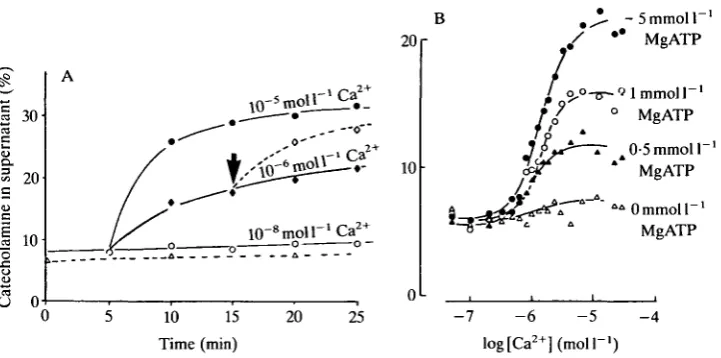
Fig. 2. Ca - and MgATP-dependence of secretion from electropermeabilized chromaffin cells. (A) Submaximal levels of Ca2+, i.e. 10~6moll~', affect mainly the extent of secretion rather than simply the rate. Further secretion is triggered when l O ^ m o i r ' C a2* is added after 10"6moir'Ca2 + (arrow). (B) Electropermeabilized chromaffin cells equilibrated with various concentrations of MgATP before being challenged with 10~5moll"1Ca2+.
as the same biochemical criteria used to determine exocytosis from intact cells were also satisfied by the leaky cells. Essentially this was that both catecholamine and dopamine-/3-hydroxylase were released into the extracellular medium at the same rate and in the same proportions as found in the soluble component of the secretory granule, whereas the cytosolic enzyme lactate dehydrogenase was only released into the extracellular medium at a much slower rate that was quite independent of the Ca2+ level. Very little catecholamine was released from leaky cells held at Ca2+ levels close to 0-1 /imol I"1, i.e. at resting levels of Ca2 +, whereas half-maximal release occurred at l ^ m o l l "1 Ca2+ and maximal secretion at lOjUmol l~l Ca2+ (Fig. 2A). The shape of the Ca2+ activation curve suggested that two calcium ions could be involved with each exocytotic reaction (Baker & Knight, 1981; Knight & Baker, 1982).
In the hope of shedding some light on the underlying mechanism of secretion, the effects of various agents introduced into the leaky cell were investigated. Although many agents had little effect, it was very clear that MgATP had to be present for Ca2+-dependent secretion to proceed (Fig. 2B).
Electropermeabilized platelets
14 T. J. RINK AND D. E. KNIGHT
that in intact platelets, low levels of the natural agonist thrombin triggered the release of the contents of one type of secretory granule (serotonin from the amine-storage granules) but not the contents of another type of granule (acid hydrolases from lysosomes), whereas higher levels of thrombin triggered the release of both serotonin and acid hydrolase. This example of differential secretion from the same cell could have been explained in terms of the exocytotic machinery of the first type of granule having a higher sensitivity to Ca2+ than that of the other type of secretory granule. When platelets were rendered leaky, however, and the intracellular Ca2+ levels clamped, it was found that their sensitivities to Ca2+ were the same. The differential secretion triggered by thrombin in intact cells therefore provided evidence for a transduction pathway that was more effective on one granule type than on the other and led us to consider the involvement of a 'non-Ca2 +' factor involved in the triggering and control of exocytosis (Knight, Hallam & Scrutton, 1982) - a suggestion that was entirely compatible with the quin2 results outlined below.
Fluorescent calcium indicators
Just after these developments came Roger Tsien's invention of the fluorescent Ca2 + indicator, quin2, and a way of introducing it into intact cells by means of ester permeation and cytosolic hydrolysis and trapping of the tetracarboxylic acid dye (Tsien, Pozzan & Rink, 1982a). The technique was developed first in lymphocytes and then rapidly applied to many other cells starting with the blood platelet (Rink, Smith & Tsien, 1982). Resting [Ca2+]; measured by this method
was close to lOOnmolF1 in nearly all cell types, including platelets (Rink etal. 1982) and chromaffin cells (Knight & Kesteven, 1983). In quin2-loaded platelets, step changes in [Ca2+]j could be imposed by application of increasing concen-trations of Ca2+ ionophore. Secretion could be measured simultaneously, by measuring release of radioactive 5-hydroxytryptamine (5-HT) or, in parallel, by following secretion of the ATP also contained in the amine-storage granules by luciferin-luciferase (Rink etal. 1982; Rink & Hallam, 1984). These experiments showed an apparent threshold for secretion near l^moll"1 [Ca2+]j, with maximal effects at several micromolar beyond the range of resolution of quin2. These results were consistent with the data from permeabilized platelets. Interestingly, shape change, another important functional response, was much more sensitive to Ca2 +, being activated between 300 and 800 nmol 1~'. These studies were the first in which secretion was measured along with quantitation of [Ca2+]j in an intact cell, and provided the data to support what had been supposed from other more indirect measurements.
The role of protein kinase C
Stimulus-secretion coupling
B
15
100
3 40
S« 0
o 0-2 — a.
U
S- 50
2min
Diacylglycerol
oo
-o—0-0-6
I
[image:15.451.47.406.62.230.2]001 0-1 1-0 10-0
Fig. 3. Experiments showing apparent Ca2+-independent secretion and its possible explanation. (A) Collagen, lOmgml"1 added at the arrow, promotes secretion of ATP from intact platelets without a rise in Ca2+ level as measured with quin2. (B) In electrically permeabilized platelets, diacylglycerol (which is produced following exposure to collagen) brings about a marked increase in the Ca2+-sensitivity of the secretory process.
threshold needed when Ca2+ ionophore translocated Ca2+ into the cytosol bypassing receptor-mediated events (Rink, Sanchez & Hallam, 1983; Rink & Hallam, 1984) (see Fig. 3A). Just at this time we began to appreciate the importance of diacylglycerol production from receptor-mediated phosphoinosit-ide hydrolysis and its role in activation of protein kinase C. Nishizuka, with Castagna and others (Castagna et al. 1982; Nishizuka, 1984), had also just shown that a particular phorbol ester, phorbol myristate acetate (PMA), could substitute for diacylglycerol in activating protein kinase C. We had previously observed that PMA caused platelet aggregation with no rise in quin2 fluorescence (Tsien et al. 19826) but could not work out what this meant. Now all fell into place and we showed that PMA, and an exogenous diacylglycerol which also activated kinase C in platelets, could stimulate extensive, if slow, secretion from both classes of granules (Rink et al. 1983). In these experiments the retention of cytosolic quin2 served as an in-built control for selective secretion from the granular store. We proposed therefore that, in the particular conditions we used, thrombin and collagen were stimulating secretion at basal [Ca2+]j by their ability to promote diacylglycerol formation and activate kinase C. These findings, incidentally, also strongly suggested that receptor-mediated phosphoinositide secretion preceded and was thus independent of, any elevation of [Ca2+]j, at least in this system. We also confirmed and extended the previous observations of synergy between elevated [Ca2+]j and diacylglycerol in accelerating the exocytotic process.
The ability of surface ligands to stimulate exocytosis without promoting any
16 T. J. RINK AND D. E. KNIGHT
process did not call for a specific role for this divalent cation and, in any case, many other examples of membrane fusion, including that of constitutive secretory exocytosis, clearly did not need elevated [Ca2+]; (Judah & Quinn, 1978; Shoback, Thatcher, Leombruno & Brow, 1984; DiVirgilio, Lew & Pozzan, 1984; Pozzan et al. 1984). Nonetheless, it is clear that in many secretory processes, particularly those in which speed is important, elevated [Ca2+]j under the plasma membrane, just where it is needed, is the major and probably requisite trigger in stimulus-secretion coupling.
Support for a role of protein kinase C in secretion came from the finding that in electropermeabilized chromaffin cells PMA could increase the Ca2+-sensitivity (albeit modestly) of the exocytotic machinery (Knight & Baker, 1983a). This experiment paved the way for the crucial experiments a year later which showed that PMA or diacylglycerol increased the Ca2+-sensitivity of serotonin secretion from electropermeabilized platelets to such an extent that PMA could elicit appreciable secretion at resting [Ca2+]j levels, i.e. lOOnmoll"1 (Fig. 3B; Knight & Scrutton, 1984a,b). We had earlier shown that the signal transduction process involving the thrombin receptor remained functional in electropermeabilized platelets (Knight & Scrutton, 1983, 1984a; Haslam & Davidson, 1984a), and also shown that it operated by increasing the Ca2+-sensitivity of serotonin secretion. These two events were firmly linked when it was shown that activation of the thrombin receptor led to phospholipase C activity in the electropermeabilized cells and hence an elevated diacylglycerol level (Haslam & Davidson, 1984a,b).
Another interesting finding was that although PMA, diacylglycerol or activation of the thrombin receptor increased the Ca2+-sensitivity of serotonin release, their effect on secretion from another type of granule (i.e. secretion of acid hydrolase) was quite different. Here the extent of secretion was increased without the Ca2+ -sensitivity being altered. This demonstration that two distinct populations of secretory granules in the platelet (i.e. serotonin-containing amine-storage gran-ules and enzymes in lysosomes) could exhibit quite different responses to activators of kinase C provided a mechanism for effecting differential release of secretory products. Thus, at lowcytosolic Ca2+ levels, a rise in diacylglycerol level induced by either thrombin or collagen would trigger the release of serotonin but have little effect on lysosomal release, whereas higher levels of thrombin, which elevated the cytosolic Ca2+ still further, would trigger secretion of both (Knight, Niggli & Scrutton, 1984).
Are different classes of protein kinase C responsible for different secretory responses?
Stimulus-secretion coupling 17
Ca2+-independent. The second type of response is characterized by a fairly modest increase in the sensitivity to Ca2+ and this is typified by the secretion of catecholamine from chromaffin cells (Knight & Baker, 1983a). The third type of response is where activators of the kinase enhance the extent of secretion without altering the Ca2+-sensitivity. This is shown in the case of lysosomal enzyme secretion from platelets (Knight et al. 1984). The different types of response may result from different forms of kinase C (Knopf et al. 1986; Kariya & Takai, 1987; Ido, Kazuo, Kikkawa & Nishizuka, 1987). Alternatively, if the enzyme has a preferred order of binding Ca2+ and diacylglycerol (Baker, 1986), then the amount of enzyme associated with Ca2+ and diacylglycerol as a function of [Ca2+] will depend on the order of binding (i.e. diacylglycerol followed by Ca2+, or Ca2+ followed by diacylglycerol, or when there is no preferred order of binding). The relationship between the amounts of enzyme associated with Ca2+ and diacylgly-cerol for the three orders of binding is strikingly similar to Ca2+ activation curves of the three types of secretory response. Therefore, the three different secretory responses may result from similar enzymes with different preferred orders of substrate binding (Baker, 1986).
Theories and models of exocytosis
Sub-maximal calcium levels
A rather interesting finding from experiments with electropermeabilized cells was that suboptimal levels of Ca2+ affected mainly the extent of secretion rather than the rate (Fig. 2A). If the kinetics of release from these intact cells resembled that from intact cells then the significance of this finding was that a maintained elevated Ca2+ level would trigger a transient secretory response rather than a maintained one. Although secretion from leaky cells ceased after a few minutes in response to a maintained Ca2+ challenge (say l ^ m o l P1) , further secretion could be triggered by this same Ca2+ concentration if the Ca2+ level was briefly lowered to resting levels (i.e. 0-1/imoir1) before being raised again (Knight & Baker, 1982). These results suggest that oscillating Ca2+ levels would lead to a greater secretory response than that due to a maintained elevated Ca2+ challenge. If brief bursts of nervous impulses, interspersed by quiet periods, result in an oscillation of [Ca2+]j, whereas a maintained firing pattern gives rise to a maintained elevated [Ca2+]j, then these observations with leaky cells provide an explanation of why bursts of nervous activity give rise to more secretion than do tonic firing patterns.
Sites of action of calcium
18 T . J. R I N K AND D . E. K N I G H T
where it has been shown that the Ca2+-sensitivity of secretion is altered by activators of the kinase, and there is an absolute requirement for MgATP.
However, other possible receptors should not be discounted and include synexin (Creutz, Pazoles & Pollard, 1978), chromobindins (Creutz et al. 1983) and calelectrins (Sudhof, Walker & Fritsche, 1985). Just how these Ca2+-binding proteins could trigger membrane fusion leading to exocytosis is not clear, but a clue could come from the method by which certain enveloped viruses are able to fuse with membranes. The method seems to involve a special pH-sensitive spike protein that the viral membrane has projecting from its surface (White, Kielan & Helenius, 1983). Viruses attach to the cell surface, become internalized into endosomes and, as the H+ concentration inside the endosomes rises to about 10jUmoll~L, the spike protein undergoes a conformational change revealing a hydrophobic sequence that buries itself into the neighbouring wall of the endosome. It is this step that seems to lead to fusion of the viral and vesicle membranes, permitting the escape of the viral contents into the cytosol. Although it seems most improbable that such a pH-sensitive process is involved in exocytosis, the underlying principle may well be the same, i.e. that in association with either the secretory vesicle or plasma membrane there is a protein specialized for effecting fusion, Ca2+ being the trigger that reveals some hydrophobic sequence.
Calmodulin has been suggested as another receptor for Ca2+ in its control over secretion (Steinhardt & Alderton, 1982; Llinas et al. 1985). Support for an involvement of calmodulin also comes from experiments which show that calmodulin antibodies introduced into chromaffin cells block Ca2+-dependent secretion (Trifaro & Konigsberg, 1983).
Calmodulin- and protein kinase-C-dependent processes could both play a part in the control of exocytosis. For example, rather than protein kinase C being the integral part of the machinery controlling exocytosis, the enzyme could be involved as a modulator of an underlying calmodulin-dependent process. If this were the case, it should be possible to separate the two processes pharmacologi-cally. Unfortunately, we have been unsuccessful in showing this as, using a range of putative inhibitors, we have been unable selectively to remove the PMA-induced shift of the Ca2+ activation curve without also removing the underlying (PMA-insensitive) Ca2+ activation curve. Either the agents we have used are not specific enough to disentangle the different pathways, or protein kinase C may well operate as the integral part of the exocytotic machinery.
Role of MgATP
Stimulus-secretion coupling 19
chromaffin cells or platelets failed to identify a single phosphoprotein uniquely associated with Ca2+-dependent secretion, a very large number being phosphoryl-ated (Niggli etal. 1984; Knight et al. 1984). There is good evidence that in some tissues the phosphoprotein, synapsin 1, may be involved in exocytosis, although this protein is apparently absent from chromaffin cells (Llinas etal. 1985). The MgATP dose-response curve obtained from electropermeabilized chromaffin cells was best fitted by a model in which a chemical reaction closely associated with exocytosis involved one molecule of MgATP per round of exocytosis. The data therefore provided no evidence for cooperative sites of action of MgATP involved in secretion. The kinetics of catecholamine release studied under conditions of clamped intracellular Ca2+ and MgATP levels suggested that if phosphorylation or dephosphorylation was a key step in exocytosis, then the rate of phosphorylation/ dephosphorylation would be the controlling factor in secretion rather than the absolute levels of protein phosphorylated (Knight & Baker, 1982). Some recent studies favour phosphatase activity to be the regulatory step (Zeiseniss & Plattner, 1985).
Although MgATP is certainly required for secretion from bovine chromaffin cells, its role is still somewhat unclear. For example, two sets of data lent some support to the idea that one role of MgATP is simply to protect or prime the secretory system. First, the millimor concentrations of nucleotide needed for a secretory response could be reduced by over an order of magnitude if the leaky cells were incubated and challenged in a medium of low ionic strength. Although this can be explained in terms of the affinity for MgATP being dependent on the ionic strength, another interpretation is that the nucleotide protects a system otherwise destabilized by anions, the order of potency of the anions following the lyotopic series. Second, the cells respond better if incubated continuously in a medium containing ATP, rather than one in which the nucleotide is added just prior to the Ca2+ challenge.
20 T. J. R I N K AND D . E . KNIGHT
prime a component involved in the granule/plasma membrane interaction process rather than in the exocytotic mechanism itself. Similarly Ca2+ may exert its control of secretion by a troponin-type molecule rather than by a phosphorylation/ dephosphorylation step as seen in smooth muscle.
Osmotic forces
Stimulus—secretion coupling 21
The cytoskeleton
Baker & Whitaker gave evidence that the cytoskeleton was not intimately associated with exocytosis of cortical granules in sea urchin egg plaques (Whitaker & Baker, 1983). In this case the cortical granules were fixed in position and not expected to move. In electropermeabilized chromaffin cells, where secretory granules can move towards the periphery of the cell, agents that might be expected to perturb the cytoskeleton also had no effect on Ca2+-dependent secretion. Recent work on leaky (Perrin, Langley & Aunis, 1987) and intact (Cheek & Burgoyne, 1986) chromaffin cells, however, suggests a role for the cytoskeleton in secretion. In the latter studies it was shown that actin filaments were disassembled around the periphery of the cell in response to nicotinic stimulation and over a time course that closely followed secretion. A potassium challenge was reported to cause a much smaller amount of secretion and, as it did not lead to actin disassembly, the higher secretion with nicotine was linked with the breakdown of the filament network. [In our early experiments we also observed that nicotinic stimulation led to a larger amount of catecholamine release than a potassium challenge, but we only observed this at room temperature when the secretory response due to nicotine was more maintained (Knight & Baker, 19836). At 37°C we saw very little difference in the amounts secreted in response to these two stimuli, whether from freshly isolated cells, cultured cells or a perfused gland. We interpreted our data in terms of the acetylcholine receptor not desensitizing as quickly at the lower temperature, rather than the result of another second messenger possibly affecting the cytoskeleton.]
The finding that forskolin, an agent that elevates cyclic AMP level, not only reduces the nicotine-evoked release but also blocks the disassembly of the actin filaments suggested that cyclic AMP could modulate secretion (Burgoyne & Cheek, 1987). The earlier results from electropermeabilized cell preparations did not support this idea, as the Ca2+-dependent secretion observed from the electropermeabilized cell was unaffected by cyclic nucleotides or by agents that should have either disrupted or prevented disassembly of the cytoskeleton. One of the ways of reconciling these data is if the act of rendering the cell leaky by electric fields causes the dissassembly of the actin filaments. Another possibility is that an ingredient essential for cyclic nucleotide effects was lost from the cytosol of the leaky cell.
Guanine-nucleotide-binding proteins
22 T. J. RINK AND D. E. KNIGHT
possibility that kinase from the medulla was not activated by this particular diacylglycerol, that the lipid could not get into the cell or that it was being rapidly metabolized. (We later found that other diacylglycerols, e.g. 1,2-dioctanoylgly-cerol and other activators of the kinase, e.g. mezerein, did increase the Ca2+ sensitivity in the same way as did PMA.) Shortly after these early experiments Richard Haslam and Monica Davidson showed that the thrombin-induced production of diacylglycerol in the permeabilized platelet was enhanced by GTP or GTPyS and they suggested that phospholipase C was under guanine-nucleotide-binding-protein control (Haslam & Davidson, 1984c). A similar control over phospholipase C activity was later shown in other preparations (Cockcroft & Gomperts, 1985; Merritt, Taylor, Rubin & Putney, 1986).
As phospholipase C in platelets could be stimulated by GTPyS in the absence of an agonist, we looked for an effect of this nucleotide on Ca2+-dependent secretion from chromaffin cells. We hoped that endogenous diacylglycerol would be produced in the cell by this nucleotide, and that this would increase the Ca2+ -sensitivity of the secretory process in a way similar to PMA. As predicted, we found that GTPyS stimulated Ca2+-dependent catecholamine release from chicken chromaffin cells. It was a surprise, however, to find that GTPyS inhibited the extent of secretion from leaky bovine adrenal medullary cells (Knight & Baker, 19856). We could not interpret these data in terms of GTPyS inhibiting endogenous diacylglycerol production as the effect could not be overcome by direct activation of protein kinase C with PMA. We suggested, therefore, that the site of action of the GTPyS in the bovine chromaffin cell was downstream of the sites of action of phospholipase C and protein kinase C, and perhaps was at the site of exocytosis itself (Knight & Baker, 19856). Quite independently, this hypothesis was also proposed by Bastien Gomperts and his colleagues (Barrowman, Cockcroft & Gomperts, 1986) who showed in neutrophils that the site of exocytosis might be under the control of a GTP-binding protein. However, in their systems GTP was stimulatory. A stimulatory effect of GTPyS on leaky bovine adrenal medullary cells was found by Ronald Holz and his colleagues (Bittner, Holz & Neubig, 1986). One explanation for this difference might lie in the different method of making the cells leaky, i.e. electropermeabilization versus digitonin treatment.
Stimulus-secretion coupling 23
only do micromolar levels of GTPyS and GTP enhance the thrombin effect on Ca2+-dependent secretion from electropermeabilized platelets, but the same enhancement is also seen by micromolar levels of GDT, GMP or cyclic GMP.)
Other second messengers
The evidence so far is that the main messengers acting on exocytosis are Ca2+ and diacylglycerol. Cyclic AMP has also been postulated to act as a second messenger for secretion (Gardner & Jensen, 1981). The experiments that have been directed towards clarifying this, however, show that where there is an effect it appears to be expressed through a Ca2+-dependent process. This may result either as a modulation of the Ca2+-sensitivity of the secretory process (Jones, Fyles & Howells, 1986) or by modulating the production of another second messenger, e.g. Ca2+ or diacylglycerol (Knight & Scrutton, 1984a,b). Secretion can be triggered in some systems not only by an increase in Ca2+ level, but also by GTyS acting in a seemingly Ca2+-independent manner. Whether this nucleotide is operating on a pathway that is truly separate from the Ca2+-dependent pathway, or whether it operates downstream of the site of action of Ca2+ is not yet clear (Barrowman etal. 1986; Fernandez et al. 1984; Oetting et al. 1986; Wollheim, Ullrich, Meda & Vallar, 1987; Penner, Pusch & Neher, 1987).
Identifying the proteins associated with exocytosis
One method of identifying some of the proteins essential for secretion is to allow the cytosolic proteins to diffuse out of the leaky cell and see at what point the cell becomes refractory. This approach has recently had some success when it was shown that associated with the onset of refractoriness was the appearance of proteins in the extracellular fluid, and that these proteins, when concentrated and added back to the cells, could restore Ca2+-dependent secretion (Sarafian, Aunis & Bader, 1987).
24 T. J. RINK AND D. E. KNIGHT
botulinum toxins are, like cholera toxin, internalized by the cell before they can express their inhibitory effect. Another possibility, however, is that the toxin acts on a protein positioned on the outside of the cell surface, and which is revealed to the toxin when the cell is depolarized.
Post-secretory events - endocytosis
With the exception of perhaps cortical granule discharge from the sea urchin egg and trychocyst discharge from Paramecium, the constancy of cell size means that any increase in the cell surface resulting from exocytosis must in the long term be balanced by endocytosis of an equivalent area of membrane (Linng, Fischer-Colbrie, Schmidt & Winkler, 1983; Phillips, Burridge, Wilson & Kirshner, 1983). We showed that endocytosis, as measured by horseradish peroxidase uptake, occurred in electropermeabilized chromaffin cells alongside exocytosis (Baker & Knight, 1981). Therefore, if secretion included a cycle of events involving both exocytosis and endocytosis, we reasoned that the intracellular conditions control-ling secretion might not necessarily reflect those exclusively for exocytosis but could also include those for endocytosis. Experiments designed to differentiate the intracellular conditions controlling these two pathways have only recently begun. Peter Baker and his colleagues showed that horseradish peroxidase could be taken up into intact chromaffin cells after catecholamine release had been stopped (von Grafenstein, Roberts & Baker, 1986). In this experiment, cells were stimulated with carbamylcholine and then the secretory stimulus was blocked by the addition of hexamethonium; at the same time, the extracellular marker was added. Uptake of this marker continued for several minutes even though exocytosis had stopped. They showed that after exocytosis had been stopped, the subsequent endocytosis could be interrupted by lowering the temperature, only to resume as normal when the temperature was raised again. Removal of extracellular calcium during this interrupted period did not alter the subsequent uptake of horseradish peroxidase, strongly suggesting that endocytosis was [Ca2+]rindependent. This result has
recently been supported, and the study extended, using electropermeabilized cells. In these experiments in which exocytosis is blocked by lowering the Ca2+ level after a few minutes, horseradish peroxidase uptake continues independently of Ca2+ level and, as in the intact cells, can be interrupted if the temperature is lowered. Endocytosis seems to continue normally when the temperature is raised only if micromolar levels of MgATP are present, and completely ceases in the presence of nonhydrolysable analogues, e.g. ATPyS (von Grafenstein, 1988).
Ways forward
Stimulus—secretion coupling 25
defined conditions (Davey, 1987; Crabb, Morden & Jackson, 1987). Other advances may come from techniques that allow the synthesis of foreign secretory proteins within a model cell or from the use of secretory mutants. It is a great loss to us all that Peter Baker is not here to help pursue and unravel these challenging problems.
Derek Knight's work is supported by the MRC and Wellcome Trust. We thank M. Bowden, M. Fitzgerald and B. Leigh for help in producing the manuscript.
References
ADAM-VIZI, V., AKTORIES, K., KNIGHT, D. E. & ROSENER, S. (1988). Botulinum toxin induced ADP-ribosylation and inhibition of catecholamine secretion may be unrelated events. /. Physiol., Lond. (in press).
AHNERT-HIGLER, G. & GRATZL, M. (1987). Further characterisation of dopamine release by permeabilised PC12 cells. J. Neurochem. 49, 764-770.
AKTORIES, K., WELLER, U. & CHATWAL, G. S. (1987). Clostridium botulinum type C produces a novel ADP-ribosyltransferase distinct from botulinum C2 toxin. FEBS Letts 212, 109-113.
ASHLEY, C. C. & RIDGWAY, E. B. (1970). On the relationship between membrane potential, calcium transient and tension in single barnacle muscle fibres. J. Physiol., Lond. 209, 105-130.
BAKER, P. F. (1972). Transport and metabolism of calcium ions in nerve. Prog. Biophys. molec. Biol. 24, 177-223.
BAKER, P. F. (1986). Protein kinase C and exocytosis. Prog. Zool. 33, 265-274.
BAKER, P. F., BLAUSTEIN, P., HODGKIN, A. & STEINHARDT, R. A. (1969). Influence of calcium on sodium efflux in squid axons. J. Physiol., Lond. 200, 431-458.
BAKER, P. F. & CRAWFORD, A. C. (1975). A note of the mechanism by which inhibitors of the sodium pump accelerate spontaneous release of transmitter from motor nerve terminals. J. Physiol., Lond. 247, 209-226.
BAKER, P. F. & DIPOLO, R. (1984). Axonal calcium and magnesium homeostasis. Curr. Topics Membr. Transport 22, 195-247.
BAKER, P. F., HODGKIN, A. L. & RIDGWAY, E. B. (1971). Depolarisation and calcium entry in squid giant axons. J. Physiol., Lond. 218, 709-755.
BAKER, P. F. & KNIGHT, D. E. (1978). Calcium-dependent exocytosis in bovine adrenal medullary cells with leaky plasma membranes. Nature, Lond. 276, 620-622.
BAKER, P. F. & KNIGHT, D. E. (1981). Calcium control of exocytosis and endocytosis in bovine adrenal medullary cells. Phil. Trans. R. Soc. Ser. B 296, 83-103.
BAKER, P. F., KNIGHT, D. E., PIDDINGTON, R. W. & Ross, D. A. (1977). Laser measurement of particle size and movement in cells. J. Physiol., Lond. 266, 6-8P.
BAKER, P. F., KNIGHT, D. E. & WHITAKER, M. J. (1980). The relation between ionised calcium and cortical granule exocytosis in eggs of the sea urchin Echinus esculentus. Proc. R. Soc. Ser. B 207, 149-161.
BAKER, P. F. & MCNAUGHTON, P. A. (1978). The influence of extracellular calcium binding on the calcium efflux from squid axons. J. Physiol., Lond. 276, 127-150.
BAKER, P. F., MEVES, H. & RIDGWAY, E. B. (1973a). Effects of manganese and other agents on the calcium uptake that follows depolarization of squid axons. J. Physiol., Lond. 231, 511-526.
BAKER, P. F., MEVES, H. & RIDGWAY, E. B. (1973ft). Calcium entry in response to maintained depolarization of squid axons. J. Physiol., Lond. 231, 527-548.
BAKER, P. F. & RINK, T. J. (1975). Catecholamine release from bovine adrenal medulla in response to maintained depolarization. J. Physiol., Lond. 253, 593-620.
BAKER, P. F. & WHITAKER, M. J. (1978). Influence of ATP and calcium on the cortical reaction in | sea urchin eggs. Nature, Lond. 276, 513-515.
26 T. J. RINK AND D. E. KNIGHT
BITTNER, M. A., HOLZ, R. W. & NEUBIG, R. R. (1986). Guanine nucleotide effects on catecholamine secretion from digitonin-permeabilised adrenal chromaffin cells. J. biol. Chem. 261,10182-10188.
BLASCHKO, H. & WELCH, A. D. (1953). Localisation of adrenaline in cytoplasmic particles of the bovine adrenal medulla. Nannyn-Schmiedebergs Arch. exp. Path. Pharmak. 219, 17-22.
BLAUSTEIN, P. & HODGKIN, A. L. (1969). The effect of cyanide on the efflux of calcium from squid axon.7. Physiol., Lond. 200, 497-527.
BRECKENRIDGE, L. J. & ALMERS, W. (1987). Final steps in exocyosis observed in a cell with giant secretory granules. Proc. natn. Acad. Sci. U.S.A. 84, 1945-1949.
BURGOYNE. R. D. & CHEEK, T. R. (1987). Reorganisation of peripheral actin filaments as a prelude to exocytosis. Bioscience Rep. 7, 281-288.
CASTAGNA, M., TAKAI, Y., KAIBUCHU, K., SANO, K., KIKKAWA, U. & NISHIZUKA, K. (1982). Direct activation of calcium-activated, phospholipid-dependent protein kinase by tumour promoting phorbol esters. J. biol. Chem. 257, 7847-7851.
CHEEK, T. R. & BURGOYNE, R. D. (1986). Nicotine-evoked disassembly of cortical actin filaments in adrenal chromaffin cells. FEBS. Letts 207, 110-114.
COCKCROFT, S. & GOMPERTS, B. D. (1985). Role of guanine nucleotide binding proteins in the activation of polyphosphoinositide phosphodiesterase. Nature, Lond. 314, 534-536. COLE, K. S. (1935). Electric impedance of Hipponoe eggs. J. gen. Physiol. 18, 877-887.
CRABB. J. H., MORDEN, P. A. & JACKSON, R. C. (1987). In vitro reconstitution of exocytosis from sea urchin egg plasma membrane and isolated cortical vesicles. Bioscience Rep. 7, 399-409.
CRAWFORD, A. C. (1975). Lithium ions and the release of transmitter at the frog neuromuscular junction. 7. Physiol., Lond. 246. 109-142.
CREUTZ, C. E., DOWLING, L. G., SANDO, J. J., VILAR-PALASI, C , WHIPPLE, J. H. & ZAKS, W. J.
(1983). Characterisation of the chromobindins: soluble proteins that bind to the chromaffin granule membrane in the presence of Ca2+. J. biol. Chem. 258, 14664-14674.
CREUTZ, C. E., PAZOLES, C. J. & POLLARD, H. B. (1978). Identification and purification of an adrenal medullary protein (synexin) that causes the calcium-dependent aggregation of isolated chromaffin granules. J. biol. Chem. 253, 2858-2866.
DAVEY, J. (1987). A cell free analysis of the endocytotic pathway. Bioscience Rep. 7, 299-306.
DEL CASTILLO, J. & KATZ, B. (1956). Biophysical aspects of neuro-muscular transmission. Prog. Biophys. 6, 121-170.
DE ROBERTIS, E. D. P. & VAZ FERRERIA, A. (1957). Electron microscope study of the excretion of catechol-containing droplets in the adrenal medulla. Expl Cell Res. 12, 568-574.
DIPOLO, R. & BEAUGE, C. (1979). Physiological role of ATP-driven calcium pump in squid axon. Nature, Lond. 278, 271-273.
DIVIRGILIO, F., LEW, D. & POZZAN, T. (1984). Protein kinase C activation of physiological processes in human neutrophils at vanishingly small cytosolic Ca2+ levels. Nature, Lond. 310, 691-693.
DOUGLAS, W. W. (1968). Stimulus-secretion coupling: the concept and clues from chromaffin and other cells. Br. J. Pharmac. Chemother. 34, 451-474.
DOUGLAS, W. W. & RUBIN, R. P. (1961). The role of calcium in the secretory response of the adrenal medulla to acetylcholine. J. Physiol., Lond. 159, 40-57.
DUNN. L. A. & HOLZ, R. W. (1983). Catecholamine secretion by digitonin treated adrenal medullary chromaffin cells. J. biol. Chem. 258. 4989-4993.
EDWARDS. C , DOLEZAL, V.. TUCEK, S., ZEINKOVA, H. & VYSKOCIL, F. (1985). Is an acetylcholine transport system responsible for non quantal release of acetycholine at the rodent myoneuronal junction? Proc. natn. Acad. Sci. U.S.A. 82, 3514-3518.
FERNANDEZ, J., NEHER, E. & GOMPERTS, B. (1984). Capacitance measurements reveal stepwise fusion events in degranulating mast cells. Nature, Lond. 312, 453-455.
FINKELSTEIN. A., ZIMMERBERG. J. & COHEN, F. S. (1986). Osmotic swelling of vesicles: Its role in the fusion of vesicles with planar phospholipid bilayer membranes and its possible role in exocytosis. A. Rev. Physiol. 48. 163-174.
GARDNER. J. D. & JENSEN. R. T. (1981). Regulation of pancreatic exocrine secretion in vitro: the action of secretagogues. Phil. Trans. R. Soc. Ser. B 296. 1-193. A
Stimulus-secretion coupling 27
GOMPERTS. B. D. & FERNANDEZ. J. M. (1985). Techniques for membrane permeabilisation. Trends biochem. Sci. 10. 414-417.
HARVEY. A. M. & MACINTOSH. F. C. (1940). Calcium and synaptic transmission in a sympathetic ganglion. J. Physioi, Lond. 97, 408-416.
HASLAM, R. J. & DAVIDSON, M. M. L. (1984a). Potentiation by thrombin of the secretion of
serotonin from permeabilised platelets equilibrated with Ca2+ buffers. Biochem. J. 222, 351-361.
HASLAM, R. J. & DAVIDSON, M. M. L. (19846). Receptor induced diacylglycerol formation in permeabilised platelets; possible role for a GTP binding protein. J. Receptor Res. 4, 605-629.
HASLAM, R. J. & DAVIDSON, M. M. L. (1984C). Guanine nucleotides decrease the free Ca2+ required for secretion of serotonin from permeabilised blood platelets. FEBS Letts 174, 90-95.
HEUSER, J. E. (1978). Quick-freezing evidence in favour of the vesicular hypothesis. Trends Neurosci. 1, 80-82.
HEUSER, J. E. & REESE, T. S. (1973). Evidence for recycling of synaptic vesicle membrane during transmitter release at the frog neuromuscular junction. J. Cell Biol. 57, 315-344.
HODGKIN, A. L. & KATZ, B. (1949). The effect of calcium on axoplasm of giant nerve fibres. J. exp. Biol. 26. 292-294.
HODGKIN, A. L. & KEYNES. R. D. (1957). Movements of labelled calcium in squid giant axons. J. Physioi., Lond. 138, 253-281.
HOLZ, R. W. & SENTER, R. A. (1986). Effects of osmolarity and ionic strength on secretion from adrenal chromaffin cells permeabilised with digitonin. J. Neurochem. 46, 1835-1842.
HOLZ, R. W., SENTER, R. A. & SHARP, R. R. (1983). Evidence that the H+ electrochemical gradient across membranes of chromaffin granules is not involved in exocytosis. J. biol. Chem. 258, 7506-7513.
Hu, G.-Y., HVALBY, O., WALAAS, S. I., ALBERT, K. A., SKJEFLO, P., ANDERSEN, P. &
GREENGARD, P. (1987). Protein kinase C injection into hippocampal pyramidal cells elicits features of long term potentiation. Nature, Lond. 328, 426-429.
IDO, M., KAZUO, S., KIKKAWA, U. & NISHIZUKA, Y. (1987). Phosphorylation of the EGF receptor from A431 epidermoid carcinoma cells by three distinct types of protein kinase C. FEBS Letts 219, 215-218.
ISRAEL, M., MEUNIER, F. M., MOREL, N. & LESBATS, B. (1987). Calcium induced desensitisation of acetylcholine release from synaptosomes or proteliposomes equipped with mediatophore, a presynaptic membrane protein. J. Neurochem. 49, 975-982.
JONES, P. M., FYLES, J. M. & HOWELL, S. L. (1986). Regulation of insulin secretion by cAMP in rat islets of Langerhans permeabilised by high-voltage discharge. FEBS Letts 205, 205-209.
JUDAH, J. D. & QUINN, P. S. (1978). Calcium ion-dependent vesicle fusion in the conversion of prealbumin to albumin. Nature, Lond. 271, 384-385.
KARIYA, K. & TAKAI, Y. (1987). Distinct functions of down-regulation sensitivity and resistant types of protein kinase C in rabbit aortic smooth muscle cells. FEBS Letts 219, 119-124. KATZ, B. (1969). The Release of Neuronal Transmitter Substances. Liverpool: Liverpool
University Press
KATZ, B. & MILEDI, R. (1967). The timing of calcium action during neuro-muscular transmission./. Physioi., Lond. 189, 535-544.
KATZ, B. & MILEDI. R. (1969). Tetrodotoxin-resistent electric activity in presynaptic terminals.
J. Physioi., Lond. 203,459-487.
KATZ, B. & MILEDI, R. (1977). Suppression of transmitter release at the neuro-muscular junction. Proc. R. Soc. Ser. B 196, 465-469.
KNIGHT, D. E. & BAKER, P. F. (1982). Calcium dependence of catecholamine release from bovine adrenal medullary cell after exposure to intense electric fields. J. Membr. Biol. 68, 107-140.
KNIGHT, D. E. & BAKER, P. F. (1983a). The phorbol ester TPA increases the affinity of exocytosis for Ca in leaky adrenal medullary cells. FEBS Letts 160, 98-100.
KNIGHT, D. E. & BAKER, P. F. (19836). Stimulus secretion coupling in isolated bovine adrenal medullary cells. Q. J. exp. Physioi. 68, 123-143.
28 T. J. RINK AND D . E. KNIGHT
KNIGHT, D. E. & BAKER, P. F. (19856). Guanine nucleotides and Ca-dependent exocytosis: Studies on two adrenal preparations. FEBS Letts 189, 345-349.
KNIGHT, D. E., HALLAM. T. J. & SCRUTTON, M. C. (1982). Agonist selectivity and second messenger concentration in Ca2+-mediated secretion. Nature, Lond. 296, 256-257.
KNIGHT, D. E. & KESTEVEN, N. T. (1983). Evoked transient intracellular free Ca2+ changes and secretion in isolated bovine adrenal medullary cells. Proc. R. Soc. Ser. B 218, 177-199.
KNIGHT, D. E., NIGGLI, V. & SCRUTTON, M. C. (1984). Thrombin and activators of protein kinase C modulated the secretory response of permeabilised human platelets induced by Ca2+. Em. J. Biochem. 143, 437-446.
KNIGHT, D. E. & SCRUTTON, M. C. (1980). Direct evidence for a role for Ca2+ in amine storage granule secretion by human platelets. Thromb. Res. 20, 437-446.
KNIGHT, D. E. & SCRUTTON, M. C. (1983). Secretion induced by Ca2+ and thrombin in platelets subjected to dielectric membrane breakdown. Thrombosis and Haemostasis 50, 93.
KNIGHT, D. E. & SCRUTTON, M. C. (1984a). The relationship between intracellular second messenger and platelet secretion. Biochem. Trans. 12, 969-972.
KNIGHT, D. E. & SCRUTTON, M. C. (1984ft). Cyclic nucleotides control a system which regulates the Ca sensitivity of platelet secretion. Nature, Lond. 309, 66-68.
KNIGHT, D. E. & SCRUTTON, M. C. (1987). Secretion of 5-hydroxytryptamine from electropermeabilised human platelets: Effects of GTP and cyclic 3',5'-AMP. FEBS Letts 223, 47-52.
KNIGHT, D. E., TONGE, D. A. & BAKER, P. F. (1985). Inhibition of exocytosis in bovine adrenal medullary cells by botulinum toxin type D. Nature, Lond. 317, 719-721.
KNOPF, J. L., LEE, M. H., SULTZMAN, L. A., KRIZ, R. W., LOOMIS, C. R., HEWICK, R. M. & BELL, R. M. (1986). Cloning and expression of multiple protein kinase C cDNAs. Cell 46, 491-502.
LINGG, G., FISCHER-COLBRIE, R., SCHMIDT, W. & WINKLER, H. (1983). Exposure of an antigen of chromaffin granules on cell surface during exocytosis. Nature, Lond. 301, 610-611.
LLINAS, R. J., BLINKS, R. & NICHOLSON, C. (1972). Calcium transients in presynaptic terminal of squid giant axon. Detection with aequorin. Science 176, 1127-1129.
LLINAS, R., MCGUINESS,T., LEONARD, D.,SUGIMORI,M. &GREENARD, P. (1985). Intraterminal injection of synapsin I or calcium/calmodulin dependent protein kinase II alters the neurotransmitter release at the squid giant synapse. Proc. natn. Acad. Sci. U.S.A. 83, 3035-3039.
MERRITT, J. E., TAYLOR, C. W., RUBIN, R. P. & PUTNEY, J. W. (1986). Evidence suggesting that a novel guanine nucleotide regulatory protein couples receptors to phospholipase C in exocrine pancreas. Biochem. J. 236, 337-343.
NEHER, E. & MARTY, A. (1982). Discrete changes of cell membrane capacitance observed under conditions of enhanced secretion in bovine adrenal chromaffin cells. Proc. natn. Acad. Sci.
U.S.A. 79, 6712-6716.
NIGGLI, V., KNIGHT, D. E., BAKER, P. F., VIGNY, A. & HENRY, J. P. (1984). Tyrosine
hydroxylase in leaky adrenal medullary cells: evidence for in situ phosphorylation by separate Ca2+ and cyclic AMP-dependent systems. J. Neurochem. 43, 646-658.
NISHIZUKA, Y. (1984). The role of protein kinase C in cell surface signal transduction and tumour promotion. Nature, Lond. 308, 693-698.
NORDMANN, J. J., DREIFUSS, J. J., BAKER, P. F., RAVAZZOLA, M., MALAISSE-LAGAE, F. & ORCI,
L. (1974). Secretion-dependent uptake of extracellular fluid by the rat neurohypophysis. Nature. Lond. 250, 155-157.
OETTING, M., LEBOFF, M., SWISTON, L., PRESTON, J. & BROWN, E. (1986). Guanine nucleotides are potent secretagogues in permeabilised parathyroid cells. FEBS Letts 208, 99-104.
OHASHI, Y. &NARUMIYA, S. (1987). ADP-ribosylation of a MT 21,000 membrane protein by type
D botulinum toxin. J. biol. Chem. 1&1, 1430-1433.
PARSONS, S. M. & KOENIGSBERGER, R. (1980). Specific stimulated uptake of acetylcholine by Torpedo electric organ synaptic vesicles. Proc. natn. Acad. Sci. U.S.A. 77, 6234-6238.
PENNER, R., NEHER, E. & DREYER, F. (1986). Intracellularly injected tetanus toxin fragment B inhibits exocytosis in bovine adrenal chromaffin cells. Nature, Lond. 324, 76-78. A