J. Richard Steele 1 James C. Hoffman 1
Received April 11, 1980; accepted after revi-sion June 24, 1980.
'Department of Radiology, Emory University Clinic, 1365 Clifton Rd. N.E., Atlanta, GA 30322. Address reprint requests to J. R. Steele.
This article appears in November/December
1980 AJNR and February 1981 AJR.
AJNR 1 :521-526, November/December 1980 0195-6108/80/0016-0521 $00.00
© American Roentgen Ray Society
Brainstem Evaluation with
CT Cisternography
521
Seventy-eight positive contrast computed tomography (CT) cisternograms were reviewed to assess the normal anatomy of the brainstem and its surrounding cisterns. Normal brainstem and cisternal anatomy was found to be constant and symmetrical. The review included six patients with brainstem gliomas and five patients with extraaxial masses. In these patients CT cisternography accurately identified mass formation and permitted the confident distinction of extraaxial from intra-axial masses. CT cistern-ography is a safe and accurate method for evaluating the anterior compartment of the posterior fossa. This procedure is particularly applicable to those cases where conven-tional CT yields insufficient diagnostic information.
Before the advent of computed tomography (CT), patients suspected of har-boring a brainstem glioma were subjected to angiography and pneumoenceph-alography. In most instances CT is now capable of identifying a brainstem mass [1, 2], although angiography and pneumoencephalography are frequently con-sidered necessary to support the diagnosis [1 -4]. The reports of early investi-gators concerning the potential value of positive contrast CT cisternography encouraged us to use this technique for brainstem evaluation [5-7]. This report discusses our experience with CT cisternography as it relates to the normal and abnormal brainstem, and the application of this technique to those cases in which conventional CT of the brainstem is negative or insufficiently diagnostic for patient management.
Materials and Methods
522 STEELE AND HOFFMAN AJNR:1 , November/December 1980
TABLE 1: Indications for CT Cisternography
Diagnosis Normal pressure hydrocephalus Empty sella
Pituitary tumor Optic chiasm lesion Brainstem lesion Cerebrospinal fluid leak . Foramen magnum lesion Miscellaneous:
Arachnoid cyst .
Chordoma . . . .
Arnold-Chiari malformation, type 1 Cholesteatoma .
Craniopharyngioma
Hypoglossal neuroma
Metastasis to cerebellopontine angle
Subtotal
Total
Results
No. Patients 15 17 9 7 13 7 3 7 78
The basal cisterns of the posterior fossa were imaged in
about one-half of these cases. For this study, all patients who were examined for reasons unrelated to the brainstem
or posterior fossa were considered" normal."
Normal Brainstem
The axial surface anatomy of the normal brainstem is
constant and symmetrical (figs. 1 -6). The posterior cere··
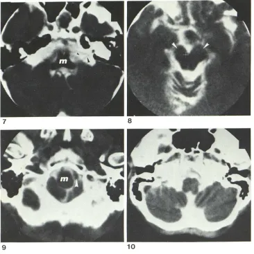
bral, basilar, and vertebral arteries are routinely identified. The posterior communicating arteries and cranial nerves 3 and 5 are often seen. Occasional scans demonstrated cra -nial nerves 7 and 8 (fig. 7). Usually these two cranial nerves cannot be separated.
In about 10% of normal cases, a minor degree of brain-stem asymmetry was noted, particularly in the cerebral peduncles (fig. 8). In most cases the patient's head was not symmetrically positioned. Minor asymmetry or brainstem rotation in the pons and medulla may occur due to an ectatic
vertebral or basilar artery (fig. 9). Minor asymmetry may also occur in the medulla without apparent explanation, but
the surface anatomy is preserved (fig. 10).
The transverse diameters across the medulla, pons, and
cerebral peduncles were measured in all cases where pos-sible. The distance from the anterior pons to the floor of the
fourth ventricle was also measured (fig. 11). Transverse
measurements were easily made except at the level of the brachium pontis, where the lateral pontine borders are not readily identified. At this location transverse measurement
of the brainstem was made 5 mm posterior to the anterior
pontine margin.
The various normal brainstem measurements are found in
table 2. There were three normal patients under 10 years of
age. Brainstem measurements in this group tended to be at
the lower end of the normal range (defined as ±2 SO from
the mean). Elderly patients sometimes exhibited brainstem size at the lower end of the normal range with prominence of the surrounding cisterns. Otherwise, brainstem diameters
varied widely without regard to age. In 11 "normal" adult patients, a part of the brainstem was 1-2 mm above the 2 SO range. In each of these patients, the brainstem was generally large, but normal-appearing at all levels.
Table 2 indicates a rather large standard deviation in the measurement of each brainstem diameter among normal patients. This broad range of values is due to several factors. For practical reasons, measurements were made from hard copy and corrected for minification. The pons is best seen where its sides blend with the diverging limbs of the
bra-chium pontis and reproducible transverse measurement is difficult. Transverse measurement of the cerebral peduncles is difficult because this dimension tends to decrease as the peduncles descend to the pons. Incomplete mixing of me-trizamide with cerebrospinal fluid in the fourth ventricle prevented adequate delineation of the ventricular floor in
several patients. Finally, it is difficult to precisely control the scan angulation and slice level. Despite the above technical pitfalls, we were impressed by the wide range of normal brainstem size.
Fourth Ventricular Filling
In our department metrizamide is delivered to the basal
cisterns in the prone position. Nevertheless, the fourth ven-tricle filled with contrast material in all normal patients. Conversely, in this small series posterior fossa pathology was present in all cases where fourth ventricular filling was not achieved (table 3).
Abnormal Brainstem
In the 2 year period of review, nine patients with progres-sive brainstem and/or cranial nerve disease had radio-graphic studies demonstrating brainstem enlargement. Brainstem masses are not biopsied at our institution so histologic confirmation is unavailable in these cases. All patients were still living at the time of this study, but the interval of follow-up was short. In most, symptoms had progressed.
Six of the nine patients were studied with CT c isternog-raphy; in two conventional CT scanning was entirely nega-tive. The fourth ventricle failed to fill with metrizamide in two cases. In the first instance, mild hydrocephalus was present,
and in the second, tumor mass went into the cisterna magna. In all six cases brainstem enlargement was recognized by disproportionate size of the affected structure and alteration
of the normal symmetrical surface anatomy (figs. 12 and 13). A recognizable increase in the distance from the
ante-rior pons to the fourth ventricle was often observed. In five out of the six patients, measurements of the af-fected brainstem were greater than 2 SO above the mean,
but in three cases the brainstem diameter exceeded the 2
SO range by less than 3 mm. In the sixth patient, the medulla was of borderline size, but quite asymmetrical. The patient's symptoms corresonded to the larger side. In each case brainstem enlargement was appreciated from simple
[image:2.612.57.298.96.281.2]1
5
/~--~~~~~~~~~~6
--~~~~~~~--~++-5
--~--~-+~~~~~L-4
----~~~~~~~~~3 ---~~~~~~L---2
Fig. 1 .- Approximate levels of figs. 2-5.
2
4
6
Fig. 2.- Pyramids (p), olivary nuclei (0), cerebellar tonsils (t), and basilar artery (b) at level of midmedulla (M).
Fig. 3.- Rostral margin of medulla. Posterior columns (arrowheads), which contain lower cranial nerve nuclei, diverge 10 blend with inferior cerebellar peduncles. Foramen of Luschka (L) and laterally cerebellar flocculus (F) projecting inlo cerebellopontine angle cistern.
Fig. 4.-Level of midpons (P). Fourth ventricle (v), brachium pontis (bp), and often fifth cranial nerve (5) are seen. Inferior cerebellar vermis (arrowhead) indents posterior fourth ventricle.
Fig. 5.- Cut through upper pons (P) usually demonstrates temporal lobe uncus (U), suprasellar cislern (s), upper fourth ventricle (v), and superior cerebellar peduncle (sp). Posterior communicating arteries (arrowhead) are seen occasionally.
Fig. 5.-Appearance of cerebral peduncles (x) varies slightly with level of scan. Ambient cistern (peri mesencephalic) (a) encircles midbrain to enter quadrigeminal cistern (C), the anterior boundary of which is formed by the colliculi. Aqueduct (large white arrowhead) may be seen when metrizamide enters this structure. Third cranial nerves (black arrowhead) are frequenlly
[image:3.612.104.501.40.649.2]524 STEELE AND HOFFMAN AJNR: 1 , November/December 1980
7
8
9
10Pneumoencephalography was not used in any of the nine
brainstem glioma patients. Six patients with positive
conven-tional CT scans were studied with angiography. Brainstem enlargement was confirmed in each case, and in one other case where conventional CT had been negative.
Extraaxial Masses
This review included five patients with extraaxial masses contiguous with the brainstem. Each CT cisternogram
con-firmed the extraaxial location of the mass. Coronal scanning
was sometimes required. The brainstem may be
com-pressed, displaced, and sometimes rotated by an extraaxial
mass. If small, an extraaxial mass tends to widen the in-volved cistern. If sufficiently large, the mass may entirely fill the cistern. In either event, a pointed contrast interface
between the mass and the brainstem indicates the extraaxial
location of the disease process (fig. 14). Bone destruction,
seen in three of our five cases, also indicates an extraaxial
mass. In one patient, whose conventional CT examination
had demonstrated a low density prepontine mass suggestive
of cholesteatoma, CT cisternography supported this
pre-operative diagnosis (fig. 15).
Morbidity
Fig. 7.-At level of upper medulla (m). Cranial nerves 7 (small arrowhead)
and 8 (large arrowhead) course through cerebellopontine angle cistern to enter internal auditory canal.
Fig. 8.-Asymmetry of cerebral pe-duncles (arrowheads) due to positioning
error.
Fig. 9.-Deformity of lower medulla (m) due to adjacent dominant vertebral artery (arrowhead).
Fig. 1 D.- Medulla exhibits mild asymmetry. No clinical evidence of brainstem disease.
A retrospective review indicates that about 50% of the patients developed a headache after cisternography. About one-third developed nausea. These symptoms were
gener-ally mild and of short duration. There were no recorded
seizures. This degree of morbidity is comparable with the
experience of others [5, 6].
Discussion
Brainstem gliomas are most prevalent in childhood and
adolscence, but they also occur during adult life [8-10].
About 50% of these are high-grade astrocytomas or glio-blastomas [11]. The accepted treatment is radiation therapy, in many cases without biopsy confirmation [8]. The typical progressive symptoms of brainstem and cranial nerve dis-ease may be mimicked by an extraaxial mass [12].
Radio-graphic evaluation is therefore critical to patient manage-ment in that most extraaxial masses are amenable to surgery
and relatively unresponsive to radiation therapy.
CT scanning is currently the screening procedure of
choice in patients with symptoms of progressive cranial
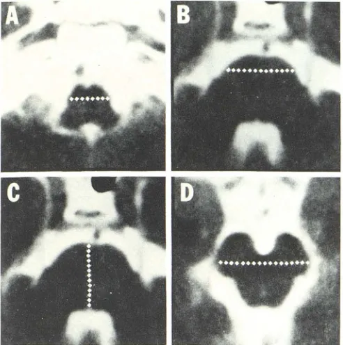
[image:4.612.56.422.85.452.2]AJNR: 1 , November /Oecember 1980 CT CISTERNOGRAPHY OF BRAINSTEM 525
Fig. 11.-Locations at which transverse medullary (A), transverse pontine
(B), pons-fourth ventricle (e), and transverse peduncular (0) dimensions
were obtained.
Fig. 12.-Mass within cerebral
pe-duncle (M) distorts normal symmetry and
effaces adjacent uncus (U).
Fig. 13.-Metrizamide outlines
en-larged medulla (arrowheads). (Conve
n-tional CT scan was negative.)
12
gliomas are quite variable [1 -3, 13]. They may appear
isodense, hyperdense, or hypodense on noncontrast CT. If
isodense, the tumor may indicate its presence by
oblitera-tion or displacement of the fourth ventricle and compression
of the adjacent cisterns. Low-density lesions are seen, in
some cases indicative of cyst formation [13]. High density
lesions most likely indicate foci of hemorrhage although
calcification has been reported [14]. Hydrocephalus, usually
mild, is present in one-third to one-half of cases. Contrast
enhancement mayor may not occur. A definite relation
between enhancement and tumor grade has not been
es-tablished, although an early trend has been reported [3].
Despite its efficacy in the diagnosis of brainstem masses,
sometimes conventional CT is falsely negative, equivocal, or
unable to distinguish between an intra- and extraaxial
pro-TABLE 2: Normal Brainstem Measurements
Location No. Cases Mean (mm) SO (0) Transverse
peduncu-lar 41 25.4 3.1
Transverse
pontine 43 24.8 2.2
Transverse
medullary 29 13.8 1.9
Pons-4th
ventricle 39 22.8 2.3
TABLE 3: Nonfilling of the Fourth Ventricle
Diagnosis
Brainstem glioma
Hydrocephalus (communicating) Chordoma
Arnold-Chiari malformation (type 1) Hypoglossal neuroma
Total
13
Range (±20) (mm)
19.5-31.5
20.4-29.2
10.0-17.6
18.1-27.4
No. Cases (n = 78)
2
6
cess [5, 13, 15-17]. Most authors have recommended
angiography or pneumoencephalography for such circ um-stances.
Initial experience has documented the safety and efficacy of CT cisternography for imaging the basal cisterns [6, 7]. Brainstem gliomas have been diagnosed [5, 18], but a
discussion of normal high-resolution CT cisternographic
anatomy of the brainstem has not been published. Our experience suggests that this procedure is the safest and most accurate means of brainstem evaluation. CT
cistern-ography in combination with a current generation CT
scan-ner can match or exceed the precision of
pneumoenceph-alography. The brainstem and fourth ventricle can be
pre-cisely imaged, and cranial nerves often seen without thin
[image:5.612.53.299.85.333.2] [image:5.612.314.555.98.199.2] [image:5.612.53.560.144.563.2] [image:5.612.57.560.370.564.2]526 STEELE AND HOFFMAN AJNR:1, November/December 1980
14 15
brainstem enlargement can be appreciated readily Trom simple inspection of the images. Borderline cases are more easily evaluated by visual assessment than by brainstem measurement. CT cisternography is also of benefit in those circumstances where conventional CT is unable to distin-guish between an intra-and extraaxial mass. In the case of cholesteatoma, CT cisternography offers a specific diag-nosis.
CT cisternography promises to further reduce the need for angiography. Nevertheless, angiography retains legiti-mate indications. If clinical or conventional CT findings suggest the possibility of a vascular lesion (aneruysm, ar-teriovenous malformation, hemangioblastoma) angiography should be performed. The neurosurgeon may find it useful to know the extent of displacement or involvement of major vessels by an extraaxial mass. Occasionally angiography may be necessary to distinguish an intra- from an extraaxial mass.
The high accuracy and low morbidity of CT cisternography should render pneumoencephalography obsolete for the detection of brainstem enlargement except for unusual cir-cumstances. As with any new technique one must approach minimal findings with caution. Minor asymmetry is not rare. Pending further experience one must also assume that such nonneoplastic processes as inflammatory disease, multiple sclerosis, and Leigh disease, may cause brainstem swelling sufficient to be detected by CT cisternography.
REFERENCES
1. Baker HL, Houser OW. Computed tomography in the diagnosis
of posterior fossa lesions. Radiol Clin North Am 1976;14:
129-147
2. Gado MG, Huete I, Mikhael M. Computerized tomography of
infratentorial tumors. Semin Roentgeno/1977;12: 1 09-120
3. Bilaniuk L T, Zimmerman RA, Littman P, et al. Computed
to-Fig. 14.-Hypoglossal neuroma (N).
Contralateral displacement of medulla
and poinled contrast interface (arrow
-head), indicating extraaxial location of
mass.
Fig. 15.-Diagnosis of choles
tea-toma (verified at surgery) made pr
eop-eratively with CT cisternography when
contrast was demonstrated within inte r-sticies of mass.
mography of brain stem gliomas in children. Radiology
1980; 1 34: 89-95
4. Davis KR, Taveras JM, Roberson GH, Ackerman RH. Some
limitations of computed tomography in the diagnosis of
neuro-logical diseases. AJR 1976;127: 111-123
5. Drayer BP, Rosenbaum AE, Reigel DB, Bank WO, Deeb ZL.
Metrizamide computed tomography cisternography: pediatric
applications. Radiology 1977;124:349-357
6. Drayer BP, Rosenbaum AE. Studies of the third circulation. J
Neurosurg 1978;48: 946-956
7. Greitz T, Hindmarsh T. Computer assisted tomography of
intra-cranial CSF circulation using a water-soluble contrast medium.
Acta Radiol [Diagn] (Stockh) 1974;15 :497-507
8. Lassman LP, Arjona VE. Pontine gliomas of childhood. Lancet
1967;1 :913-915
9. Panitch ES, Berg BO. Brain stem tumors of childhood and
adolescence. Am J Dis Child 1970; 11 9: 465-471
10. White HH. Brain stem tumors occurring in adults. Neurology
(Min neap) 1963;13: 292-300
11. Golden G"S, Ghatak NR, Hirano A, French JH. Malignant glioma
of the brain-stem: a clinicopathological analysis of 13 cases.
J Neural Neurasurg Psychiatry 1972;35: 732-738
12. Seeger JF, Gabrielsen TO. Angiography of eccentric brain
stem tumors. Radiology 1972;105: 343-351
13. Berger PE, Kirks DR, Gilday DL, Fitz CR, Harwood-Nash DC.
Computed tomography in infants and children: intracranial
neoplasms. AJR 1976;127: 129-137
14. Duffner PK, Klein DM, Cohen ME. Calcification in brain stem
gliomas. Neurology (Minneap) 1978;28: 832-834
15. du Boulay GH, Radu EW. How should one investigate the
posterior fossa? Neuroradiology 1978;15: 253-261
16. Miller EM, Newton TH. Extra-axial posterior fossa lesions
sim-ulating intra-axial lesions on computed tomography. Radiology
1978;127: 675-679
17. Naidich TP, Lin JP, Leeds NE, et al. Computed tomography in
the diagnosis of extra-axial posterior fossa masses. Radiology
1976;120: 333-339
18. Glanz S, Geehr RB, Duncan CC, Piepmeier JM.
Metrizamide-enhanced CT for evaluation of brainstem tumors. AJNR