Jrinted in Great Britain © The Company of Biologists Limited 1989
RESPONSE FEATURES OF VISUAL UNITS IN THE LOWER
MIDBRAIN OF THE RAINBOW TROUT
BY NICO A. M. SCHELLART AND WALTHER E. I. RIKKERT
Laboratory of Medical Physics, University of Amsterdam, Academic Medical Centre, Meibergdreef 15, 1105 AZ Amsterdam, The Netherlands
Accepted 20 March 1989
Summary
In the mesencephalic torus semicircularis of fish, visual processing is carried out in addition to acousticolateral processing, and takes place in unimodal and bimodal units. To study the encoding of visual stimuli in the torus of the rainbow trout, unit recordings made in the torus were compared with recordings made in the tectum.
The receptive fields (RFs) of the torus units in the rainbow trout {Salmo
gairdneri) mostly measured more than 80°, generally had no well-defined borders
and showed non-X type behavior. As in the tectum, the majority of the units showed On-responses and the RFs of On units were smaller than those of On/Off and Off units. From single unit and evoked potential data, obtained up to an eccentricity of 45°, it appears that the rostral visual field projects to the rostral and central part of the torus. In contrast to the features of tectal units, the RFs lacked spatial antagonism, wavelength coding was practically absent and directional selectivity rare. The responses, generally with latencies between 40 and 160ms, were often erratic. The most striking neurophysiological feature of the torus units was habituation to repetition of the stimulus, e.g. to an on-off flashing spot. The visuo-auditory interaction of most bimodal units showed algebraic summation, but for about 20% the visual modality enhanced the sensitivity to auditory stimu-lation.
The data from the trout confirm that the tectum plays an important role in visual functions such as acuity, pattern vision and vision of movement. The common feature of habituation in the torus suggests that the torus may be involved in the signalling of sudden changes in the visual scenery.
Introduction
358 N. A. M . SCHELLART AND W . E. I. RlKKERT
of the mammalian colliculus inferior, has scarcely been studied (Page & Sutterlin, 1970; Schellart, 1983). Yet, visual sensitivity is abundantly present among torus neurons (Schellart et al. 1987). The torus obtains its visual input bilaterally from the tectum and probably also from the nucleus dorsolateralis tegmenti (De Wolf et al. 1983), which is innervated by the tectum (Grover & Sharma, 1979; De Wolf et al. 1983). In goldfish, carp and Sebastiscus the direct tectal projection is provided by type XII neurons (for the classification of tectal neurons see Meek & Schellart, 1978) in the stratum album centrale and type XIII neurons in the stratum griseum centrale (Grover & Sharma, 1979; Echteler, 1984). About one-fifth of the torus units show sensitivity to visual as well as acousticolateral (acoustic plus lateral line) stimulation (Schellart et al. 1987). Therefore, integration of both sensory modalities and interaction between them should be an important function of the torus. For some of the bimodal neurons visual stimulation changes the acousticola-teral sensitivity (Page & Sutterlin, 1970; Schellart, 1983). Knowledge about the visual input/output relationship of the bimodal units as well as of purely visual units is, however, still fragmentary.
In the first place this study addresses the processing of visual stimulus features on the basis of single-unit recordings from the torus of the trout. To provide a better insight into the extent to which information processing changes along the visual pathway from the tectal level to the torus, this study compares the processing features of units in the torus with those in the tectum. To evaluate the general validity of the data obtained from the trout, a comparison is made with data obtained from the goldfish (e.g. Page & Sutterlin, 1970; N.A.M. Schellart, unpublished data). These data enhance our understanding of the sensory role of these large nuclei in fish, and of the interaction along the visual pathway between visual and acousticolateral processing.
Materials and methods
Experimental animals
Rainbow trout (Salmo gairdneri) 22-30 cm in length were deeply anesthetized (250 mg I"1 MS 222) directly before and during surgery, and immobilized during the experiments with intraperitoneally injected pancuronium bromide (0-15 mg) or D-tubocurarine (0-6mg). The fish was placed in an experimental tank within a dark cage. The eyes of the submerged fish were just below the meniscus of the water, which was below the opening in the skull. Details of surgery and handling of the fish during the experiments have been described before (Nederstigt & Schellart, 1986). After the experiments the animal was killed immediately by destruction of the brain.
Recording from the torus semicircularis and tectum opticum
the right tectum opticum. The electrode was advanced by a micromanipulator and, for fine displacements, by a remotely controlled stepping motor microelectrode drive. By measuring the relative x-y coordinates with respect to the tectum, the angle of inclination and the depth of the microelectrode, the position of its tip was calculated, using the dorsal tectal surface as a reference. Occasionally horseradish peroxidase (HRP) was injected to mark the tip position. Comparison of the stereotactically calculated and the histologically measured positions showed that in the mediolateral direction there was an inaccuracy of 5 % (N = 12). However, the recording depth showed an offset: the HRP marks were found 19 ± 13 % (mean ± S.D.) deeper than expected owing to denting of the tectal surface after some hours. The depth and dorsocaudal positions of all recordings were accordingly corrected (by 550 and 28/0/jm, respectively). The recordings were obtained from 123 single units in the torus region and 62 single units in the tectum. As concluded from the calculated positions of the tip of the micropipette, some of the recordings were located just outside the torus within the nucleus dorsolateralis tegmenti, the nucleus lateralis valvulae and the fasciculus longitudinalis lateralis. In addition, from the torus and tectum some tens of multi-unit recordings were obtained. In the tectum we did not obtain visual responses at a recording depth of less than 510/zm but, as this figure is biased by adhesion of the electrode to the tissue, the unresponsive superficial layer - which contains the 140 fmi thick stratum marginale - is actually thinner.
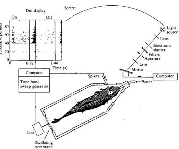
A search for visual sensitivity was made by moving a bright flickering spot (720ms on and 720ms off, i.e. a flicker frequency of 0-7Hz), projected onto a screen in front of the left eye. In general, both visual and acoustic searching stimuli were presented simultaneously and in-phase. Fig. 1 is a diagram of the experimen-tal set-up. Tone bursts, whose frequency was swept within 720 ms from 45 to 550 Hz, were used to test whether the units were also sensitive to acousticolateral stimulation. The bursts were alternated with silent periods of equal duration.
Sensory stimulation
360
N . A . M . SCHELLART AND W. E . I. RlKKERTDot display Screen
•3 o
•c
8.
y
1/5On
' • * - - .
-t...
-'ft- •
".---• i " / ' . ".---•*:-";
Off
• ,j ' • ~ ~ ~
: . c'. ~
.
:
"•
1
0-72'
Computer Tone burst sweep generator
1-44 Time (s)
Spikes A\
Coil
Computer -Water
[image:4.451.45.409.62.364.2]Oscillating membrane
Fig. 1. Schematic view of the experimental set-up for recording responses from the trout to visual (and acousticolateral) stimulation. The fish was placed in a holder and fixed with Velcro strips. See text for further details. In the (on-line)-generated dot display each dot represents a spike, plotted with the time along the abscissa and the successive on/off stimulus periods along the ordinate. The histogram in the middle denotes the number of spikes elicited during the 720 ms light-on periods and the histogram on the right that of the dark intervals.
rectangle. In this way, every point within this rectangle had approximately the same chance of being illuminated by the spot of light. The visual and acousticola-teral stimuli could be applied separately or simultaneously.
Evoked potentials from the torus were recorded during stimulation with an immobile spot of 5 or 10° in diameter the intensity of which was sinusoidally modulated with a modulation depth of 70% and a frequency of 1-5-4 Hz. These frequencies were chosen since the evoked potentials show low-pass filtering with a cut-off frequency of 5 Hz and a high-frequency attenuation of 6 dB per octave. The spot was subsequently projected in the middle (0°, 0°) and at the corners (±35°,
±25°) of the screen and again in the middle. The responses were measured from a depth of 2250/im from the tectal surface up to a depth of 3225 ^m. For each stimulus position and depth the peak-to-peak response size was measured by averaging the response 32 or 64 times. The non-linear distortion of the response was generally limited to 10 %.
The acousticolateral stimuli were generated by an oscillating membrane mounted in the rear wall of the experimental tank (Fig. 1). Details about the generation of the acousticolateral stimulus can be found elsewhere (Nederstigt & Schellart, 1986). The strength of this stimulus, as measured with a hydrophone, was 2-5 Pa. The kinetic component of the stimulus was measured with an accelerometer and a seismic velocity meter. Within the water of the tank the acoustic displacement waves accompanying this stimulus had amplitudes of several micrometers in the 40- to 100-Hz range, enough to stimulate the lateral line organs (Kroese & Schellart, 1987). The fish body itself made oscillations of 0-2/an (at most), well above the sensitivity threshold of the otolith organs of salmonids (Hawkins & Johnstone, 1978).
Registration and classification of the units
The spikes, shaped by a window discriminator, were fed into a computer (PDP 11/60 or 11/23), registering the instant of firing of the unit in relation to the onset and cessation of the light stimulus (and the tone bursts). The responses to a sequence of on-off stimulus periods were processed on-line, yielding dot displays (Fig. 1). Dot displays were not always used to study bimodal interaction. The standard procedure involved counting the number of spikes elicited in the 720 ms on and off periods during both unimodal and bimodal stimulation. The units were classified as purely visual units or bimodal units, the latter being sensitive to visual as well as acousticolateral stimulation. With respect to the visual modality, the units were distinguished, as is commonly done, as On, On/Off and Off units.
Results
Temporal characteristics of torus units
362 N. A. M . SCHELLART AND W . E. I. RlKKERT
About 35 % of the units showed predominantly tonic responses. The On/Off units generally had a phasic response to both the stimulus onset and cessation. In the goldfish all units show predominantly phasic behavior (Page & Sutterlin, 1970).
The latency of the first spike of the response to light stimulation showed a large variation among the units. Half the units had latencies less than 75 ms, with a lower limit of 40 ms. A latency more than 175 ms was observed twice. The latency of the response to the cessation of the light stimulus was often different from the latency of the response to the onset. The response of the unit depicted in Fig. 2A shows an onset latency twice as long as the latency to light-off. For several units it was found that the latency was dependent on the position of the stimulus within the receptive field. This is illustrated by Fig. 2B, which shows the responses of a phasic On/Off unit to a flickering spot in the rostral (section 5 of the dot display) and caudal (section 4) visual field. That the latency is indeed position-dependent and not dependent on response strength, is obvious, since the strongest phasic responses do not show the shortest latencies.
Visual responses of the torus were often erratic (e.g. Fig. 3), so that averaging of the spike discharges was needed to establish the response. The units showed brief as well as long-lasting fluctuations in responsiveness. The former could result in stimulus cycles without a response, preceded or followed by a cycle with a vigorous response. Such erratic responses have also been found in other central visual structures (see figs 11 and 14 of Friedlander, 1983). The long-term component is more typical for the torus and the deep tectal layers.
Visual processing in the trout midbrain
On Off
[image:7.451.38.389.50.513.2]363
364
A
N . A . M . SCHELLART AND W . E . I. RlKKERT
B
120
3 100
to
Successiv
e period
s
8
S
40
1 20
On
-.
' •-••••
•
- 1
1
•
"3
Off
• - • i • . I / ,
/ • * ; . '
:>,!• •'. , ' V . • •-••.•.'•.
11
f
1
On Off
0-72 1-44
Time (s)
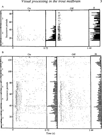
Fig. 3. Units with weak (A) and strong (B) habituation. (A) Spontaneously active visual Off unit. (1) No stimulus; (2) stationary flickering spot; (3) flicker stimulus with random movement (40° s"1). (B) Spontaneously active visual unit with weak habitua-tion to on-off flicker and erratic responses. (1) No stimulus; (2) on-off flicker; (3) flicker with random movement (30°s"1).
three units had much longer time constants. One minute after the first stimulus period the response strength of the majority of the units stabilized at a level of 0-30 % of the response strength of the first period. The value of the time constant and the (relative) final response strength were not related. Visual and bimodal units could not be distinguished with respect to the occurrence and the quality of the habituation.
Spatial and other characteristics of torus units
Spatial coding (distinct On and Off regions within the RF) was observed too rarely to justify any attempt at classification further than the on-off classification. Fig. 2A gives an example of a spatially coded unit. Comparison of sections 2 and 3 of this dot display shows that the on component of the RF of this silent, phasic On/Off unit did not extend to the caudal part of the visual field. This is not due to habituation of the RF, since repositioning of the spot in the rostral part again gave a good, but habituating, response. Occasionally attempts were made to elicit a surround response by adapting the RF with a stationary spot of light. In the goldfish tectum, in most cases such an adapting light has been found to induce a surround response (Schellart et al. 1979). However, in the trout torus this method was not successful.
Visual processing in the trout midbrain
B Torus
>100 >99
365
A Tectum
25"
T
1
On
N=U
Off 11
On/off
5
On 18
Off 8
On/off
[image:9.451.117.334.67.261.2]12
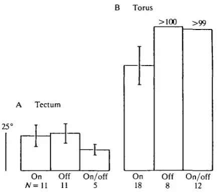
Fig. 4. Receptive field size distinguished by the response sign of units in the tectum (A) and the torus (B). The height of the histograms give the mean size, the bars the S.D. The RF size of all torus Off and On/Off units (except one) is greater than 100°. The mean size of the tectal RFs (irrespective of coding) is 23-4° ± 13-0° (mean ± S.D.,
N = 21), that of the torus RFs is at least 88°.
observe red-green antagonism, the most common wavelength antagonism in the teleost visual system. For some other units, red-green antagonism could be elicited by chromatic adaptation. In addition, some units were found which appeared to lack either the red or the green wavelength input.
Directional selectivity, the ability of a unit to respond exclusively to an object moving in a well-defined direction, was tested with a non-flickering spot moving at various speeds. Although well-developed directional selectivity was never found, five units showed directional preference. An example of the responses of such a unit are illustrated in Fig. 2B (section 6 and 7). A minority of the units responded well to a moving (non-flickering) stimulus. Repetition of movement can result in habituation, as happens with all types of repetitions.
366
N . A . M . SCHELLART AND W. E . I. RlKKERT(35°, 25°)
respond as a whole (X-type behavior), as holds in general for tectal cells (Schellart et al. 1979), but appear to be composed of subunits and can, therefore, be compared with the non-X-type retinal ganglion cells of the goldfish (Shapley & Gordon, 1978; Levine & Shefner, 1979).
Visuotopy in the torus
Visual processing in the trout midbrain 367
Fig. 5. Visuotopy of the torus. (A) Relationship between RF position and site of single-unit recording. Each recording site with its associated RF (if less than 100°) is represented by an arrow. The foot of each arrow indicates the location of the recording site in the 'x-y plane' of the torus. The orientation and length of the arrow represent the polar coordinates of the middle of the RF within the visual field, such that the orientation indicates the direction of the RF within the visual field and the length the space angle (eccentricity) relative to the center of the visual field, as estimated by the projection of the optical axis. The solid arrow at the top gives the anatomical orientation of the torus and the rostral direction of the visual field. It also gives the calibration of the length of the arrows. (B) Dependency of evoked potentials on the stimulus location within the visual field. The histogram bars denote peak-to-peak amplitude as a function of recording depth. The five amplitude-depth profiles were obtained with the sine-wave-modulated stimulus (modulation depth 70%) on the five screen positions. An averaged (64 times) response made at one of the recording depths is depicted for each stimulus position. (C) Topographic relationship between the location within the visual field - yielding the largest evoked potentials - and the site of recording within the torus for seven different animals (1-7). The way in which the stimulus position, which evoked the largest response for a chosen recording site, was determined is described in the text. This position is represented in polar coordinates (see A). The cross indicates that for this particular electrode location the five stimulus positions could not be distinguished by response amplitude. (D) Vector addition of the combined data of A and C performed for each of the four quadrants into which the torus is divided. The length of the resultants is weighed with the number of vectors used, with the resultant in the first quadrant as reference.
368 N . A . M . SCHELLART AND W . E . I. RlKKERT
Interaction between the visual and acousticolateral systems in the torus When an on-off flashing, moving spot and a sequence of tone burst were given simultaneously and in phase, the strength of the response for 14 of the 19 bimodal units was approximately the algebraic sum of the two unimodally elicited responses. Summation was also found when both modalities were applied in anti-phase. The algebraic mode of interaction is known in the goldfish for units in the deep torus in the vicinity of the fasciculus longitudinalis lateralis (Page & Sutterlin, 1970). Interaction was not only examined for bimodal units, but also for unimodal units, since the ineffective modality may still influence the response to the other modality. The majority of the unimodal visual and acousticolateral units in the trout did not change their response when additional stimulation by the ineffective modality was applied. However, two acousticolateral units showed strong en-hancement of the response by an additional visual stimulus, whereas the units were not sensitive to visual stimulation alone. Similar behavior was found for two visual units. Also, bimodal units could show a strong deviation from the algebraic mode. Four bimodal units exhibited strong enhancement and one showed strong attenuation to bimodal stimulation, interaction types also found in the goldfish torus (Page & Sutterlin, 1970) and in the torus of Xenopus between the acoustic and lateral line system (Zittlau etal. 1985; Lowe, 1987). One unit was found which gave a response only when both modalities were presented simultaneously.
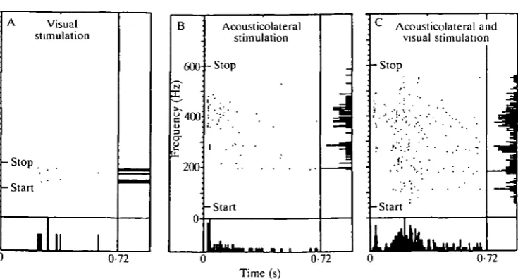
The responses of one of the bimodal units to uni- and bimodal stimulation with non-algebraic behavior are presented in Fig. 6, which shows the response to: a stationary on-off flashing stimulus, which evoked a weak, already habituated response (Fig. 6A); a tone-burst which was increased in frequency by about 2 Hz each time it was presented again (Fig. 6B); and both stimuli presented simul-taneously with the onsets and cessations in register (Fig. 6C). The dot displays of the dark and silent intervals are not shown. Within this interval hardly any spikes were elicited in this non-spontaneous unit. The response to light shows the usual long latency (about 220 ms), whereas the response to sound stimulation starts within 24 ms. The simultaneous presentation produced a response approximately twice as strong as the sum of both individual responses.
Temporal characteristics of tectal units
Nearly all visual units encountered showed sensitivity to the flickering stimulus spot. A sample of 62 tectal units yielded 42% On, 30% Off and 28% On/Off units, findings in general agreement with other tectal studies on teleost fish (e.g. Jacobson & Gaze, 1964; Schellart et al. 1979).
Visual processing in the trout midbrain
369Visual stimulation
- Stop -Start
Acousticolateral stimulation 600 -Stop
!400
200--Start
0-72
- - Stop
Acousticolateral and visual stimulation
-Start ;
0-72 0-72
[image:13.451.41.409.67.265.2]Time (s)
Fig. 6. Visuo-acousticolateral non-linear interaction in a bimodal, non-spontaneous torus unit. The spike responses are depicted in dot display fashion. The light-on periods of 720 ms were alternated with dark periods (not shown) of the same duration. The tone bursts also lasted 720 ms and were followed by silent intervals of the same length (not shown). The successive stimulus-on periods are depicted along the ordinate. Start and stop along the scaled ordinate indicate the presentation of the first and last stimulus period (a flickering spot of light or a tone burst alternated with silent periods), respectively. In A only 40 periods of flicker were presented. The vertical histograms denote the number of spikes elicited during a single light-on or tone-burst-on period. The horiztone-burst-ontal histograms denote the number of spikes elicited in the successive time bins along the abscissa, added for all periods. All six histograms were normalized to their own maximum.
constant of 4 s and an ultimate response level of 36 %. For most habituating units the response was generally stabilized after 1 min at a level of 0-30 % of the strength of the response in the first period.
Nearly half the tectal visually sensitive units had a spontaneous spike rate of more than 1 spikes"1, with a population mean of 4-9 spikess"1. In the goldfish tectum only 23% of the units (N=160) are spontaneously active (N. A. M. Schellart, unpublished data), with a lower occurrence in the superficial than in the deep tectal layers. In the torus of the trout, spontaneous activity was more common (69%, N = 86) than in the tectum (46%, N = 37). In the tectum and especially in the torus (see Fig. 3A section 1) irregular spontaneous firing is more common than regular firing. Moreover, the spontaneous activity occurred in irregular periods. The purely visual units in the trout torus have a population mean spike rate of 4-7 spikess"1 (Schellart et al. 1987), a value close to that of the tectum.
370 N . A . M . SCHELLART AND W . E . I . RlKKERT On
20
16 "5 "S 12
3
4-0 5
• Present bsent
W/W//////////.
11 14 17 20 Depth (xlOOfim)
60
o
o.
iv
e
Succes
s
l
I:
= • :
. " — • •
/
=
=
[image:14.451.38.417.68.272.2]Time (s) 0-72
Fig. 7. Habituation in the tectum. (A) Habituation in the tectum related to recording depth. In the deep tectal layers (recording depths >1100^m) habituation occurs significantly more frequently than in the central and superficial layers. (B) Time course of the habituation of a tectal unit (RF size 40°, not spontaneously active). The arrow indicates the instant that the stimulus was switched on.
superficial layers of the tectum appeared to be on average smaller than in the lower layers. This is also found for single units in the goldfish (N. A. M. Schellart, unpublished data). However, in the deeper layers of the trout tectum the latencies do not deviate from those in the torus.
Spatial and other characteristics of tectal units
The size of the tectal RFs had a lower limit of 7° and only seven of the 66 units had a field larger than 80°. The tectal RFs were on average at least four times smaller than the torus RFs, as is obvious from a comparison of the histograms of Fig. 4A and B. As in the goldfish (Schellart & Spekreijse, 1976) and other vertebrates (e.g. Hughes & Pearlman, 1974), the circular RFs were smaller than the elongate RFs (f-test, P<0-01). Their mean sizes were 21° and 35°, respectively. The majority (60 %) of the units had a circular RF. In the tectum and optic nerve of the goldfish, RFs of circular On units are on average smaller than those of circular Off units (Schellart & Spekreijse, 1976). The trout data show the same tendency.
which were only studied occasionally (8 and 15 units, respectively), occurred in about half and one-quarter of the units, respectively. This compares well with the data obtained from the cyprinid tectum (e.g. Riemslag & Schellart, 1978; Schellart etal. 1979).
Discussion
The visuotopic organization of the teleost tectum is well described anatomically and neurophysiologically (see Guthrie, 1983; Vanegas, 1983, for reviews). The first descriptions (Jacobson & Gaze, 1964; Schwassmann & Kruger, 1965) were based on multi-unit recordings made with metal electrodes, which mainly recorded the activity of the arborizations of the optic nerve fibers. This resulted in a point-to-point topographic mapping of the visual field onto the tectum. At single-unit level, however, the visuotopy is less precise, with offsets up to 12° (N. A. M. Schellart, unpublished data). The visuotopy of the torus is even more global, as shown in Fig. 5. It could not be established whether the (whole) visual field is projected in as orderly a manner to the torus as to the tectum. In reality, the projection may be better, since the figure is based on pooled data. An additional indication of (global) visuotopy is based on the data of HRP track-tracing (N. A. M. Schellart, unpublished data). When the torus is labeled, fibers leaving and penetrating the torus (at its dorsolateral pole) run a roughly transverse course to and from the ipsilateral tectum. This would result in the same rostrocaudally oriented topography of the torus as in the tectum. Many nuclei connected directly to the retina, and nuclei obtaining their visual input from the tectum also have visuotopy to some extent (e.g. Ito & Vanegas, 1983; Presson et al. 1985; Springer & Mednick, 1985a,b). The torus, although not directly connected to the retina, is the largest central structure with visual processing after the tectum. Therefore, the finding of a visuotopic organization within the torus, albeit weakly developed, is not a complete surprise.
The lack of any type of spatial coding in the torus, even when an adapting central spot is used, the generally huge RFs and the rare occurrence of directional selectivity indicate this nucleus does not process shape and contrast, precise position, or direction of movement. In the tectum of trout (this study) and other teleosts (e.g. Guthrie & Banks, 1978; Schellart et al. 1979) the RF properties are amazingly variable. The RF size and shape, and the occurrence of distinct On and Off parts within the RF, can be either completely separated or overlap more or less. Therefore, it is hypothesized that spatial stimulus features are analyzed in the tectum.
Behavioral studies indicate that the trout has well-developed color vision (Douglas, 1983). In the tectum of the trout, antagonistic wavelength coding seldom occurs, whereas in the goldfish it is more common (21 %, N = 149) and is even shown by the majority of the units during chromatic adaptation (Schellart,
372 N . A . M . SCHELLART AND W . E . I. RlKKERT
demonstrate wavelength antagonism (e.g. Schellart, 1973). Apparently color processing is primarily a function of retinal circuitry and to a lesser extent a task of the tectum.
The most striking neurophysiological feature of the torus is the frequently observed habituation of unit responses. In the goldfish, visually habituating units have been found dorsolaterally in the fasciculus longitudinalis lateralis (Page & Sutterlin, 1970), presumably in the ventral torus and the nucleus dorsolateralis tegmenti. These units, and also those in the trout torus (Schellart et al. 1987), include visually habituating bimodal units which often do not show habituation to auditory stimuli. The different responses to these two modalities suggest that visual habituation is a characteristic that arises at a level more distal along the visual pathway than the point of bimodal convergence. Habituation is also a common feature of the deep tectal layers of trout (this study) and goldfish (O'Benar, 1976; N. A. M. Schellart, unpublished data). Therefore, plausible candidates for generating habituation are visual units in the torus and the efferents in the deep tectum.
Improvement of visual or acousticolateral detection, leading to higher sensi-tivities and therefore to easier perception of an object, can be effected by two different mechanisms: algebraic interaction (e.g. dendritic summation) of both sensory inputs or multiplicative interaction between both inputs (e.g. presynaptic facilitation). Improvement of localization of an object is achieved by sensory spatiotopic mapping in register. Visuo-acoustic spatiotopy has been established for various higher vertebrates and a visuo-lateral spatiotopy has recently been described for the Xenopus tectum (Claas et al. 1989). Such maps have not been found in teleosts, but a rather coarse spatiotopy of the lateral line projection has been demonstrated in the subtectum of elasmobranchs (Bleckmann et al. 1987). It is doubtful whether well-developed spatial mapping in register exists in the torus, owing to its very crude visuotopy. The present discussion of the visuo-acousticola-teral integration at unit level within the subtectum will, therefore, be focused on the interaction without considering the spatiotopic aspects.
abscissa were close to each other. This indicates that multiplicative interaction played a role.
The experiment depicted in Fig. 6 clearly shows non-algebraic interaction between the two modalities. The strong increase in spike density in Fig. 6C, also shown in the bottom histogram of this panel, is found from the instant that the light response is expected. The first part of the response to combined stimulation is very similar to that of the response to sound stimulation alone. The vertical histogram in Fig. 6C lacks a frequency-independent 'background' of spike firing and this also holds for responses to bimodal stimulation with 3-15 times stronger light stimuli. As a further confirmation of this lack, repetition of the interaction experiment with this unit sometimes showed gaps of spike firing along the frequency axis. The absence of such a background rules out algebraic interaction with a dominating threshold after the location where the visual and the acousticolateral pathways come together. The shapes of the vertical histograms in Fig. 6B and C are alike, although between 50 and 150 Hz a new peak is found in the vertical histogram of Fig. 6C. The emergence of this new peak, at frequencies that are subthreshold for sound stimulation alone, and the frequency-dependent increase of spike firing for the other frequencies, can most easily be understood on the assumption that the light stimulus enhances the acousticolateral sensitivity. The explanation that the acousticolateral stimulus proportionately enhances the visual sensitivity requires that the 50-150 Hz acousticolateral stimulus, although by itself subthreshold, nevertheless achieves this enhancement. This is a much more complicated mechanism.
As the horizontal histogram in Fig. 6C indicates, the enhancement, which can be described formally by a multiplicative type of interaction, has a very instantaneous nature. From the functional point of view, this active visuo-acousticolateral interaction can be interpreted as a visually induced acousticola-teral arousal with switch-over times of less than 0-1 s. Since the torus projects to the reticular formation and spinal cord, this fast arousal or alerting behavior may possibly play a role in sensorimotor control. The enhancement of the acousticola-teral sensitivity could be achieved by presynaptic facilitation, for example. The visuo-acousticolateral interaction in the torus is, however, dominated by sum-mation, the simplest form of neuronal interaction.
374 N. A. M . SCHELLART AND W . E. I. RlKKERT
scenery. A second important functional characteristic of the torus is the visuo-auditory interaction which promotes visuo-auditory processing.
The critical comments and helpful suggestions of Drs Rob J. A. Buwalda and Alfons B. A. Kroese on how to improve the manuscript are gratefully acknowl-edged. The majority of the data were obtained in collaboration with L. J. A. Nederstigt, who was supported by a grant from the Netherlands Organization for Scientific Research (NWO-BION).
References
BLECKMANN, H., BULLOCK, T. H. & JORGENSEN, J. M. (1987). The lateral line mechanoreceptive mesencephalic, diencephalic, and telencephalic regions in the thornback ray, Platyrhinoidis
triseriata (Elasmobranchii). J. comp. Physiol. 161, 67-84.
CLAAS, B., MONZ, H. & ZITTLAU, K. E. (1989). Direction coding in the central parts of the lateral line system. In The Mechanosensory Lateral Line. Neurobiology and Evolution (ed. S. Coombs, P. G6rner & H. Miinz), pp. 410-419. Berlin: Springer-Verlag.
DE WOLF, F. A., SCHELLART, N. A. M. & HOOGLAND, P. V. (1983). Octavolateral projections to the torus semicircularis in the trout, Salmo gairdneri. Neurosci. Lett. 38, 209-213.
DOUGLAS, R. H. (1983). Spectral sensitivity of rainbow trout {Salmo gairdneri). Rev. Can. Biol.
Exp. 42, 117-122.
ECHTELER, S. F. (1984). Connections of the auditory midbrain in a teleost fish, Cyprinus carpio.
J. comp. Neurol. 230, 536-551.
FRIEDLANDER, M. J. (1983). The visual prosencephalon of teleosts. In Fish Neurobiology, vol. 2
(ed. R. E. Davis & R. G. Northcutt), pp. 91-115. Ann Arbor: University of Michigan Press.
GROVER, B. G. & SHARMA, S. C. (1979). Tectal projections in the goldfish (Carassius auratus): A degeneration study. /. comp. Neurol. 184, 435-454.
GUTHRIE, D. M. (1983). Visual central processing in fish behaviour. In Recent Advances in
Vertebrate Neuroethology (ed. J. P. Ewert, R. R. Capranica & D. I. Ingle), pp. 381-412. New
York: Plenum Press.
GUTHRIE, D. M. & BANKS, J. R. (1978). The receptive field structure of visual cells from the optic tectum of the freshwater perch (Perca fluviatilis). Brain Res. 141, 211-225.
HAWKJNS, A. D. & JOHNSTONE, A. D. F. (1978). The hearing of the Atlantic salmon Salmo salar.
J. Fish Biol. 13, 655-673.
HUGHES, C. P. & PEARLMAN, A. L. (1974). Single unit receptive fields and the cellular layers of the pigeon optic tectum. Brain Res. 80, 365-377.
Pro, H. & VANEGAS, H. (1983). Cytoarchitecture and ultrastructure of nucleus prethalamicus, with special reference to degenerating afferents from optic tectum and telencephalon, in a teleost (Holocentrus ascensionis). J. comp. Neurol. 221, 401-415.
JACOBSON, M. & GAZE, R. M. (1964). Types of visual response from single units in the optic tectum and optic nerve of the goldfish. Q. Jl exp. Physiol. 49, 199-209.
KROESE, A. B. A. & SCHELLART, N. A. M. (1987). Evidence for velocity- and
acceleration-sensitive units in the trunk lateral line of the trout. J. Physiol, Lond. 394,13P.
LEVINE, M. W. & SHEFNER, J. M. (1979). X-like and not x-like cells in goldfish retina. Vision
Res. 19, 95-97.
LOWE, D. A. (1987). Single-unit study of lateral line cells in the optic tectum of Xenopus laevis: evidence for bimodal lateral line/optic units. /. comp. Neurol. 257, 396-404.
MEEK, J. & SCHELLART, N. A. M. (1978). A Golgi study of the goldfish optic tectum. /. comp.
Neurol. 182, 89-358.
NEDERSTIGT, L. J. & SCHELLART, N. A. M. (1986). Acousticolateral processing in the torus
semicircularis of the trout Salmo gairdneri. Eur. J. Physiol. Pflilgers Arch. 406, 151-157.
O'BENAR, J. D. (1976). Electrophysiology of neural units in goldfish optic tectum. Brain Res.
Bull. 1, 529-541.
PAGE, C. H. & SUTTERLIN, A. M. (1970). Visual-auditory responses in the goldfish tegmentum.,
Visual processing in the trout midbrain 375
PRESSON, J., FERNALD, R. D. & MAX, M. (1985). The organization of retinal projections to the diencephalon and pretectum in the cichlid fish, Haplochromis burtoni. J. comp. Neurol. 210, 37-48.
RIEMSLAG, F. C. C. & SCHELLART, N. A. M. (1978). Evoked and spike responses to moving
stimuli in the optic tectum of goldfish. /. comp. Physiol. 128,13-20.
SCHELLART, N. A. M. (1973). Dynamics and statistics of goldfish retinal ganglion cells. PhD
thesis, University of Amsterdam.
SCHELLART, N. A. M. (1980). Receptive field organization of tectal neurons in the goldfish.
Photobiol. Bull. 1, 286.
SCHELLART, N. A. M. (1983). Acousticolateral and visual processing and their interaction in the torus semicircularis of the trout, Salmo gairdneri. Neurosci. Lett. 42, 39-44.
SCHELLART, N. A. M., KAMERMANS, M. & NEDERSTIGT, L. J. A. (1987). An electrophysiological study of the topographical organization of the multisensory torus semicircularis of the rainbow trout. Comp. Biochem. Physiol. 88A, 461-469.
SCHELLART, N. A. M., RIEMSLAG, F. C. C. & SPEKREJJSE, H. (1979). Center-surround
organization and interactions in receptive fields of goldfish tectal units. Vision Res. 19, 459-467.
SCHELLART, N. A. M. & SPEKREUSE, F£. (1976). Shapes of receptive field centers in optic tectum of goldfish. Vision Res. 16, 1018-1020.
SCHWASSMANN, H. O. & KRUGER, L. (1965). Organization of the visual projection upon the optic tectum of some freshwater fish. /. comp. Neurol. VIA, 113-126.
SHAPLEY, R. M. & GORDON, J. (1987). Ganglion classes and spatial mechanisms. J. gen. Physiol. 71,139-155.
SPRINGER, A. D. & MEDNICK, A. S. (1985a). Topography of the retinal projection to the
superficial pretectal parvicellular nucleus of goldfish; a cobaltous-lysine study. /. comp.
Neurol. 237, 239-250.
SPRINGER, A. D. & MEDNICK, A. S. (1985fc). A quantitative study of the relative contribution of different retinal sectors to the innervation of various thalamic and pretectal nuclei in goldfish. /. comp. Neurol. 242, 369-380.
VANEGAS, H. (1983). Organization and physiology of the teleostean optic tectum. In Fish
Neurobiology, vol. 2 Higher Brain Areas and Functions (ed. R. E. Davis & R. G. Northcutt),
pp. 43-90. Ann Arbor: University of Michigan Press.