Shiwei Yu1 Victor M. Haughton1 Lowell A. Sether2 Marvin Wagner2
Received September 30, 1988; revision re -quested November 17, 1988; revision received Jan-uary 17, 1989; accepted January 18, 1989.
This work was supported by NIH grant 5 R01 AR33667-03.
1
Department of Radiology, Froedtert Memorial Lutheran Hospital, Medical College of Wisconsin, 9200 W. Wisconsin Ave., Milwaukee, WI 53226. Address reprint requests to V. M. Haughton.
2
Department of Anatomy and Cellular Biology, Medical College of Wisconsin, Milwaukee, WI 53226.
0195-6108/89/1005-1077
© American Society of Neuroradiology
Comparison of MR and
Diskography in Detecting Radial
Tears of the Anulus:
A Postmortem
Study
1077
Radial tears of the anulus fibrosus, which anatomic studies suggest are a primary event in disk degeneration, can be detected by diskography or MR imaging. We compared the sensitivity of MR and diskography in the detection of anular tears. MR, diskography, and cryomicrotomy anatomic sectioning were performed in eight cadaver lumbar spines. Diskography demonstrated 15 radial tears in 36 intervertebral disks. MR demonstrated 10 of the 15, a sensitivity of 67%. MR (T2-weighted images) in each of the diskographically normal disks showed the high signal intensity characteristic of normal disks. Thirteen of 15 disks from which contrast medium extravasated at diskog-raphy had diminished signal intensity in MR images.
We conclude that although MR may demonstrate some radial tears of the anulus, and associated changes in the disk, it cannot be used as effectively as diskography to visualize a radial tear_
AJNR 10:1077-1081, September/October 1989
The radial tear of the anulus may be a primary event in disk degeneration [1, 2]. A radial tear is a necessary condition for the development of a herniated nucleus pulposus. It may also occur regularly with other degenerative changes in the disk. A radial tear of the anulus can be found in most disks with reduced height, or with reduced signal intensity in MR images [1]. Disks without a radial tear, even in older subjects, have normal height and signal intensity. Another study showed that the anular tear was consistently associated with bulging of the anulus fibrosus [3]. Experimentally, disk degeneration has been produced by surgically creating a tear of the anulus. After incision of the anulus, the disk loses height and spondylotic changes occur. Some investigators have suggested that radial tears cause pain.
The high frequency of radial tears in cadaver disks suggests that they are not consistently symptomatic.
Although MR has been shown to demonstrate radial tears of the anulus, its accuracy in demonstrating such tears has not been documented. Therefore, we compared the sensitivity of diskography and MR in demonstrating radial tears.
Materials and Methods
Six fixed and two fresh cadavers were selected for study. Among the eight cadavers were one newborn, one 2-year-old, and six adults (54, 66, 68, 72, 78, and 87 years old). In the case of fixed cadavers, the spine was removed from the body by dissection prior to diskography and imaging. In each case, MR imaging, diskography, CT, and cryomicrotome sectioning were performed.
MR imaging was performed on a 1.5-T GE imager. A 4-in. butterfly or other specially designed surface coil* was used as a radiofrequency receiver. Images were obtained with a 256 x 256 matrix, 3-mm section thickness, and 4000/20,70/2 and 800/20,40/2 (TRfTE/ excitations), spin-echo sequences in the sagittal plane.
For diskography, a solution of iodinated contrast medium (300 mg ljml) mixed with green dye (40:1) was prepared. Each lumbar nucleus pulposus was cannulated with a 21-gauge
1078 YU ET AL. AJNR:10, September/October 1989
spinal needle from the posterolateral (in fresh cadavers) or a
nterola-teral approach (in fixed spine specimens) under fluoroscopic
monitor-ing. After the needle tip was verified to be in the nucleus pulposus
the contrast medium was injected until resistance was felt. The
volume of solution injected varied from 0.3-0.5 ml in the newborn to
0.5-1.0 ml in adults. Anteroposterior and lateral plain films of the
lumbar spine were obtained. After MR imaging and diskography were
completed, the cadaver was frozen and embedded in carboxymethyl
cellulose solution in a styrofoam box. A series of contiguous 1.5
-mm-thick CT images of the block was obtained in a GE 9800 scanner in
the sagittal plane. Lines were marked on the surfaces of the s
tyro-foam box by means of the localizer lights to identify exactly the plane and location of the CT slices. The blocks were sectioned on a
cryomicrotome in the same plane as was used in MR and CT imaging.
As each millimeter (in newborns) or 2 mm (in adults) of tissue were removed, the surface of the specimen was photographed.
The disks were classified on the basis of their diskographic
ap-pearance into the categories described by Adams et al. and Kieffer
et al. (4, 5]. They were called type 1 if the contrast medium collected
in a regular ovoid-shaped nucleus pulposus, type 2 if the contrast medium had an irregular ovoid shape, type 3 if the contrast medium
collected in two or more lobular-shaped collections, and type 4 (anular
tear) if the contrast medium escaped from the nucleus through a
fissure in the anulus. The disks were classified on the basis of the
cryotome appearance into immature, transitional, adult, early degen
-erated, and late degenerated, on the basis of criteria previously
described [1]. In brief, the immature disk was characterized by a translucent nucleus sharply differentiated from the anulus; transi-tional, by ingrowth of fibrous tissue around the equator of the nucleus;
A
8
adult, by a fibrous nucleus and a poorly defined border between
nucleus and anulus; early degenerated, by slight loss of disk height
and a radial tear in the anulus fibrosus; and severely degenerated, by complete collapse of the disk space. The MR images were examined without reference to the radiographs or cryotome sections
for evidence of radial tears of the anulus. On the MR images an anular tear was diagnosed if a tongue of high signal intensity was evident in the periphery of the disk, where normally a low signal
intensity was present. Detection of anulus tears by MR was compared to detection by diskography (the standard of comparison).
Results
Thirty-six disks (eight L 1-L2, eight L2-L3, seven L3-L4, seven L4-L5, and six L5-S1) were studied with diskography, MR, and cryomicrotomy. Technical difficulties prevented can-nulation of one disk. Three disks in the 2-year-old were intentionally not injected so they could be saved for other experiments.
Seven disks with a type 1 diskographic appearance were identified (Fig. 1 A). Five were found in the newborn and two in the 2-year-old. On cryotome sections in each case of type 1 (Fig. 1 B), the nucleus appeared homogeneously stained with dye, except in the equator. The band of less translucent tissue, which failed to stain with dye, contained remnants of primitive notochord with degenerating syncytial cells. The seven disks with type 1 diskograms conformed to the
"im-c
Fig. 1.-A-C, The L 1-L2 and L2-L3 disks of a 2 year old shown in a lateral diskographic image (A), sagittal cryotome section (8), and sagittal MR
image (C). The diskograms are classified as type 1. An ovoid contrast medium collection (arrowheads in A) corresponds to the dye-stained nucleus
pulposus (arrowheads in 8). The plate of degenerating notochordal cells (long thin arrows in 8) in the equator of the nucleus, the anulus fibrosus (straight
black arrow in 8), and Sharpey's fibers (curved open arrow in B) are labeled. In the T2-weighted MR image (4000/70), the high signal intensity region
[image:2.612.53.561.400.687.2]AJNR:1 0, September/October 1989 MR AND DISKOGRAPHY OF ANULAR TEARS 1079
mature" classification. Long TR and TE MR imaging in each
case showed a homogeneous, high signal intensity region that corresponded to the nucleus and the anulus, and low
signal intensity corresponding to Sharpey's fibers (Fig. 1 C).
The term Sharpey's fibers is used to refer to the broad band
of collagenous fibers in the periphery of the disk that have an
osseous origin and insertion in the ring apophysis.
Five disks, all in adult cadavers, had a type 2 diskographic
appearance (Fig. 2A, L5-S1 disk). On cryomicrotomy, the
nucleus was stained irregularly with dye (Fig. 2B). The disks
appeared on the anatomic sections as typical "adult" disks
with the anulus intact. Long TR and TE MR imaging in most
of these disks showed a region of moderately high signal
intensity corresponding to the nucleus and anulus (Fig. 2C).
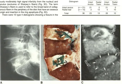
There were nine type 3 diskograms, all in adults (Fig. 3A,
L 1-L2 and L2-L3). On cryomicrotomy there were irregular
spaces within the disk stained densely with dye, which
cor-responded to the contrast medium collections demonstrated
on diskography (Fig. 3B). On anatomic sections, the disks
appeared as typical "adult" with the anulus intact in each case. Long TR and TE MR imaging showed an inhomogene-ously moderately high signal intensity from the nucleus and anulus (exclusive of Sharpey's fibers) (Fig. 3C). The term
Sharpey's fibers is used to refer to the broad band of
collag-enous fibers in the periphery of the disk that have an osseous origin and insertion in the ring apophysis (Fig. 3C).
There were 15 type 4 diskograms showing a fissure in the
A
B
anulus fibrosus through which contrast medium escaped into
the spinal epidural space (Fig. 2A, L4-L5; Fig. 3A, L3-L4;
and Fig. 4A). Cryomicrotomy showed a dye-stained fissure in
the anulus in 13 of the 15 cases (Fig. 2B, L4-L5; Fig. 3B,
L3-L4; and Fig. 46). The cryomicrotome appearance was
that of "degenerated" disks; that is, reduced disk height,
discoloration of the nucleus, and loss of fibrocartilage. At L4
-L5 in two of the 15 cases, diskography showed a small tear
of the anulus that was not evident in the anatomic sections
of the disk. On T2-weighted MR images, 10 of the 15 fissures
were identified as high signal intensity regions in the normally low signal intensity portion of the disk (Fig. 2C, L4-L5 and Fig. 4C). In 13 of the 15 cases, the signal intensity of the disk
appeared reduced. The sensitivity of MR for radial tears
(compared with diskography) was 67% (see Table 1 ).
TABLE 1: Cryomicrotomy and Dlskography Compared for
Detection of Anular Tears
Cryomicrotome MR Diskogram Intact Radial Intact Radial
Anulus Tears Anulus Tears
Intact anulus (n = 21) 21 0 21 0
Radial tear (n = 15) 2 13 5 10
Total 36 23 13 26 10
c
Fig. 2.-A-C, The L4-L5 and L5-S1 disks of a 72 year old shown in a lateral diskographic image (A), sagittal cryotome section (B), and sagittal MR image (C). A type 2 diskogram is illustrated at L5-S1, a type 4 at L4-L5. At L5-S1, the irregular contrast medium collection in (A) corresponds to the dye-filled region in the disk (bent black arrow in B). At L4-L5, the contrast medium escaped from the nuclear region through a fissure in the anulus and
[image:3.613.55.564.319.671.2]1080 YU ET AL. AJNR:1 0, September/October 1989
A
8
c
Fig. 3.-A-C, lateral diskographic image (A), sagittal cryotome section (8), and sagittal MR image (C) of lumbar spine of a 68-year-old. The diskograms were classified as type 3 at l1-l2 and l2-l3, type 4 at l3-l4. The lobular contrast medium collections at l1-l2 and l2-l3 (A) correspond to dye-stained regions (8). The diskogram (A) and cryotome section (8) show contrast medium and dye in the fissure of the radial tear of the anulus and Sharpey's fibers at l3-l4 (curved open arrows in A and 8). Nucleus and anulus have high signal intensity in MR image (4000/70) at l1-l2 and l2-l3, and lower signal intensity at l3-l4. The radial tear at l3-l4 shown on diskogram and cryotome section is not well demonstrated by MR (C). The intact Sharpey's fibers
(straight open arrows in 8) appear as homogeneously low signal intensity regions (straight open arrows in C).
A
8
c
Fig. 4.-A-C, Type 4 diskograms at l4-l5 and l5-S1 in lumbar spine of a 68-year-old. lateral diskogram (A) and sagittal cryotome section (8) show contrast medium and dye in fissures (arrowheads in A and 8) in radial tears of anulus and Sharpey's fibers. In MR image (4000/70), the fissures have high signal intensity (arrows in C). The signal intensity of the l5-S1 disk is reduced.
Discussion
In this study MR was less sensitive than cryomicrotomy or
diskography in demonstrating anular tears. In the two L4-L5
disks with diskographic but no anatomic evidence of a tear,
the cryomicrotome sectioning was assumed to be falsely
negative. Since the cryotome sections were at 2.0-mm inter
-vals in the sagittal plane, a small defect in the anulus
theoret-ically could be missed.
MR had a sensitivity of 67% for detecting radial tears of the anulus in this series. Because of partial volume averaging
or because defects may contain insufficient fluid or mucoid
material to produce a discernible high signal intensity, the 3-mm-thick MR sections may fail to demonstrate some small defects in the anulus. The defects identified in cadavers may not be representative of defects detected in a patient popu-lation referred for diskography. That population could have larger defects, more acute defects, or other discrepancies
from the cadaver population.
In other studies, diminished signal intensity from the disk
was a reliable sign of a radial tear of the anulus [1, 6). Signal
intensity from the disk was reduced (long TR, long TE) in all
cases of radial tears. We did not attempt to evaluate absolute
[image:4.614.56.563.86.270.2] [image:4.614.57.561.346.550.2]AJNR:1 0, September/October 1989 MR AND DISKOGRAPHY OF ANULAR TEARS 1081
fresh cadavers were used. The two disks that lacked the expected low signal intensity in association with a radial tear were fixed cadavers.
The clinical significance of radial tears is debatable. Radial tears have not been thoroughly investigated. Fernstrom [7] showed that a disk with a tear of the anulus (simple disk rupture) and a herniated disk have exactly the same disko
-graphic appearance and that patients with anular tear with or without herniation cannot be distinguished on the basis of their signs or symptoms or their outcomes from laminectomy.
His study suggests that the anular tear may produce pain in the back or radiating into the legs. The high frequency of anular tears in older cadavers suggests that they are not always symptomatic.
We conclude that MR imaging with SE acquisitions dem-onstrates radial tears in the anulus with less precision than diskography. MR may produce indirect evidence of radial
tears (reduced signal intensity from the disk) even when the
tears are not directly demonstrated.
ACKNOWLEDGMENTS
The unflagging assistance of Debbie Bauer, Sue Madden, and
Jane Worzalla is gratefully acknowledged.
REFERENCES
1. Yu S, Haughton VM, Ho PSP, Sether LA, Wagner M, Ho KC. Progressive
and regressive changes in the nucleus pulposus: Part II: the adult. Radio/· ogy 1988;169:93-97
2. Hirsch C, Schajowikz F. Studies on structural changes in the lumbar anulus
fibrosus. Acta Orthop Scand 1952;22: 185-231
3. Yu S, Haughton VM, Sether LA, Wagner M, Haughton VM. Anulus fibrosus
in bulging intervertebral disks. Radiology 1988;169:761-763
4. Adams MA, Dolan P, Hutton WC. The stages of disc degeneration as revealed by discograms. J Bone Joint Surg 1986;688:36-41
5. Kieffer SA, Stadlan EM, Mohandas A, Peterson HO.
Discographic-anatom-ical correlation of developmental changes with age in the intervertebral disc. Acta Radio/ [Diagn) 1969;9:733-739
6. Buckley GJ, Mawhinney R, Mulholland RC, Worthington BS. Magnetic resonance and discography in the diagnosis of degeneration. J Bone Joint Surg 1986;688:369-373
7. Fernstrom V. A discographic study of ruptured lumbar intervertebral discs.