D. Dormont1 F. Gelbert2 E. Assouline2 D. Reizine2 A. Helias2 M. C. Riche2 J. Chiras1
J.
Sories1J. J. Merland
2Received November 23, 1987; accepted after revision March 13, 1988.
1 Service de Neuroradiologie, Hopital de la Sal-petriere, 47 bd de I'Hopital, 75013, Paris, France.
2 Service de Neuroradiologie, Hopital Lariboi-siere, 2 rue Ambroise Pare, 75010, Paris, France. Address reprint requests to F. Gelbert.
AJNR 9:833-838, September/October 1988 0195-6108/88/0905-0833
© American Society of Neuroradiology
MR Imaging of Spinal Cord
Arteriovenous Malformations at
0.5 T:
Study of 34 Cases
833
Thirty-four patients with angiographically proved arteriovenous malformations of the spinal cord were studied between May 1986 and July 1987. Examinations were per-formed on a CGR 5000 Magniscan 0_5-T scanner with a surface coil in all cases, and multislices in both T1- and T2-weighted sequences were obtained in sagittal and axial planes. The results showed that MR can accurately diagnose all cases of intramedullary arteriovenous malformations, since transverse images were able to precisely locate the nidus within the spinal cord_ Other types of spinal cord arteriovenous malformations were also studied (perimedullary fistulae and dural arteriovenous fistula), and the findings confirmed the value of MR in the characterization of these lesions as well.
The routine diagnostic protocol for vascular malformations of the spinal cord includes myelographic and angiographic investigations. Only a few reports [1-4] based on a small number of cases have been published concerning the MR imaging aspects of spinal cord arteriovenous malformations (AVMs).
We report on 34 cases of angiographically proved AVMs studied with MR. The purpose of our study was to establish the value of MR in the diagnosis and characterization of these lesions.
Materials and Methods
MR imaging was performed on 34 patients with proved spinal cord AVMs. Angiography was done during the same week in all cases and the results of the two diagnostic methods were correlated.
MR was performed before embolization in 20 cases of intramedullary AVMs (10 women and 10 men; mean age, 25 years); four cases of extramedullary AVMs (three women and one man; mean age, 30 years); three cases of perimedullary fistula (two women and one man; mean age, 40 years); and seven cases of dural arteriovenous fistula (AVF) with perimedullary drainage (all men; mean age, 58 years). In five cases MR was also repeated after embolization. These included three cases of AVMs 48 hr after embolization and two cases of dural AVFs 3 months after embolization.
All studies were performed between May 1986 and July 1987 on a 0.5-T CGR 5000 Magniscan imaging unit. A surface coil was used in every case. A multislice program was available, and both T1- and T2-weighted images were obtained with spin-echo sequences (TR = 400-2000, TE = 28-60). An image acquisition matrix 256 x 256 was used. In our routine examination no gated sequence was performed. Slice thickness varied from 6 to 8 mm contiguous in sagittal and axial planes. Average duration of examination (including sagittal T1-weighted images and transverse T1- and T2-weighted images) was 60 min.
Results
Intramedullary A VMs
834 DORMONT ET AL. AJNR:9. September/October 1988
reported cases, the posterior spinal arteries participated in
forming the malformation [5-7].
MR was performed in 20 patients with intramedullary AVM.
In all, T1-weighted sagittal sequences showed an
intramed-ullary low signal associated with a focal enlargement. This abnormal portion of the spinal cord also contained areas of serpiginous vascular flow void (Fig. 1). These abnormalities characterized the nidus. The nidus was demonstrated in all cases, but it was seen better on axial images, which showed its intramedullary location with some anatomic detail: it ap-peared as an area of low signal intensity surrounded by normal cord tissue. Vascular signal void corresponding to arterial feeders and venous drainage surrounded the nidus and was seen over and under the nidus in the subarachnoid space
(Fig. 1 C). T2-weighted sequences in the axial plane were
obtained in all cases. Vascular signal voids were better delin-eated, because of the surrounding CSF signal hyperintensity. Sometimes, the nidus presented with a heterogeneous signal intensity (Fig. 2). In some cases high signal was observed in the spinal cord over and under the nidus in T2-weighted
sequences.
Some special findings are worth pointing out.
1. Postembolization examination: MR was repeated in three cases 48 hr after embolization and no significant change was
detected.
2. Five patients displayed a Cobb syndrome with a vertebral angioma (Fig. 3). According to Riche et al. [7], a Cobb syndrome is defined as a metameric angiomatosis associated with intramedullary A VM and/or epidural A VM and/or verte-bral and/or superficial angiomas. On T1-weighted images the vertebra involved by the angioma showed a low signal inten-sity.
A
B
3. Three patients had complications closely related to the
intramedullary AVM. In one case, hemorrhage revealed the
AVM, and a sagittal T1-weighted image clearly demonstrated
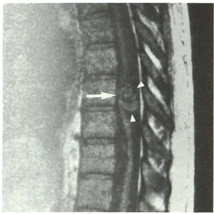
the high signal of a subacute subdural hematoma (Fig. 4). 4. Atrophy of the spinal cord (Fig. 5) was noticed in one case. Major scoliosis and syrinx cavity were observed in another patient.
Extramedullary A VMs
Extramedullary A VMs are a very rare type of spinal cord A VM [8). Clinical data are similar to those that characterize
intramedullary AVMs. In four cases, the extramedullary AVM
was diagnosed by angiography (angiotomomyelographic ex-amination). In two of these cases MR disclosed a previous erroneous angiographic diagnosis of extramedullary lesion by showing both intra- and extramedullary angiomatous
com-ponents. Sagittal T1-weighted images clearly demonstrated
a localized enlargement of the spinal cord with a central low signal corresponding to the intramedullary portion of the nidus. The extramedullary portion of the nidus was also well visualized in axial T1- and T2-weighted sequences (Fig. 6).
In the two other cases, the malformation was strictly
extra-medullary. On sagittal T1-weighted images, it was not
possi-ble to distinguish between low signal intensity due to CSF and flow-void phenomenon due to extramedullary vessels. The spinal cord was of normal size and appearance and demonstrated a normal signal at the level of the angiographic
abnormalities. Extramedullary vessels were only identified on
transverse T1- and T2-weighted images. For those two
pa-tients, the normal aspect of the spinal cord confirmed the diagnosis of strictly extramedullary AVM.
c
Fig. 1.-21-year-old woman with cervical intramedullary arteriovenous malformation.A, Sagittal T1-weighted (400/28) image shows characteristic MR findings of intramedullary vascular malformation: local enlargement and signal void are seen within spinal cord (arrow).
B, Corresponding selective vertebral angiogram: note the nidus (arrow).
[image:2.612.55.562.482.673.2]AJNR:9. September/October 1988 MR OF SPINAL CORD AVMs 835
Fig. 2.-Cervical intramedullary AVM in a 30-year-old man. Transverse T2-weighted (2000/120) image. Nidus within spinal cord appears as an area of low intensity with signal void images (arrow) and extramedullary vessels outlined well by high-intensity CSF (arrowhead).
Fig. S.-Sagittal T1-weighted (400/28) image shows upper thoracic intramedullary arteriovenous malformation (arrow) as-sociated with cervical spinal cord atrophy (arrowhead) in 28-year-old patient.
Fig. 3.-Cobb syndrome with tho-racic (T7) intramedullary arteriove-nous malformation (arrow) in 18-year-old boy. Sagittal T1-weighted (400/28) image shows that vertebral angioma involves T8 and T9 with huge low-intensity signal within ver -tebral body (arrowheads).
A
Fig. 4.-Thoracic intramedullary arteriove-nous malformation in 1S-year-old boy. Preme-dullary hyperintensity on sagittal T1-weighted (400/28) image corresponds to a chronic sub-dural hematoma (arrow).
B
Fig. G.-Extramedullary thoracic arteriovenous malformation in 30-year-old woman. The identification is difficult on sagittal T1-weighted (400/28) image behind the cord.
A, Slight area of low intensity is seen (arrow). Spinal cord signal and size are normal (arrowhead).
[image:3.615.51.557.89.310.2] [image:3.615.53.556.440.708.2]B36 DORMONT ET AL. AJNR:9. September/October 1988
Perimedullary Fistulae
Peri medullary fistulae fed by radiculomedullary arteries rep-resent another rare type of vascular malformation of the cord affecting young adults [9, 10]. MR was performed in three
cases. One of the patients presented with a microfistula of
the anterior spinal artery. The MR was normal. The two other
patients had a giant fistula of the conus. Huge signal-void
images corresponding to the aneurysmal dilatation of the
veins were well seen (Fig. 7).
Dural AVFs with Perimedullary Drainage
Dural AVFs with perimedullary drainage consist of a direct,
microscopic fistula between a dural branch of a radicular
artery and the perimedullary veins. The fistula is located between 03 and S2. Whatever the level of the fistula, the clinical result is a progressive myeloradiculopathy (often a conus medullaris syndrome) that occurs in patients who are
60 years old or older [11].
MR exploration of the conus medullaris was performed in seven patients. Sagittal T1-weighted images showed a slight enlargement of the conus (Fig. BA). Axial T1-weighted images
define this slight enlargement with a central area of
hypoin-tensity, but axial T2-weighted images were more informative. On these T2-weighted images the conus had a high signal in all cases. Careful observation also revealed subarachnoidal signal-void images that could be correlated with enlarged
perimedullary veins. After treatment, all patients improved
dramatically. In two of them a follow-up MR study showed a significant decrease of the pathologic signal of the conus. And in one patient a strictly normal MR appearance correlated well with the clinical cure (Fig. BD).
Discussion
Very few reports of the MR appearance of spinal cord
AVMs have been published in the literature. Six cases were reported in a study by Di Chiro et al. [1] in which MR was performed without a surface coil. MR was informative in three of four cases of intramedullary A VM, and the authors identified
the nidus as an intramedullary low signal surrounded by spinal
cord tissue. In two cases of dural AVF, the examination
focused on the level of the fistula and was not able to
demonstrate the lesion. A retrospective study showed
vas-cular signal void that could correspond to the venous drain-age.
Kulkarni et al. [2] examined a pregnant patient with spinal
hemorrhage using a O.S-T magnet with sagittal and coronal
slices (spin echo and inversion recovery). The results showed a vascular Signal void that probably corresponded to the feeding vessels.
Recently, Masaryk et al. [3] reported on signal abnormali-ties of the conus medullaris in three patients with dural AVF.
The purpose of our study was to test the value of MR as a
noninvasive procedure for diagnosing AVMs of the spinal
Fig. 7.-Perimedullary fistula above conus medullaris in a 38-year-old patient. Sagittal Tl-weighted (400/28) image. In thoracic area, spinal cord is displaced forward (arrow) by huge abnormal signal structures (arrowheads) corresponding to pos-terior venous drainage.
cord. Today, routine examination of spinal cord AVMs in-cludes myelography and arteriography. Myelography gives an indirectimage of enlarged vessels that are defined by selective
angiography. The combination of these examinations with
anteroposterior and lateral views demonstrates the
relation-ship between the nidus and the spinal cord. In our study, MR
diagnostic protocol (TE, TR, image planes) was determined
by the performance of our magnet.
Intramedullary A VMs
We confirmed the ability of MR to easily recognize
intra-medullary AVMs, or the intramedullary component of an
extra-medullary lesion. The association of spinal cord enlargement
with intramedullary signal changes corresponding to the nidus
was a constant and easily recognizable finding in every
se-quence. Vascular flow-void images do not permit
differentia-tion between arterial feeders and high-flow venous drainage because they produce the same signal. Postembolization aspects are difficult to analyze. We speculate that the disap-pearance of high-flow signal void and the signal changes within the nidus may correspond to intravascular embolic
material and/or consequent progressive thrombosis. Finally,
interesting information is also available on such complications as atrophy and chronic hematoma with the latter easily diag-nosed on sagittal T1-weighted images.
Extramedullary A VMs
In our experience these malformations were not identified
[image:4.613.338.548.91.301.2]AJNR:9, September/October 1988 MR OF SPINAL CORD AVMs 837
Fig. 8.-Dural arteriovenous malformation with peri medullary drainage in a 60-year-old pa-tient.
A, Sagittal T1-weighted (400/28) image shows that conus medullaris is slightly enlarged (ar· row).
B, Axial T1-weighted (400/28) image. Anterior venous drainage can be identified as areas of signal void (arrow).
C, Transverse T2-weighted (2000/120) image shows characteristic MR finding suggesting
chronic ischemia. Note anterior signal void cor-responding to perimedullary veins (arrow) and global hyperintensity of conus (arrowhead).
D, 3 months after treatment patient has im-proved clinically and axial T2-weighted (2000/
120) image shows significant decrease in abnor-mal signal (arrow) of conus medullaris.
angiographic examinations, and axial sequences were per-formed at the level of angiographic abnormalities. Therefore, it is difficult to assess the ability of MR as a diagnostic first step. It must be emphasized, however, that MR seems to be the best examination for defining the site of the nidus, which is important for indicating the course of therapy [12].
Dura/ AVFs
In dural AVFs, the MR slice thickness we had to use (6 mm) seemed to us inappropriate to show the microfistula directly. For this, thinner slices would be necessary. We therefore focused the MR exploration on the conus medul-laris, which appeared to be the site of clinical findings (path-ophysiological hypothesis suggested a chronic venous high pressure). Conus enlargement and intramedullary high signal were observed in all cases. These abnormalities are compat-ible with chronic venous ischemia as reported by Masaryk et al. [3]. Clinical improvement after successful endovascular or surgical treatment corresponded to total or partial regression of the MR abnormalities in the two patients reexamined.
Conclusions
MR is helpful in the diagnosis of AVMs involving the spinal cord as it provides accurate information as to the exact location of the nidus. As far as extramedullary malformations are concerned, improvement of the MR resolution is still necessary before MR can represent the first diagnostic step. Therefore, myelography might be used only in cases of neg-ative MR examinations. In any case, arteriography cannot yet be discarded as a method for studying the vascular architec-ture of the malformation.
REFERENCES
1. Di Chiro G, Doppman JL, Awyer AJ, et al. Tumors and arteriovenous
malformations of the spinal cord: assessment using MR. Radiology
1985;156: 689-697
2. Kulkarni MV, Burks DO, Price AC, et al. Diagnosis of spinal arteriovenous malformation in a pregnant patient by MR imaging. J Comput Assist Tomogr
1985;9: 171-173
3. Masaryk T J, Ross JS. Modic MT, Ruff RL, Selman WR, Ratcheson RA. Radiculomeningeal vascular malformations of the spine: MR imaging.
[image:5.612.222.555.87.451.2]838
DORMONT ET AL. AJNR:9, September/October 19884. Modic MT, Masaryk T J, Paushter D. Magnetic resonance imaging of the
spine. Radiol Clin North Am 1986;24:229-245
5. Djindjian R. Angiographie de la moelle epiniere. Paris: Masson Edit., 1970 6. Hurth M, Houdart R, Djindjian R, Rey A, Djindjian M. Arteriovenous
malformations of the spinal cord. Prog Neurol Surg 1978;9:238-266 7. Riche MG, Reizine D, Melki JP, Merland JJ .. Glassification of spinal cord
malformations. Radial Med 1985;3(1): 1, 17-24
8. Djindjian M, Djindjian R, Rey A, Hurth M, Houdart R. Intradural extramed-ullary spinal arteriovenous malformations fed by the anterior spinal artery.
Surg Neuro/1977;8:85-94
9. Gueguen B, Merland JJ, Riche MG, Rey A. Vascular malformation of the
spinal cord: intrathecal perimedullary arteriovenous fistulas fed by
medul-lary arteries. Neurology 1987;37: 969-979
10. Merland JJ, Riche MG, Ghiras J. Les fistules arterio-veineuses
intracana-laires extra-medulintracana-laires
a
drainage veineux m€ldullaire. J. Neuroradiol1980;7:271-320
11. Kendall BE, Logue V. Spinal epidural angiomatous malformations draining into intrathecal veins. Neuroradiology 1977;13: 181-189