Jay S. Tsuruda
1Ann Shimakawa
2Norbert J
.
Pelc
3David Saloner-4
Received August 27, 1990; revision requested November 13, 1990; final revision received Decem -ber 27, 1990; accepted December 27, 1990.
1 Department of Radiology, Division of Diagnos -tic and lnterventional Neuroradiology, Box 0628, University of California, San Francisco, 505 Pamas -sus Ave., San Francisco, CA 94143-0628. Address reprint requests to J. S. Tsuruda.
2 General Electric Medical Systems, Milwaukee,
Wl53188.
3 Department of Radiology, Stanford University,
Stanford, CA 94305.
'Department of Radiology, San Francisco Vet -erans Administration Medical Center, and University of California, San Francisco, San Francisco, CA 94143-0628.
0195-6108/91/1203-0481
<0 American Society of Neuroradiology
Dural Sinus Occlusion:
Evaluation
with Phase-Sensitive Gradient-Echo MR
Imaging
The purpose of this study was to evaluate the usefulness of limited-flip-angle, phase-sensitive velocity imaging with gradient-recalled-echo (VIGRE) MR when combined with spin-echo MR in the diagnosis of dural sinus thrombosis. The VIGRE sequence consists of a rapid single-slice acquisition, 50/15/2 (TR/TEfexcitations), and 30° flip angle. At each slice position, a total of four images were reconstructed; these consisted of one magnitude image and three images sensitive to proton motion in each orthogonal direction. The flow direction and flow velocity (emf sec) were obtained from each of the phase images, and results were correlated with data obtained from a phantom experi-ment. In normal controls, dural sinus velocities ranged from a mean of 9.9 to 14.4 emf
sec for the transverse and superior sagittal sinuses, respectively. Three patients with proved dural sinus occlusion were studied with spin-echo images at 1.5 T. Three-dimensional time-of-flight MR angiography was also performed in one patient. The presence of dural sinus occlusion was determined by the lack of flow void on the spin-echo images, the absence of phase shift on the VIGRE study, and the presence of retrograde flow on the phase image in the sinus proximal to the occluded segment. Time-of-flight angiography overestimated the extent of the thrombosis caused by spin saturation. Follow-up VIGRE studies detected the formation of collateral flow in one patient and recanalization with the establishment of normal antegrade sinus flow in the other.
We conclude that phase-sensitive MR imaging is helpful in establishing the diagnosis and extent of dural sinus occlusion. Because of the short acquitition time, selected images can be obtained whenever the flow within the dural sinus is in question on a routine spin-echo study. Retrograde sinus flow appears to be a sensitive indicator of distal sinus occlusion or high-grade obstruction
AJNR 12:481-488, May/June 1991; AJR 157: July 1991
Dural venous sinus thrombosis has been associated with several pathologic
conditions [1
].
Its clinical diagnosis may be difficult because of the wide variety of
nonspecific
findings
[2].
Earl
y
recognition of
this condition
may
be
necessary in
order
to institute prompt therapy [3]. In the
past, cerebral angiography or
venog-raphy was required to establish th
is diagnosis
.
Less
invasive techniques such as
CT may indicate the presence of dural sinus occlusion with the findings of the cord
and delta signs
, congested deep subcortical veins, or increased tentorial or gyral
enhancement
[4, 5].
These CT signs may be subtle and easily overlooked
.
In one
clinical seri
es on septic
lateral sinus thrombosis, the majority of
CT
examinations
were completely normal [6].
482
TSURUDA ET AL.
AJNR:12, MayfJune 1991Because of these potential pitfalls, the use of phase-shift
imaging with SE techniques [1 0-15]
,
which yields direct
measurements of blood velocity as well as flow direction, may
contribute additional MR specificity. More
recently,
prelimi-nary experience with rapid phase-sensitive, limited-flip-angle
gradient-recalled-echo pulse sequences has been described
as a method for evaluating portal hypertension, discriminating
arterial
from venous flow as well as clot from slow flow
,
and
quantifying blood flow within the carotid arteries [16, 17]. Our
experience
with the use of this method in proved cases of
dural sinus
occlusion is described.
Materials, Subjects, and Methods
A velocity-sensitive technique based on phase contrast and gra-dient-recalled echoes, VIGRE (velocity imaging with gragra-dient-recalled echoes, General Electric Medical Systems), was initially tested with a flow phantom. The theory behind phase-shift velocity imaging and methods of image reconstruction using this technique are described in greater detail elsewhere [17]. Briefly, in the most basic mode, VIGRE requires two separate gradient schemes resulting in two views that are acquired for each phase-encoding increment. The first view is a flow-compensated sequence that results in zero phase shift for both stationary spins and spins moving with constant velocity during TE. This first view is the equivalent of a conventional GRASS study, and the data can be used to reconstruct a magnitude image (Fig. 1 A). The second view is a flow-encoded sequence that uses a slightly altered gradient waveform such that the constant-velocity spins ac-cumulate a phase shift related to that component of the flow vector that lies along the flow-encoding gradient axis. Again, the stationary spins do not accumulate any phase shifts. The TR and TE are kept constant for both views. From the data acquired with both views, a single-phase image is constructed such that the degree of phase shift is directly related to the flow velocity at each voxel (Fig. 1 B). The flow sensitivity for this single-phase image is limited to the vector com-ponent parallel to the flow-encoded gradient, which can be selected to lie along any one of the three orthogonal directions. Stationary voxels in the phase image correspond to the special case of zero velocity and appear as intermediate (gray) intensity, representing 1 024-magnitude units. Spatial resolution of the phase-reconstructed image is similar to that of a standard MR image.
A
B
In our clinical examples, the basic VIGRE acquisition described above is modified such that the flow-compensated and flow-encoded view is repeated three times in an interleaved fashion during each phase-encoding increment as a single data set. The purpose for this more complex set is to acquire flow information along all three orthogonal directions corresponding to the frequency, phase, and slice-select axes. As a result, the imaging time is increased by a factor of three. The rationale for obtaining separate velocity measure-ments in all three axes during a single imaging session is to ensure that velocities in vessels that are obliquely oriented to the plane of section can be analyzed. The sequences are interleaved to minimize motion between flow encodings. The resulting data are then com-bined to produce four separate images: a conventional-magnitude or GRASS image, a flow image in which pixel intensity is related to the flow velocity along the frequency-encoding direction, a flow image in which pixel intensity is related to the flow velocity in the phase-encoded direction, and a flow image in which pixel intensity is related to flow velocity along the slice-select direction. The imaging param-eters include 35/15/2 (TRfTEjexcitations), 30° flip angle, 4-mm slice thickness, 24-cm field of view (FOV), and 192 x 256 matrix. Two excitations are performed in order to improve the signal-to-noise ratio. Because of the short TR, data acquisition for a complete single slice study is 55 sec. Multiple single-slice images can also be pre-scribed depending on the area of coverage required.
Phase-sensitive techniques are sensitive to aliasing artifact if the actual velocity within the vessel is greater than the expected velocity range. This artifact results in a misrepresentation of flow such that the flow direction and actual velocity are displayed incorrectly. To avoid this problem, one can specify a peak velocity that controls the flow-encoding gradient lobes. In the flow-encoded experiment, the gradient lobes are designed so that a spin moving at the specified peak velocity accumulates a phase shift of +pi. A spin moving in the opposite direction at the specified peak velocity would accumulate a phase shift of -pi. This peak velocity specifies the maximum velocity that can be resolved without aliasing. On the basis of preliminary work (Mattie H, et al. Presented at the annual meeting of the Radio-logical Society of North America, November 1989), the peak velocity for our patients was set at 50 cmjsec for the analysis of dural sinus velocity. Because the sign of the phase shift is recorded, flow directionality is displayed by either relative hypointensity or hyperin-tensity with respect to the background stationary spins set at 1 024-intensity units, which is midscale on the phase-reconstructed image. The magnitude of the difference between the intensity of the moving
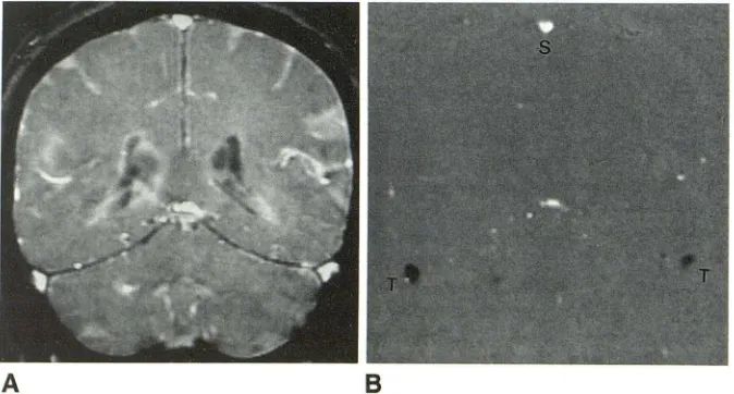
Fig. 1.-Example of VIGRE images of a nor-mal volunteer with acquisition parameters as described in the text.
A, Conventional-magnitude or GRASS image through posterior aspect of cranium in coronal plane. Hyperintensity within superior sagittal and transverse sinuses is due in part to flow-related enhancement.
[image:2.612.53.390.551.732.2]and stationary spins will allow a direct calculation of the velocity within the dural sinus.
VIGRE Calibration and Normal Controls
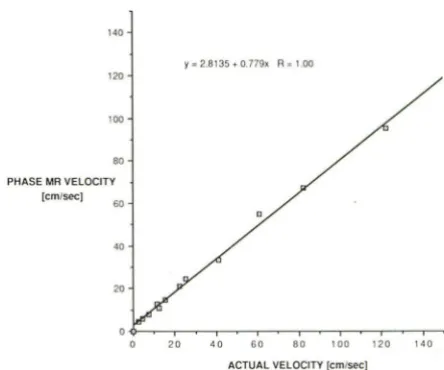
An in vitro phantom was constructed with two parallel Plexiglas
tubes with 3-and 7 -mm inner diameters placed in a water bath filled
with tap water. Tap water circulated through the two tubes by means
of a nonpulsatile gravity-fed device with mean and peak velocities calculated from a timed collection in a graduated cylinder. Actual
velocities ranged from 2.3 to 122 cmjsec. An assumption was made
that laminar flow was present in the phantom. Appropriate adjust-ments in the peak velocity settings were made for the in vitro VIGRE
studies. VIGRE imaging was performed with the long axis of the tube
oriented perpendicular to the imaging plane. A mean velocity was calculated from the phase image by specifying a region of interest
across the entire cross section of the tube and measuring the mean
voxel intensity. A peak velocity was also obtained by measuring the central voxel with the greatest deviation from 1 024.
Direct coronal images were obtained perpendicular to the superior and transverse sinuses in normal volunteers (n = 5; mean age, 30). On the phase image, a region of interest was drawn to include the entire cross section of the dural sinus, and a mean intensity and velocity were obtained.
Studies in Patients
Over a 6-month period, we had the opportunity to use the VIGRE
technique with a 1.5-T imager (General Electric Signa Performance Plus, Milwaukee, WI) to examine three inpatients proved to have
dural sinus occlusion. All patients were initially imaged with a
multi-slice T1-weighted sagittal sequence, SE 600/20/2, with 5-mm-thick slices, a 1-mm interslice gap, a 22-cm FOV, and a 192 x 256 matrix. This was followed by a multislice, multiecho T2-weighted sequence, 2800/30,80/1, with 5-mm-thick slices, a 2.5-mm interslice gap, a 20-cm FOV, and a 192 x 256 matrix. Gradient-moment nulling (Flow Compensation, General Electric Medical Systems) was used on the
long TR images. Additional coronal and axial T1-weighted images were obtained in selected cases for further anatomic detail.
After completion of the SE acquisitions, the major dural sinuses were viewed on the monitor for the possible use of VIGRE images.
The criteria for their use in these patients included atypical flow
features in the dural sinus, such as the lack of flow void; the presence of subacute thrombus; replacement or compression of the dural sinus; the lack of normal rephasing with the application of gradient-moment
nulling; and poor visualization of the dural sinus. A midline sagittal plane for the VIGRE study was selected to evaluate the entire course of the superior sagittal sinus, straight sinus, and torcular Herophili. The coronal plane through the posterior fossa was selected to evaluate the transverse and superior sagittal sinuses. An axial VIGRE
image was used to evaluate the distal superior sagittal sinus, torcular
Herophili, jugular bulb, and occasionally the transverse sinus if this
vessel was parallel to the axial plane of section. Usually, one to four single-slice VIGRE images were obtained during a total acquisition time ranging from 55 sec to 3:40 min.
One patient was also studied with time-of-flight MR angiography [18]. This sequence consisted of a flow-compensated, three-
dimen-sional, spoiled gradient-echo sequence (50/7 /1/15° flip angle) con-sisting of 64 0.9-mm-thick partitions, a 23-cm FOV, and a 256 x 256 matrix. IV gadopentetate dimeglumine was not administered. The
partitions were oriented in the axial plane. Selective maximum- inten-sity-projection reconstructions were performed through the dural sinuses.
140
120
100
80
PHASE MR VELOCITY
(em/sec)
60
40
20
y"' 2.8135 + 0.779x A"' 1.00
o~~.-~.-~.-~-.-.-.-.-.~-.~
0 20 40 60 80 100 120 140
ACTUAL VELOCITY [em/sec]
Fig. 2.-ln vitro comparison between actual and observed flow velocities with VIGRE sequence. Because of velocity range tested, peak velocity was set higher than velocity range used in clinical imaging.
Results
Phantom Experiments and Normal Controls
The MR velocity data obtained from the phantom
experi-ment was compared with the actual flow velocities (Fig. 2). A
nearly linear relationship was
noted
between actual and
meas-ured velocities. In the normal controls, the calculated mean
velocity was 14.4 cmjsec
(range
,
10.6-17.5 cmjsec) in the
superior sagittal sinus and 9.9 cmjsec (range
, 2.7-14.2
emf
sec) in the transverse sinus.
The greater range of velocities
in the transverse sinuses was due to developmental
asym-metry in one patient. In all normal controls, anterior to
poste-rior flow was
demonstrated in
the superior sagittal
sinus and
posterior to anterior flow in the transverse sinus
.
Studies in Patients
The clinical
information
and imaging
findings in the three
patients are summarized in Table 1.
Representative
SE and
VIGRE images from the
three
patients are depicted in Figures
3-5
.
Discussion
MR imaging has become the noninvasive method of
choice
in the evaluation of dural
sinus
occlusion. Characteristi
cally
,
direct visualization of
thrombus within
the
sinus and
loss
of
normal
flow
void
on
SE
imaging
have
been reported
i
n clinical
examples (1
, 5, 8, 9, 19, 20]
. Initially,
acute
thrombosis
may
appear as a
region
of isointensity
relative to normal brain
parenchyma on T1-weighted
images (Fig
.
48)
,
becoming
pro-gressively hyperintense
corresponding to the formation o
f
methemoglobin
(Fig.
3E)
(20]
.
In
acute thrombosis
,
t
he use
of T2-weighted images
a
lone
may be potentially misleading
[image:3.615.324.546.77.262.2]im-484
TSURUDA ET AL.
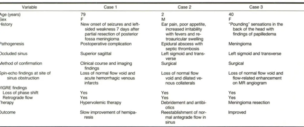
AJNR:12, MayfJune 1991TABLE 1: Summary of Patients with Dural Sinus Occlusions Studied by Velocity Imaging with Gradient-Recalled Echoes (VIGRE)
Variable Case 1
Age (years) 79
Sex F
History New onset of seizures and
left-sided weakness 7 days after
partial resection of posterior
fossa meningioma
Pathogenesis Postoperative complication
Occluded sinus Superior sagittal
Method of confirmation Clinical course and imaging findings
Spin-echo findings at site of Loss of normal flow void and
sinus obstruction acute hemorrhagic venous
infarcts
VIGRE findings
Loss of phase shift Yes
Retrograde flow Yes
Therapy Hypervolemic therapy
Outcome Slow improvement of hemipa
-res is
aging at high field strengths
,
can produce hypointense signal
mimicking flow void (20
,
21 )
.
The absence of flow void on SE
imaging may not necessarily represent thrombosis
,
since the
lack of flow void may be confused with flow-related
enhance-ment or rephasing of spins caused by even-echo
rephasing
andjor gradient-moment nulling [5
,
15]
.
As a result
,
the
sus-pected area should
be evaluated in
multiple planes with both
long and short TR
SE
techniques (1).
The potential pitfalls of SE imaging can be partially
over-come
by the use of enhancement with gadopentetate dime
-glumine
,
which may show nonenhancing thrombus within
enhancing
dural sinus
as well as improved delineation of
venous collaterals [1). One should also be aware that contrast
enhancement in the
presence of slow-flow states
may
lead
to some pitfalls
in
interpretation (Boyko 0
.
Presented at the
annual
meeting of the Radiolog
i
cal Society of North America,
November 1990).
RF presaturation pulses (22) also may aid
in identifying flow voids without
the presence of flow-related
enhancement
,
which on occasion may mimic thrombus.
More
rece
ntly
,
single-slice
gradient-recalled-echo MR has proved
t
o be
valuable in
the assessment of sinus patency [7).
Reli-ance on gradient-echo techniques alone may be misleading
,
since hyperintense
th
rombus may mimic
flow
(23
,
24
).
In
addition
,
the
presence of paradoxical enhancement
is
not a
quantitative measurement of flow,
and
the min
im
·
um flow rate
r
equ
ire
d to
yield hyperintensity on either SE or gradient-echo
images
needs
f
urther clinical
evaluation (7
,
1
OJ.
Phase-contrast MR angiography has been anecdotally
shown to be helpful in detecting dural sinus
occlusion
(25).
However
,
the authors
in
this report did not specify the velocity
sensitiv
it
y with their technique
.
In add
iti
on
,
their study did not
supply any
information
about flow directionality
.
In one of our
cases
,
three-dimensio
na
l
time-of-flight MR angiography
con-firmed
sinus occlusion
by a
meningioma
.
However,
t
he
MR
angiography
results
were somewhat misleading
in
that the
prox
ima
l
left transverse
sinus
appeared
to be completely
occluded
.
This contradicted
the
find
ing
on both the
VIGRE
and conventional angiograp
h
ic
studies
,
which showed a pat
-Case 2 Case 3
2 40
M F
Ear pain, poor appetite, "Pounding" sensations in the increased irritability back of the head with
with fevers and re- findings of papilledema
troauricular swelling
Epidural abscess with Meningioma
septic thrombosis
Left sigmoid and trans- Left sigmoid and transverse
verse
Surgical Surgical
Loss of normal flow Loss of normal flow void and
void and dilated ve- flow-related enhancement
nous collaterals on MR angiogram
Yes Yes
Yes Yes
Debridement and antibi- Meningioma resection
otics
Reestablishment of nor- Improved
mal antegrade flow in
sinus
ent proximal transverse sinus and retrograde filling
toward
the contralateral side. This misleading finding on MR
angiog-raphy is due
to
the saturation of spins
in
this dural sinus
secondary to the relatively long dwell time within the imaging
volume
.
A similar saturati
o
n effect was noted in the right
lateral sinus distal to partial obstruction by an arachn
o
id
granulation, in which faint flow-related enhancement of the
right
sigmoid sinus and jugular bulb was noted on MR
an-giography (Fig
.
58)
.
The VIGRE study (Fig. 50), however
,
showed high flow velocities in this saturated region
.
The use
of IV gadopentetate dimeglumine may
improve
MR time-of
-flight venoangiography (Okumura R
,
et al. Paper presented
at the annual meeting of the Radiological Society of North
America
,
November 1990)
,
and if it had been used in this
patient
,
the dural sinuses with poor flow-related enhancement
would have been visualized better
.
However
,
we elected not
to use gadopentetate dimeglumine because of the concern
that the
densely enhancing portions of a meningioma within
the
occluded sinus may simulate flow-related enhancement
on time-of-flight angiography
,
especially when the results are
viewed with the use of a maximum-intensity-projection rec
o
n-struction algorithm [26).
A strong
argument could be made that the
parameters we
used for our time-of-flight MR angiography are sensitive only
for fast flow
in arteries and major dural sinuses
.
In our
e
x
perience
,
the major dural sinuses in normal subjects are
adequately visualized with these parameter
s
.
This sequence,
however
,
is very sensitive to reduced flow states
,
and
there-fore
is
a sensitive indicator of altered flow patterns such as in
this
patient.
Multislice two-dimensional
time-of
-
flight MR an
-giography
is
more
immune
to saturation effects
.
Unfortu-nately
,
this sequence was not available to us when this patient
(case
3) was studied
.
However
,
the
VIGRE
technique
does
produce a single-slice two-dimensional magnitude
i
mage that
can be used to evaluate the presence of flow-related enhance
-men
t.
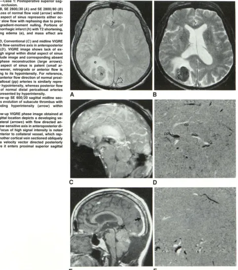
[image:4.615.59.562.89.300.2]Fig. 3.-Case 1: Postoperative superior sag-ittal sinus occlusion.
A and 8, SE 2800/30 (A) and SE 2800/80 (8)
images. Loss of normal flow void (arrow) within posterior aspect of sinus represents either oc-clusion or slow flow with rephasing due to pres
-ence of gradient-moment nulling. Portions of acute hemorrhagic infarct (h) with T2 shortening,
surrounding edema (e), and mass effect are noted.
C and D, Conventional (C) and midline VIGRE
image with flow-sensitive axis in anteroposterior
direction (D). VIGRE image shows lack of ex-pected high signal within distal aspect of sinus on magnitude image and corresponding absent
flow on phase reconstruction (large arrows).
Proximal aspect of sinus is patent (small ar
-rows); however, retrograde or anterior flow is seen owing to its hypointensity. For reference, note that anterior flow direction of normal proxi-mal pericallosal (pp) arteries is similarly repre-sented by hypointensity, whereas posterior flow direction of normal distal pericallosal arteries
( dp) is represented by hyperintensity.
E, Follow-up SE 600/20 sagittal midline sec-tion shows evolusec-tion of subacute thrombus with
corresponding hyperintensity (arrow) within
sinus.
F, Follow-up VIGRE phase image obtained at
a parasagittal location depicts a developing
ve-nous collateral (arrows) with flow directed an-teriorly (flow-sensitive axis in anteroposterior
di-rection). Focus of high signal intensity is noted
directly anterior to collateral vessel, which
rep-resents another cortical vein sectioned obliquely
that has a velocity vector directed posteriorly just before it enters proximal superior sagittal sinus.
A
c
E
relative
phase of the transverse magnetization
and the flow
velocity in
the direction of
the
magnetic fie
l
d
gradient
[11-13).
One
method
of
extracting t
his
inf
o
rm
a
tion
is
to introduce
bipol
ar
(dephasing
and rephas
ing)
gradient pulses during an
ima
ging sequence
[1
OJ.
If
the
protons
are stationary, there is
no
net change
in phase
.
If
there is net motion of protons
,
t
here will
not
be complete rephasing resulting
in
a net phase
s
hift
proportional to velocity
.
If
the
applied
field gradient
i
s
kn
own
,
a
di
re
ct
velocity
measurement
can be estimated [14)
.
B
D
F
The VIGRE
technique used in this
study is
based
on these
principles
[17).
Because of the use of a short TR and g
radi
ent-recalled echoes
,
data acquisition
is reasonab
ly
fast and can
be added to a normal
imaging sequence if the SE study is
equivocal
,
as was described
in
the Materials
,
Subjects
,
and
Method
s
section
.
In our
clin
ical
examples
,
only a
limited
number of VIGRE images were required to evaluate the
sinus
in question without appreciably increasing the
imaging time
.
[image:5.617.86.569.78.625.2]-486
TSURUDA ET AL.
AJNR:12, MayfJune 1991D
E
F
G
ion
along
all
three
orthogonal
imag
ing
p
lanes
,
which we found
helpful in analyzing
obliquely oriented vessels.
I
f
necessary,
velocity
analysis
in these
obliquely oriented
vessels
can
be
performed by vector
addition
(12].
Ove
ra
ll
,
the
VIGRE images
allowed high
contrast and conspicuity
in
identifying flowing
spins on
the clinical studies
.
The
phantom
study showed
reasonable
correspondence
between measured and
actual
velocities in the
useful
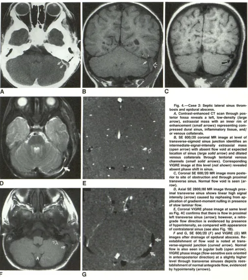
diag-Fig. 4.-Case 2: Septic lateral sinus throm-bosis and epidural abscess.
A, Contrast-enhanced CT scan through
pos-terior fossa reveals a left, low-density (large arrow), extraaxial mass with an inner rim of enhancement (small arrows) representing com-pressed dural sinus, inflammatory tissue, and/ or venous collaterals.
B, SE 600/20 coronal MR image at level of transverse-sigmoid sinus junction identifies an intermediate-signal-intensity extraaxial mass (open arr?w) with absent flow void at expected location of sinus (large solid arrow) and dilated venous collaterals through tentorial venous channels (small solid arrows). Corresponding VIGRE image at this level (not shown) revealed absent phase shift in sinus.
C, Coronal SE 600/20 MR image more poste-rior to site of obstruction and through proximal transverse sinus. Normal flow void is seen (ar
-row).
D, Axial SE 2800/80 MR image through prox-imal transverse sinus shows linear high signal intensity (arrow) caused by rephasing from ap-plication of gradient-moment nulling in presence of slow laminar flow.
E, Coronal VIGRE phase image at same level as Fig. 4C confirms that there is flow in proximal left transverse sinus (arrow); however, a retro-grade flow direction is evidenced by presence of hyperintensity, as compared with appearance of contralateral sinus (see also Fig. 18).
F and G, SE 600/20 (F) and VIGRE (G) MR images after drainage of epidural abscess. Re-establishment of flow void is noted at trans-verse-sigmoid junction (curved arrow). Normal flow is also seen in jugular bulb (open arrow). VIGRE phase image (flow-sensitive axis oriented in anteroposterior direction) at a slightly higher level through transverse sinuses depicts rees-tablishment of normal antegrade flow, evidenced by hypointensity (arrows).
nostic
range for dural
sinuses
.
The
known limitations
of
VIGRE, including gradient
imperfections
,
eddy currents
,
sen-s
itivi
ty
as a
f
unction of
the
peak
velocity settings,
resolution
,
and the distribution
of velocities
within the
im
aging voxel, may
account for the
slight discrepancies between
the
actual vs
measured velocities
(17].
Simi
l
ar
calibration curves
using
phase mapping have been
shown by other
investigators
[1
6].
[image:6.612.55.560.74.645.2]A
B
c
D
E
Fig. 5.-Case 3: Left posterior fossa meningioma with sinus occlusion.
A, Axial SE 600/20 MR image shows isointense mass (M) involving left transverse-sigmoid sinus junction. There is definite flow void in right transverse sinus (solid arrow) and possible flow void in left proximal transverse sinus (open arrow).
8, MR angiogram, from an axially oriented maximum-intensity projection, shows complete absence of left transverse sinus (small straight solid arrows). Unexpected findings are noted on right. A patent right proximal transverse sinus (large straight solid arrow) is seen, along with marked narrowing (open arrow) and incomplete flow-related enhancement distally in sigmoid sinus and jugular bulb (curved arrows).
C, Coronal VIGRE study through mid transverse sinuses shows retrograde flow (hyperintense signal) in left transverse sinus (open arrow) proximal to
occlusion and faint antegrade flow (hypointense signal) at level of right transverse sinus stricture (solid arrow).
D, Coronal VIGRE image at level of sigmoid sinus, with flow-encoding axis in superoinferior direction, depicts normal inferior flow (hypointense signal)
on right (arrow). A similar finding is expected normally on left; however, in this case there is proximal obstruction by tumor. Venous phase from a posterior fossa arteriogram (not shown) revealed identical flow patterns in dural sinuses.
E, Lateral projection, retrograde right transverse sinus venogram, confirms a segmental narrowing and filling defect (arrow) seen initially on MR angiography and presumably caused by arachnoid granulation. Pressure measurement across this stricture indicated a moderate gradient of 10 mm Hg. It was postulated that complete occlusion of left transverse sinus by a meningioma as well as partial obstruction on the right resulted in venous hypertension leading to the finding of papilledema.
that
a range of velocities can be seen within the sinuses,
particularly in
cases
of developmental asymmetry. Because
of
this range, any single measurement of
the
absolute velocity
of a dural sinus
in
a given patient may be difficult to interpret.
Measurement
of the actual flow through the
sinuses
has been
proposed
as an estimate of cerebral perfusion
(Mattie et
al.
RSNA, November 1989)
.
More work in
this
area needs
to be
performed.
In our series, the presence of dural
sinus
occlusion was
determined
on the basis of
several
imaging criteria. First
,
in
all
instances the normal flow void was not
seen
on
the
SE
i
mages. Second
,
both the magnitude
and
phase-reconstruc-tion
VIGRE
images
demonstrated
diminished
or
absent flow
at the point of obstruction owing to the lack of
flow-related
enhancement
or
phase
shift
,
respectively. Third
,
if
patency
is
seen in the proximal
segment of the
distally obstructed
sinus
,
retrograde flow
was noted.
This latter
finding makes
physio-logic
sense
,
since
normal
blood flow
is
expected to be shunted
to other collaterals
in
the
case
of
distal obstruction
.
With
further
clinical experience
,
this retrograde sign may prove to
be quite
specific
in
the
diagnosis
of sinus occlusion or
high
-grade obstruction
.
Several assumptions are required for
VIGRE
imaging to be
applicable
in
dural
sinus thrombosis
.
First
,
the absence of
phase
shift alone
is insufficient
evidence
for
complete occlu
[image:7.612.55.557.76.451.2]488
TSURUDA ET AL.
AJNR:12, MayfJune 1991sec) has not been
tested by us or others [17]. The finding of
proximal
retrograde
flow
,
therefore
,
is used as an indirect
indicator of a hemodynamically significant lesion due to either
high-grade
stenosis
or obstruction. Second
,
an assumption
was made that dural sinus velocity is nonpulsatile
,
and
there-fore a
gated acquisition was not obtained
.
We based this
assumption
on other experimental results with a
bolus-tag-ging technique
,
which demonstrated no evidence of significant
pulsatile motion due
to
the lack of dispersion of the bolus
boundaries (unpublished
results) when imaging the superior
sagittal sinus. Even
if significant pulsations exist within the
sinus,
VIGRE velocity measurements
remain
reasonably
ac-curate [17].
Third
,
the
retrograde
flow seen in the proximal
sinus represents
true reversal of flow and not an aliasing
artifact. This flow reversal was proved angiographically in one
patient. If an
aliasing artifact
were present in the other two
patients
,
the flow velocities would have been uniformly greater
than
the peak velocity of 50 cmjsec
.
It would be difficult to
explain this on
a physiological
basis
.
Also
,
in our experience,
an
aliasing
artifact is usually depicted as nonuniform and
variable signal
intensity
within the vessel. This was not the
case in
the sinuses demonstrating retrograde flow
.
Overall
,
owing
to
its quantitative nature, phase imaging
adds improved diagnostic confidence in the diagnosis of dural
sinus
occlusion
,
since the
potential pitfalls encountered in
trying to
distinguish between the loss of flow void due
to
physiological MR phenomena andjor slow flow from actual
occlusion can be reduced. An improvement in study specificity
leading
to an
earlier diagnosis may affect patient
manage-ment,
such as
with
the
institution
of heparin or urokinase
therapy [3] or prompt surgical treatment including
mastoidec-tomy, exposure
of
the
sinus
,
or
incision
and drainage in
the
case of septic lateral sinus thrombosis [27]
.
In summary
,
phase-sensitive limited-flip-angle
gradient-re-called imaging
is
an accurate
and useful adjunct to SE MR
imaging and three-dimensional time-of-flight MR angiography
in
the noninvasive
diagnosis of dural sinus thrombosis,
con-firming the development of
collateral
flow,
and determining
the results of therapy. Retrograde flow in
the proximal sinus
caused by distal
obstruction
can
be
readily
detected and
appears to be a specific indicator of sinus occlusion or
signif-icant
obstruction to antegrade
flow
.
Further work needs
to
be performed to determine the
sensit
i
vity of VIGRE imaging
in
states of very slow flow
.
Quantitative phase imaging allows
insight
into the pathophysiological changes of venoocclusive
disease and shows promise in the further evaluation of normal
and abnormal flow conditions of both intracranial arteries and
veins
.
ACKNOWLEDGMENTS
We thank Robert K. Jackler, Mark L. Rosenblum, and Charles B.
Wilson for providing the cases used for this study and Petra
Schmal-brack for providing the MR angiography software.
REFERENCES
1. Harris T, Smith R, Koch K. Gadolinium-DTPA enhanced MR imaging of septic dural sinus thrombosis. J Comput Assist Tomogr 1989;13:682-684
2. Bousser M, Chiras J, Bories J, Castaigne P. Cerebral venous thrombosis-a review of 38 cases. Stroke 1985;16:199-213
3. Di Rocco C, Iannelli A, Leone G, Moschini M, Valori V. Heparin-urokinase
treatment in aseptic dural sinus thrombosis. Arch Neural 1981;38: 431-435
4. Anderson S, Shah C, Murtagh F. Congested deep subcortical veins as a sign of dural venous thrombosis: MR and CT correlations. J Comput Assist
Tomogr 1987;11: 1059-1061
5. Hulcelle P, Dooms G, Mathurin P, Cornelis G. MRI assessment of unsu
s-pected dural sinus thrombosis. Neuroradiology 1989;31 :217-221
6. Samuel J, Fernandes C. Lateral sinus thrombosis (a review of 45 cases).
J Laryngol Oto11987;101: 1227-1229
7. Daniels D, Czervionke L, Hendrix L, et al. Gradient recalled echo MR imaging of superior sagittal sinus occlusion. Neuroradiology 1989;31:
134-136
8. Baram T, Butler I, Nelson MJ, McArdle C. Transverse sinus thrombosis in newborns: clinical and magnetic resonance imaging findings. Ann Neural
1988;24: 792-794
9. Savino P, Grossman R, Schatz N, Sergott R, Bosley T. High-field magnetic
resonance imaging in the diagnosis of cavernous sinus thrombosis. Arch
Neurol1986;43: 1081-1082
10. Ridgway J, Smith M. A technique for velocity imaging using magnetic resonance imaging. Br J Radiol1986;59:603-607
11. Wedeen V, Rosen B, Chesler D, Brady T. MR velocity imaging by phase
display. J Comput Assist Tomogr 1985;9:530-536
12. Feinberg D, Crooks L, Sheldon P, Hoenninger J, Watts J, Arakawa M. Magnetic resonance imaging of the velocity vector components of fluid flow. Magn Reson Med 1985;2:555-566
13. Axel L. Blood flow effects in magnetic resonance imaging. AJR
1984;143: 1157-1166
14. Altobelli S, Caprihan A, Davis J, Fukushima E. Rapid average-flow velocity
measurement by MR. Magn Reson Med 1986;3:317-320
15. Tavares N, Auffermann W, Brown J, Gilbert T, Sommerhoff C, Higgins C.
Detection of thrombus by using phase-image MR scans: ROC curve analysis. AJR 1989;153:173-178
16. Bendel P, Buonocore E, Bockisch A, Besozzi M. Blood flow in the carotid
arteries: quantification by using phase-sensitive MR imaging. AJR
1989;152: 1307-1310
17. Spritzer C, Pelc N, Lee J, Evans A, Sostman H, Riederer S. Rapid MR imaging of blood flow with a phase-sensitive, limited-flip-angle, gradient
recalled pulse sequence: preliminary experience. Radiology
1990;176:255-262
18. Schmalbrock P, Yuan C, Chakeres D, Kohli J, Pelc N. Volume MR angiog-raphy: methods to achieve very short echo times. Radiology
1990;175:861-865
19. Bauer W, Einhaupl K, Heywang S, Vogl T, Seiderer M, Clados D. MR of venous sinus thrombosis: a case report. AJNR 1987;8:713-715 20. Macchi P, Grossman R, Gomori J, Goldberg H, Zimmerman R, Bilaniuk L.
High field MR imaging of cerebral venous thrombosis. J Comput Assist Tomogr 1986;10:10-15
21. McMurdo S, Brant-Zawadzki M, Bradley W, Chang G, Berg B. Dural sinus thrombosis: study using intermediate field strength MR imaging. Radiology
1986;161 :83-86
22. Edelman R, Wentz K, Mattie H, et al. Intracerebral arteriovenous malfor -mations: evaluation with selective MR angiography and venography. Ra-diology 1989;173:831-837
23. Tsuruda J, Halbach V, Higashida R, Mark A, Hieshima G, Norman D. MR evaluation of large intracranial aneurysms using cine low flip angle gradient-refocused imaging. AJNR 1988;9: 415-424
24. Yousem D, Balakrishnan J, Debrun G, Bryan R. Hyperintense thrombus
on GRASS MR images: potential pitfall in flow evaluation. AJNR
1990;11 :51-58
25. Rippe D, Boyko 0, Spritzer C, Meisler W, Dumoulin C, Souza S. Demon-stration of dural sinus occlusion by the use of MR angiography. AJNR
1990;11: 199-201
26. Anderson C, Saloner D, Tsuruda J, Shapeero L, Lee R. Pictorial essay. Artifacts in maximum-intensity-projection display of MR angiograms. AJR
1990;154:623-630