THE EFFECTS OF TEMPERATURE AND pH ON THE
CONTRACTILE PROPERTIES OF SKINNED MUSCLE
FIBRES FROM THE TERRAPIN, PSEUDEMYS SCRIPTA
ELEGANS
BY GABRIEL MUTUNGI AND IAN A. JOHNSTON
Gatty Marine Laboratory, Department of Physiology and Pharmacology, University of St Andrews, St Andrews, Fife, KY16 8LB, Scotland
Accepted 11 November 1986
SUMMARY
1. Fibre types in the iliofibularis muscle of the freshwater terrapin Pseudemys
scripta elegans have been characterized on the basis of their histochemical
characteristics, nerve endings and contractile properties. Three types of focally innervated fibres are present, corresponding to the fast glycolytic (Fg), fast oxidative glycolytic (FOG) and slow oxidative (SO) fibre types of other vertebrates.
2. Single fibres or small bundles of fibres representing each histochemical type were identified on the basis of their light scattering properties under dark-field illumination. Fibres were detergent-skinned using Brij 58, and their maximum isometric tension (Po) and unloaded contraction velocity (Vo) were determined by the slack test method. At 15 °C, fast glycolytic fibres generated maximum isometric tensions of 184±5kNm~2 and Vo values of 5-5 ± 0-3 muscle lengths per second (Los~'). Slow oxidative fibres produced tensions of 70-6±3kNm~2 and had Vo values of 1-3 Los~'. Tensions and Vo values of fast oxidative glycolytic fibres were between those of Fg and SO fibres.
3. The force-velocity (P—V) characteristics of slow oxidative fibres were studied at 5° and 15°C. Points below 0'6Po on the curves could be fitted by a linear form of Hill's equation. Maximum contraction velocities (Vmax) extrapolated from the P-V relationship were 0'62Los~' at 5°C and 0-91 Los~' at 15°C. The curvature of the P—V relationship was relatively independent of temperature over the range 5 to 15°C. Values for Hill's constant a/P0 were 0-29 and 0-33 at 5°C and 15°C,
respectively.
4. The temperature dependence of Po and contraction velocity at near zero load (Vj) were studied at constant pH, and under conditions designed to simulate the changes in intracellular pH which occur with temperature in vivo (ApH/AT = —0-0186). Changes in pH in the range 6-6 to 7-8 had no effect on either tension or V, at temperatures between 0° and 20°C. However, below and above this pH range, both tension and V, were depressed.
5. It is concluded that pH changes within the normal physiological range (6*7—7-8) have no effect on the temperature dependence of Po and V;.
88 G. MUTUNGI AND I. A. JOHNSTON INTRODUCTION
The freshwater terrapin Psendemys scripta elegans remains active between 12° and 40°C, but has a preferred body temperature (PBT) of 30°C (Gatten, 1975). At this temperature, plasma and skeletal muscle pH are about 7-5 and 6-8, respectively (Malan, Wilson & Reeves, 1976). As body temperature rises, there is an increase in respiratory minute volume, oxygen consumption and carbon dioxide excretion, resulting in a decrease in plasma pH, equivalent to about 0-020pHunits/°C (Jackson, 1971). Similar changes occur in other tissues; for example, muscle pH increases from around 6-8 at 35°C to 7-5 at 5°C (Malan et al. 1976). It has been suggested that ApH/AT changes of this magnitude serve to maintain either a constant relative alkalinity ([OH~]/[H+]) or a constant charge state of the histidine groups of proteins (alphastat hypothesis, Reeves, 1972).
There is a large literature on the effects of temperature on the contractile properties of muscle in lower vertebrates (Hill, 1938; Renaud & Stevens, 1984; Johnston & Brill, 1984; Johnston & Gleeson, 1984). There is evidence that different species and classes have become specialized to match contractile function with preferred body temperature (Putnam & Bennett, 1982;. Marsh & Bennett, 1985; Johnston & Altringham, 1985). However, very few of these studies have considered the interaction of pH and temperature. In experiments on whole muscles, intra-cellular pH will depend upon the rate of temperature change and the pH of the external solution. In experiments specifically designed to investigate the effects of pH on the contractile properties of muscle, the method of changing the external pH also has a bearing on the results obtained (Hill, 1956; Caldwell, 1954; Izutsu, 1972). This can lead to apparently conflicting results; for example, acidosis has variously been reported to increase (Waller & Sowton, 1896; Pannier, Weyne & Leusen, 1970) or decrease (Creese, 1949, 1953; Hill, 1956; Renaud & Stevens, 1984) both twitch and tetanic tension of amphibian muscles.
The effects of temperature and pH on force production and contraction velocity can be studied independently by using demembranated fibre preparations (Schadler, 1967; Fabiato & Fabiato, 1978). The aim of the present study was to compare the temperature dependence of contractile properties of single fibres isolated from the iliofibularis muscle at constant pH, and under conditions where pH was allowed to vary with temperature (as occurs in vivo). Since the magnitude of the changes in intracellular pH vary somewhat between tissues (Heisler, Weitz & Weitz, 1976), it was thought of interest to compare results from the different muscle fibre types. Although this species has frequently been used in experiments on acid—base regulation and anoxic tolerance, there have been no previous study on the fibre type composition of its locomotory muscles.
MATERIALS AND METHODS
Animals
25 °C. They were provided with a photothermal gradient on a photoperiodic regime of 12h light: 12h dark. A dry concrete block placed 30-5cm below a 100-W bulb provided an area for basking on. Animals were fed daily on a diet of fresh raw beef or liver. Both sexes were used in the study, with no noticeable differences in the results obtained. The terrapins used for histochemical studies had a body mass of
10-0±0-2g (Ar = 12), while those used for mechanical experiments weighed 305±44g (;V=20) (mean±S.E.). No change in fibre type composition and distribution was noticed with size and age.
Histochemical methods
Animals were stunned with a sharp blow to the head, decapitated and pithed, and the iliofibularis muscle isolated. Small blocks of whole iliofibularis were mounted perpendicularly in embedding medium (Cryo M-Bed, Bright Ltd, Cambridge) on cryostat chucks and rapidly frozen in isopentane (2-methylbutane) cooled with liquid nitrogen to near its freezing point of — f60°C. 10-//m serial sections were cut at
— 20°C, air-dried at room temperature for at least 1 h, and stained for the activities of succinic dehydrogenase (SDH) (Nachlas et al. 1957), ar-glycerophosphate dehydro-genase (ar-GPDH) (Wattenberg & Leong, 1960) and myosin ATPase (mATPase), after acid (acid mATPase) or alkaline (alkaline mATPase) pre-incubation (Guth & Samaha, 1969, 1970). Fibres of the same type were traced on quality cartridge paper, using the drawing arm of a microscope (Labophoto, Nikon, Japan) and their relative proportions and cross-sectional areas determined by digital planimetry.
Nerve endings
Fibre types in the iliofibularis were shown to be localized in three discrete regions. Small fibre bundles (10-30 fibres) of each type were isolated under dark-field illumination, pinned to cork strips and stained for regions of high acetylcholinester-ase activity (Naik, 1963).
Determination of contractile properties Solutions
90 G. MUTUNGI AND I. A. JOHNSTON
composition was adjusted to give a pCa of 4-56, pMg of 3-00, pMgATP of 2-27 and an ionic strength of 180mmoll~'.
Isolation of fibres
Small bundles of 20-50 fibres were dissected from the intermediate and inner regions of the iliofibularis muscle in ice-cold terrapin Ringer which had the following composition (in m m o i r1) : NaCl, 75; NaHCO3) 40; KC1, 4-1; CaCl2, 2-8; MgCl2) 2-2; Na2HPO4, 1-1; pH7-4 at 10°C. Single or small bundles of FOG and SO fibres were dissected on a cooled stage in a drop of relaxing solution under silicone oil (BDH MS 550). Individual fibre types were identified by their light-scattering under dark-field illumination.
Fibre segments, 2000—2400 /im in length, were transferred to the apparatus using fine jewellers' forceps. Their ends were wrapped around two stainless steel hooks and secured using Plexiglas acetone glue (Altringham & Johnston, 1982).
Measurement of contractile properties Experimental protocol
Fibres were skinned for 15min. Sarcomere length was measured by laser diffraction and set to 2-3/im. Length and diameter were measured in situ using a high-powered microscope. Fibres were transferred to relaxing solution for 5 min prior to being maximally activated at each pH and temperature. Force was measured using a strain gauge (AE801, AME Horton, Norway), average sensitivity 0*38 V mN~' and a compliance of <4/immN~'. Experiments were carried out at 0-20°C and pH 6-0—8*0. Each fibre acted as its own control and was sequentially activated at pH7-2, the test pH, and pH7-2 again (Fig. 1). In some cases maximum isometric tension decreased with successive activations and only those fibres showing less than 5 % decline in force between the first and last activations were used. An interpolation technique was used to correct for the decline in tension. Fibres were only activated at one temperature. Attempts to activate them at a second temperature and tem-peratures above 20°C were not successful due to fibre breakage. Since fast glycolytic fibres could not be activated more than once without a >5 % decline in tension, they were not used in pH studies.
20 s
0-23 mN
7-2 7-8 7-2 6-0 7-2
Slack test determination of unloaded contraction velocity
Unloaded contraction velocity (Vo) was determined by the slack test method using a servo-controlled length transducer (Johnston & Sidell, 1984). Fibres were maxi-mally activated, and on attainment of steady tension a series of releases (<2-0ms) was given so as to abolish tension, and the time to take up slack was measured. Following each release the fibre was re-extended to its original length. Records were analysed by drawing a line parallel to and above the zero force line by an amount equivalent to the noise level of the transducer. The time to take up slack (At) was measured from the point at which the force record crossed this line. At this point, force was rapidly increasing, allowing a more accurate measurement of At. A typical record illustrating the experimental protocol is shown in Fig. 2. Unloaded contraction velocity was determined from the slope of a plot of the applied length change (AL) versus At.
Isotonic lever releases
The contraction velocity of lightly loaded (<O02P/P0) fibres was determined using an isotonic lever (Altringham &"johnston, 1982). Initial mean velocity (V;) was calculated by extrapolating the rate of shortening over the first 50 ms (Fig. 3). Contraction velocity decreased with shortening, causing the traces to be curved. The degree of curvature was greater at higher loads and in slow fibres.
Statistical analysis
All the results are reported as means ±s.E. The histochemical results were analysed using a basic quantification and statistical computer program obtained
L =
20 ms
500 —i
400
3 0 0
1 0 0
-1 10
1 20 Trme
I 30 (ms)
1
40 1 50
92
G . MUTUNGI AND I. A . JOHNSTONpH7-2
019mN
20s
\ 40/an
[image:6.451.95.357.47.380.2]50 ms
Fig. 3. Isotonic releases of a fast oxidative glycolytic fibre, 120/im in diameter, successively activated at pH 7-2 and pH 756 (at 0 C). (A) Tension records. (B) Isotonic shortening record. Note the two shortening records superimpose. Po, maximum isometric tension; P, load.
from Hewlett-Packard. A Student's t-test was used to compare the results at various test pH values with those at pH 7-2.
RESULTS
Histochemistry
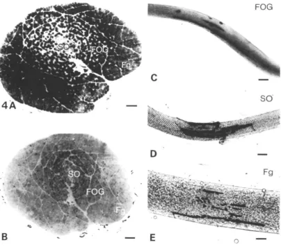
Macroscopically, the iliofibularis muscle can be differentiated into a pale outer and a red inner region. On staining fibres for acid and alkaline mATPase, SDH and cr-GPDH activities, three distinct fibre types could be differentiated (Fig. 4).
for alkaline mATPase, SDH and a"-GPDH. The fibres are intermediate in both diameter and location, forming the majority of the fibres in the intermediate region of the muscle (Figs 4, 6). The third type, which stains intensely for the activities of acid mATPase and SDH and lightly for alkaline mATPase and cr-GPDH activities, is similar to mammalian slow oxidative (SO) fibres (Fig. 5). They have the smallest mean diameter of 14-6 ± 0-4 fim and constitute 80 % of the fibres in the red region of the muscle (Fig. 6). Their proportions as a percentage of the whole cross-section of
[image:7.451.23.428.216.572.2]FOG
94 G. MUTUNGI AND I. A. JOHNSTON
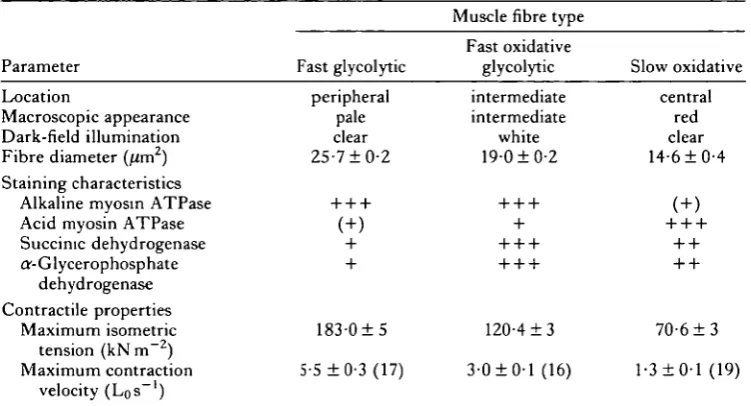
Table 1. Location, histochemical characteristics and contractile properties of the three fibre types in the iliofibularis muscle of the terrapin Pseudemys scripta elegans
Parameter
Location
Macroscopic appearance Dark-field illumination Fibre diameter (um2)
Fast glycolytic
peripheral pale clear 25-710-2
Muscle fibre type
Fast oxidative glycolytic intermediate intermediate white 19-010-2 Slow oxidative central red clear 14-610-4 Staining characteristics
Alkaline myosin ATPase Acid myosin ATPase Succinic dehydrogenase ar-Glycerophosphate
dehydrogenase Contractile properties
Maximum isometric 1 8 3 0 1 5 120-413 7 0 6 1 3 tension (kNm~2)
Maximum contraction 5-5 10-3(17) 3-010-1(16) 1-310-1(19) velocity (Los"1)
Contractile properties were studied at 15 CC.
All values represent mean 1 S.E. Mean number of fibres (N) used in mechanical studies is given in brackets.
+ + + = intense, + + = moderate, + = light, ( + ) = no staining to light staining. Lo, muscle length.
the iliofibularis were 54 % for Fg fibres, 31 % for FOG fibres and 15 % for SO fibres (Fig. 7).
Under dark-field illumination, the majority of the Fg and SO fibres scatter little or no light, making them appear transparent. In contrast, most FOG fibres scatter significant amounts of the incident light, giving them a milky appearance. The difference in their appearance under dark-field illumination, together with their location in the muscle, was used to identify individual fibre types for mechanical studies.
Nerve endings
All three fibre types in the iliofibularis muscle have one or two endplates which consist of discrete, elongated finger-like processes (Fig. 4). In Fg fibres they occurred at any point along the fibre length and most of the fibres had two endplates. However, in FOG and SO fibres endplates were located close to the fibre tendons and were usually single.
Contractile properties
183 ± 5 kN m 2. This is 2-6 times higher than that generated by the slow oxidative fibres. They also contract four times faster with an unloaded contraction velocity (Vo) of 5-5 ± 0-3 muscle lengthss"1 (Los~'). Fast oxidative glycolytic fibres have isometric tensions and unloaded contraction velocities intermediate between those of Fgand SO fibres (Table 1).
96
G . MUTUNGI AND I. A . JOHNSTON200 fan
lOO-i
80
I
60-E
4 0 2 0
-lOO-i
6 0
4 0
-" -"
lOO-i 80- 604 0 2 0
-r
o o
O <"
u.
iff O O
o ^
u.
Fig. 6. (A) A tracing of a cross-section of a whole iliofibularis showing the delineation of the three regions of predominantly one-fibre type; A, outer pale; B, intermediate; C, inner red. (B) Histograms showing the percentage of different fibre types in each region. Fg, fast glycolytic; FOG, fast oxidative glycolytic; SO, slow oxidative fibres. Bars indicate ±S.E.M.
The force-velocity (P-V) relationship of slow oxidative and fast oxidative glycolytic fibres
The force—velocity characteristics of slow fibres were determined at 5° and 15°C, while those for fast oxidative glycolytic fibres were studied at 5°C. Points below 0*6 Po on the P—V curve could be fitted to a linear form of Hill's (1938) equation:
(P + a)V = b(P0-P),
Effects of temperature and pH on tension generation and contraction velocity in fast oxidative glycolytic and slow oxidative fibres
Experiments were conducted at constant pH, and under conditions mimicking the known change in pH with temperature for terrapin skeletal muscles in vivo. Tension
60-!
4 0 -8.
2 0
-•h
[image:11.451.186.266.135.335.2]^ O ">
Fig. 7. Muscle fibre type distribution in a whole iliofibularis muscle. Fg, fast glycolytic; FOG, fast oxidative glycolytic; SO, slow oxidative fibres. Bars indicate ± S . E . M .
:hss " OO le n ju nus c OCl I 0-8 0-7 0-6 0-5 0-4 0-3 0-2 0-1 -o - %
i • d.
• < _ -o c? o • o * • • • • • * • • • • \ 1 1 o • • 1 o o
<y o
f
i
" '"".v.°
1 1 10 1 0-2 0-3 0-4 0-5 0-6 0-7 0-8 Relative load
[image:11.451.108.347.372.615.2]98
0-9 0-8 0-7 0-6 -S 0-5
1 0-4
1 0-3
>
0 2
01
-G . MUTUN-GI AND I . A . JOHNSTON
6 0 T
- ^ 4-0
C L T
^ 2 0
-_i i i 1
0-6 0-4 0-2 0-2 0-4 0-6 01 Relative load
• * •
I I
0-1 0-2 0-3 0-4 0-5 Relative load
[image:12.451.97.355.48.328.2]0-6 0-7 0-8
Fig. 9. Force-velocity relationship of fast oxidative glycolytic fibres at 5°C. Data from 10 fibres. Inset shows the same data transformed using the linear form of Hill's (1938) equation.
and V, were independent of pH in the range 6-6 to 7-8 at all temperatures. However, above and below this range, both parameters are depressed. This decline in tension and V; is not uniform in the two fibre types (Figs 10, 11).
The thermal dependence of tension and velocity can be expressed by the terms R10 and Q10, respectively (Bennett, 1984), and are obtained from the formulae:
where R! and R2 are either velocities (Qi0) or tensions (R10) at temperatures T[ and T2, respectively, T2 being greater than T]. The thermal dependence of both parameters in the, two fibre types decreased as the preferred body temperature (25-30°C) was approached. For example, in SO fibres the Rio(o-io°C) and Qio(o-io-C) ranged between 1-9 and 2-0, decreasing to 1-2-1-3 between 10° and 20°C.
DISCUSSION
Fibre types
E Z
o E E
160
120
40
6-0
A o* 20°C > * 10°C
: 0 ° C
7-2 8-4
120
40 6 0
pH
7-2 8-4
Fig. 10. Effect of temperature and pH on maximum isometric tension of fast oxidative glycolytic (A) and slow oxidative (B) fibres. Values represent mean ± S.E.M. (6—10 fibres). • Significantly different from pH7-2 at P < 0 0 5 level.
1-6
3D 1-2
-1 0-1
o o 2 0-4
o U
A I
20 °C
f* 10°C o°c
1-2
0-8
0-4
0°C
[image:13.451.64.366.51.294.2]6-0 7-2 8-4 6 0 7-2 8-4
100 G. MUTUNGI AND L A . JOHNSTON
Effects of temperature andpH on contractile properties
In a study of the iliofibularis muscle of the desert iguana, Dipsosaurus dorsalis, Johnston & Gleeson (1986) found that unloaded contraction velocities at 25 °C were l-3-l-9Los~' for multiply innervated slow fibres, 2-0-7-4 Los"1 for FOG fibres and 5-8-8-7 LQS~' for Fg fibres. While there was an overlap in the contraction speeds of fast fibres with different metabolic characteristics, slow fibres were found to constitute a distinct population. Once a correction is made for the different temperatures of study, a similar spectrum of contraction speeds is apparent for the various fibre types in the iliofibularis muscle (Table 1). This is surprising in view of the differences in slow fibre characteristics and locomotory behaviour of the two species.
There is no simple relationship between the thermal dependence of locomotion and that of contractile properties of isolated skeletal muscles (Johnston & Gleeson, 1984; Marsh & Bennett, 1985). In lizards, at temperatures above 15°C, maximum running speed and stride frequency are less temperature-dependent than either the maximum shortening velocity or power output of the iliofibularis muscle (Marsh & Bennett, 1985). It has been postulated that other relatively temperature-independent factors, such as the storage and recovery of elastic energy by muscles and tendons, serve to modulate the thermally dependent properties of muscle fibres (Bennett, 1985; Marsh & Bennett, 1985). Nevertheless, it is clear that the thermal dependence of many properties of isolated muscles, including maximum tension generation (Putnam & Bennett, 1982; Johnston & Altringham, 1985), twitch contraction times, and relaxation rates (Putman & Bennett, 1982), reflect the normal or preferred body temperatures (PBT) of the species. For example, at low temperatures skinned fibres from cold-tolerant ectotherms generate much higher tensions than homologous fibres from reptiles and mammals (Stephenson & Williams, 1984; Johnston & Altringham, 1985; Johnston & Gleeson, 1986). At temperatures approaching the PBT, maximum tetanic tension (live fibres) and maximum Ca2+-activated force (skinned fibres) generally become relatively temperature-independent (Marsh & Bennett, 1985; Stephenson & Williams, 1984). For example, values of Qio(io-20°C) for V; and PQ, in chemically skinned muscle fibres, are significantly lower in Pseudemys which has a PBT of 30°C than for comparable fibre types in the iliofibularis muscle of the desert iguana (Johnston & Gleeson, 1984) which has a PBT of 40°C (Marsh & Bennett, 1985).
102 G. MUTUNGI AND L A . JOHNSTON
Xenopus at 10°C (Lannergren, Lindblom & Johansson, 1982). The effects of temperature on the P—V relationship vary between slow and fast muscles (Ranatunga, 1982) and between species. In teleost skinned fibres a/P0 values decrease with increasing temperature (Johnston & Altringham, 1985), whereas in whole rat muscles the opposite effect occurs (Ranatunga, 1982). In contrast, for intact fibres in Dipsosaurus (Marsh & Bennett, 1985) and Xenopus (Lannergren, 1978), and skinned fibres in Pseudemys (Fig. 8), the shape of the P—V curve is similar over a wide range of temperatures. Therefore no clear picture of the significance of these results emerges.
Muscle pH in resting Pseudemys at the PBT is approximately 6-8, rising to 7-3 at 10°C, which corresponds to the minimum temperature for active locomotion (Cagle, 1946). Po and V,/Vmax for both SO and FOG fibres are relatively independent of pH change over this range. These observations are comparable to those reported for demembranated fibres from frog muscles (Schadler, 1967; Kentish & Nayler, 1977; Fabiato & Fabiato, 1978). The stability of these parameters over a wide pH range in ectotherms may be important as muscle pH varies significantly with body tem-perature (Malan et al. 1976; HeislereZ al. 1976). Outside this pH range both tension and contraction velocity in the two fibre types are depressed (Figs 10, 11). It is known that the basic pattern of acid-base regulation can be modified by other factors such as exercise (Gatten, 1975) and diving (Penney, 1974). Pseudemys scripta elegans is remarkably tolerant of anoxia (Clark & Miller, 1973). It can survive a 24-h forced dive in deoxygenated water at 22°C (Penney, 1974) and up to 2 weeks submerged in oxygenated water at 16—18°C (Robin, Vester, Murdaugh & Millen, 1964). Under these conditions, energy supply is provided by anaerobic glycolysis resulting in a lactate acidosis (Penney, 1974). For example, at 22°C a 24-h submergence results in an increase in plasma lactate concentration from 1 to 37mmoll~' (Penney, 1974) and 15 h in 100% nitrogen reduces the pH of cardiac muscle to 6-6 (Clark & Miller, 1973). The contractile performance of de-membranated fibres in this species appears to be unaffected by the levels of acidosis likely to be encountered during prolonged dives.
Our results have implications for experiments designed to investigate the thermal dependence of contractile properties using skinned muscle fibres. It is clear that at saturating Ca2+ concentrations the effects of temperature on Po and V\/Vmax are
similar under conditions of constant pH and under conditions where pH is allowed to vary with temperature within the physiological range. However, this is not likely to be the case at sub-saturating Ca2+ concentrations, because an increase in temperature or a decrease in pH are both known to shift the pCa-force relationship to higher Ca2+ concentrations (Fabiato & Fabiato, 1978; Godt & Lindley, 1982).
REFERENCES
ALTRINGHAM, J. D. & JOHNSTON, I. A. (1982). The pCa-tension and force-velocity charac-teristics of skinned fibres isolated from fish fast and slow muscles. J. Physio!., Land. 333, 421-449.
BENNETT, A. F. (1984). Thermal dependence of muscle function. Am. J. Physiol. 247, R217-R229.
BENNETT, A. F. (1985). Temperature and muscle. J . exp. Biol. 115, 333-344.
CAGLE, F. R. (1946). The growth of the slider turtle, Pseudemys scripta elegans. Am. Midi. Nat. 36, 685-729.
CALDWELL, P. C. (1954). Studies on the internal pH of large muscle and nerve fibres. J. Physiol.,
bond. 126, 169-180.
CLARK, V. M. & MILLER, A. T. (1973). Studies on anaerobic metabolism in the fresh-water turtle
(Pseudemys scripta elegans). Comp. Biochem. Physiol. 44A, 55-62.
COLE, W. V. (1955). Motor endings in the striated muscles of vertebrates. J. comp. Neurol. 102, 671-716.
CREESE, R. (1949). Bicarbonate ion and striated muscle. J . Physiol., bond. 110, 450-457. CREESE, R. (1953). Effects of carbon dioxide on muscle. J . Physiol., Land. 119, 16P.
CROWE, A. & RAGAB, A. H. M. F. (1970). The structure, distribution and innervation of spindles in the extensor digitorum brevis 1 muscle of the tortoise Testudo graeca. jf. Anat. 106, 521—538. FABIATO, A. & FABIATO, F. (1978). Effects of pH on the myofilaments and sarcoplasmic reticulum
of skinned cells from cardiac and skeletal muscles. J. Physiol., Land. 276, 233-255.
GATTEN, R. E. (1975). Effects of activity on blood oxygen saturation, lactate, and pH in the turtles
Pseudemys scripta and Terrapina ornata. Physiol. Zool. 48, 24-35.
GLEESON, T . T . (1983). A histochemical and enzymatic study of the muscle fibre types in the water monitor, Varanus salvator.J. exp. Zool. 227, 191-202.
GLEESON, T . T . & JOHNSTON, I. A. (1986). Reptilian skeletal muscle: contractile properties of identified, single fast-twitch and slow fibres from the lizard Diposaurus dorsalis. J. exp. Zool. (in press).
GLEESON, T . T., MITCHELL, G. S. & BENNETT, A. F. (1980a). Cardiovascular responses to
graded activity in the lizards Varunus and Iguana. Am. J. Physiol. 239, R174—R179.
GLEESON, T . T . , NICOL, C. J. M. & JOHNSTON, I. A. (1984). Capillarisation, mitochondrial
densities, oxygen diffusion distances and innervation of red and white muscle of the lizard
Dipsosaurus dorsalis. Cell Tissue Res. 237, 253-258.
GLEESON, T . T . , PUTNAM, R. W. & BENNETT, A. F. (19806). Histochemical, enzymatic and
contractile properties of skeletal muscle fibres in the lizard, Dipsosaurus dorsalis. J. exp. Zool. 214, 293-302.
GODT, R. E. & LlNDLEY, B. D. (1982). Influence of temperature upon contractile activation and isometric force production in mechanically skinned muscle fibres of frog. J. gen. Physiol. 80, 279-297.
GUTH, L. & SAMAHA, F. J. (1969). Qualitative differences between actinomyosin ATPase of slow and fast mammalian muscle. Expl Neurol. 25, 138-152.
GUTH, L. & SAMAHA, F. J. (1970). Procedure for the histochemical demonstration of actinomysin ATPase. Expl Neurol. 28, 365-367.
GUTHE, K. (1981). Reptilian muscle: Fine structure and physiological parameters. In Biology of
the Reptilia, vol. 13 (ed. C. Gans & T . S. Parsons). New York: Academic Press.
HEISLER, N., WEITZ, H. & WEITZ, A. M. (1976). Extracellular and intracellular pH with changes temperature in the dogfish Scyliorhinus stellaris. Respir. Physiol. 26, 249-263.
HESS, A. (1970). Vertebrate slow muscle fibres. Physiol. Rev. 50, 40-62.
HILL, A. V. (1938). The heat of shortening and the dynamic constants of muscle. Proc. R. Soc. B 126, 136-195.
HILL, A. V. (1956). The influence of external pH on the internal pH of the muscle. Proc. R. Soc. B 144, 1-22.
HOCHACHKA, P. W. (1985). Fuels and pathways as designed systems for support of muscle work.
1 0 4 G . MUTUNGI AND I . A . JOHNSTON
IZUTSU, K. T . (1972). Intracellular pH, H+ ion flux and H+ ion permeability in bullfrog todB muscle. J. Physiol, Lond. 221, 15-24.
JACKSON, D. C. (1971). The effect of temperature on ventilation in the turtle, Pseudemys scripta
elegans. Respir. Physiol. 12, 131-140.
JOHN-ALDER, H. B. & BENNETT, A. F. (1981). Thermal dependence of endurance and locomotory energetics in a lizard. Am. jf. Physiol. 241, R342-R349.
JOHNSTON, I. A. (1985). Sustained force development: specializations and variation among the vertebrates. J. exp. Biol. 115, 239-252.
JOHNSTON, I. A. & ALTRJNGHAM, J. D. (1985). Evolutionary adaptation of muscle power output to environmental temperature: force-velocity characteristics of skinned fibres isolated from antarctic, temperate and tropical marine fish. Pflugers Arch. ges. Physiol. 405, 136-140. JOHNSTON, I. A. & BRJLL, R. (1984). Thermal dependence of contractile properties of single fibres
from antarctic and various warm water marine fishes including Skipjack Tuna (Hatsuvxmus
pelamis) and Kawakawa (Euthynnus a/finis). J. comp. Physiol. B 155, 63-70.
JOHNSTON, I. A. & GLEESON, T. T. (1984). Thermal dependence of the contractile properties of red and white fibres of the desert iguana (Dipsosaurus dorsalis).jf. exp. Biol. 113, 123-132. JOHNSTON, I. A. & GLEESON, T . T . (1986). Effects of temperature on contractile properties of
skinned muscle fibres from three species of toad. Am.J. Physiol. (in press).
JOHNSTON, I. A. & SIDELL, B. D. (1984). Differences in temperature dependence of muscle contractile properties and myofibrillar ATPase activity in cold-temperate fish. J. exp. Biol. I l l , 179-189.
KENTISH, J. & NAYLER, W. G. (1977). Effect of pH on the Ca++-dependent ATPase of rabbit cardiac and white skeletal myofibrils.J'. Physiol., Lond. 265, 18P-19P.
LANNERGREN, J. (1978). The force-velocity relationship of isolated twitch and slow muscle fibres of Xenopus laevis.J. Physiol, Lond. 283, 501-522.
LANNERGREN, J., LINDBLOM, P. & JOHANSSON, B. (1982). Contractile properties of two varieties of
twitch muscle fibres mXenopus laevis. Ada physiol. scand. 114, 523-535.
LANNERGREN, J. & SMITH, R. S. (1966). Types of muscle fibres in toad skeletal muscle. Ada
physiol. scand. 68, 263-274.
MALAN, A., WILSON, T . & REEVES, R. B. (1976). Intracellular pH in cold-blooded vertebrates as a
function of body temperature. Respir. Physiol. 28, 29-47.
MARSH, R. L. & BENNETT, A. F. (1985). Thermal dependence of isotonic contractile properties of skeletal muscle and sprint performance in the lizard Dipsosaurus dorsalis. J. comp. Physiol. B 155, 541-551.
MARUM, P. & ARMSTRONG, R. B. (1978). Muscle fibre activity during locomotion in lizards. Fedn
Proc. Fedn Am. Socs exp. Biol. 37, Abstract number 1378.
MORGAN, D . L. & PROSKE, U. (1984). Vertebrate slow muscle: Its structure, pattern of innervation and mechanical properties. Physiol. Rev. 64, 103-169.
NACHLAS, M. M., TSOU, K. C , DESOUZA, E., CHENG, C. S. & SELIGMAN, A. M. (1957). Cytochemical demonstration of succinic dehydrogenase by use of a new /)-nitrophenyl substituted ditetrazole. J. Histochem. Cytochem. 5, 420-436.
NAIK, N. T . (1963). Technical variations in Koelle's histochemical method for demonstrating cholinesterase activity. Q. jfl microsc. Sci. 104, 89-100.
NICOL, C. J. M. (1985). A microcomputer program to determine the composition of solutions containing multiple metal ions and complexing ligands. J. Physiol. Lond. 367, 10P.
PANNIER, J. L., WEYNE, J. & LEUSEN, I. (1970). Effects of Pco2. bicarbonate and lactate on
isometric contractions of isolated soleus muscle of the rat. Pflugers Arch. ges. Physiol. 320, 120-132.
PENNEY, D. G. (1974). Effects of prolonged diving anoxia on the turtle, Pseudemys scripta
elegans. Comp. Biochem. Physiol. 47A, 933-941.
PETER, J. B., BARNAUD, R. J., EDGERTON, V. R., GILLESPIE, C. A. & STEMPLE, K. E. (1972). Metabolic profiles of skeletal muscle in guinea pigs and rabbits. Biochemistry 11, 2627-2633. PROSKE, U. & VAUGHAN, P. (1968). Histological and electrophysiological investigation of lizard
skeletal muscles. J. Physiol., Lond. 199, 495-509.
RANATUNGA, K. W. (1982). Temperature-dependency of shortening velocity and rate of isometric tension development in rat skeletal muscle. J. Physiol., Land. 329, 465-483.
RENAUD, J. M. & STEVENS, E. D. (1984). The extent of short- and long-term compensation to temperature shown by frog and toad sartorius muscle. J exp. Biol. 108, 57-75.
REEVES, R. B. (1972). An imida2ole alphastat hypothesis for vertebrate acid-base regulation: Tissue carbon dioxide content and body temperature in bullfrogs. Respir. Physiol. 14, 219—236. RIDGE, R. M. A. P. (1971). Different types of extrafusal muscle fibres in snake costocutaneous
muscle. J . Physiol., bond. 271, 393-418.
ROBERTSON, S. P. & KERJUC, W. G. L. (1979). The effects of pH on C a+ + activated force in frog skeletal muscle. Pflugers Arch, ges. Physiol. 380, 41-45.
ROBIN, E. D., VESTER, J. W., MURDAUGH, H. V. & MILLEN, J. E. (1964). Prolonged anaerobiosis in a vertebrate: anaerobic metabolism in the freshwater turtle. J. cell. comp. Physiol. 63, 287-297.
SCHADLER, M. (1967). Proportionale Aktivierung von ATPase-aktivitat und Kontraktionsspanung durch Calciumionen in isoliertilen Strukturen verschiedener Muskelarten. Pflugers Arch. ges.
Physiol. 296, 70-90.
SPURWAY, N. C. (1980). Histochemical typing of muscle fibres by microphotometry. In Plasticity
of Muscle (ed. D. Pette). Berlin, New York: Walter de Gruyter & Co.
STEPHENSON, D. G. & WILLIAMS, D. A. (1984). Calcium-activated force responses in fast- and slow-twitch skinned muscle fibres of the rat at different temperatures. J. Physiol., Lond. 317, 281-302.
WALLER, A. D. & SOWTON, S. C. M. (1896). Action of carbon dioxide on voluntary and on cardiac muscle..7. Physiol., Lond. 20, xvi-xvii.