932
MS
AJNR Am J Neuroradiol 21:932–938, May 2000Variations of the Superficial Middle Cerebral Vein:
Classification Using Three-dimensional CT Angiography
Yasuhiro Suzuki and Kiyoshi Matsumoto
BACKGROUND AND PURPOSE: Classification of variations of the superficial middle
cere-bral vein (SMCV) remains ambiguous. We propose a new classification system based on em-bryologic development for preoperative examination.
METHODS: Three-dimensional CT angiography was used to evaluate 500 SMCVs (in 250
patients). The outflow vessels from the SMCV were classified into seven types on the basis of embryologic development. The 3D CT angiograms in axial stereoscopic and oblique views and multiple intensity projection images were evaluated by the same neurosurgeon on two occa-sions. Inconsistent interpretations were regarded as equivocal.
RESULTS: Three-dimensional CT angiography clearly depicted the SMCV running along
the lesser wing or the middle cranial fossa. However, the outflow vessel could not be confirmed as the sphenoparietal, cavernous, or emissary type in 39 (8%) of the sides. SMCVs running in the middle cranial fossa to join the transverse sinus or superior petrosal sinus were accurately identified. SMCVs were present in 456 sides: 62% entered the sphenoparietal sinus or the cavernous sinus and 12% joined the emissary vein. Nine vessels were the superior petrosal type, 10 the basal type, 12 the squamosal type, and 44 the undeveloped type.
CONCLUSION: Three-dimensional CT angiography can depict the vessels and their
anatom-ic relationship to the bone structure, allowing identifanatom-ication of the SMCV variant in individual patients. Preoperative planning for skull base surgery requires such information to reduce the invasiveness of the procedure. With the use of our classification system, 3D CT angiography can provide exact and practical information concerning the SMCV.
The superficial middle cerebral vein (SMCV) usu-ally runs downward and forward along the sylvian fissure and flows into the sphenoparietal sinus or directly into the cavernous sinus (1–3). However, there are several common variations. The termi-nology, assumptions, and classification of such variations differ widely, and definitions remain confusing.
Information concerning the location and outflow point of the SMCV and its anatomic relationship to the bone structure is important in the preoperative planning of skull base surgery. However, such in-formation is not easy to obtain by many of the cur-rent methods of investigation. In this study, we used 3D CT angiography to analyze the character-istics of SMCVs and classified the variations into seven types on the basis of embryologic anatomy.
Received September 10, 1999; accepted after revision Decem-ber 27.
From the Department of Neurosurgery, Showa University, School of Medicine, 5-8 Hatanodai 1, Shinagawa-ku, Tokyo 142-8666, Japan.
Address reprint requests to Yasuhiro Suzuki, MD, PhD.
qAmerican Society of Neuroradiology
Methods
Two hundred fifty patients were included in the study, 132 men and 118 women, aged 28 to 85 years (mean age, 63 years). All patients were examined at our hospital between September 1996 and May 1999 for assessment of cerebrovas-cular disease, brain tumor, or abnormal findings identified at routine screening for cerebrovascular or other intracranial dis-ease. No patient had edema or midline shift on CT scans or had undergone an operation.
Three-dimensional CT angiography was initiated 35 seconds after the start of intravenous administration of nonionizing contrast material, injected at a rate of 3 mL/s for 40 seconds, for a total volume of 120 mL. The x-ray tube potential and current were 120 kV and 175 mA, respectively. The sections were 2 mm thick, the table speed was 1.3 mm/s, and the scan-ning time was 1 second. Although a section thickness of 1 mm provides better quality images than a thickness of 2 mm, it is limited in the vertical direction owing to the table transfer. Therefore, we used a thickness of 2 mm and a table transfer range of 6 cm for optimum clarity. We used the shaded surface rendering method for 3D reconstruction with a threshold of 150 to 250 HU. Axial stereoscopic and oblique scans in the bilateral anterior or posterior directions were reconstructed. The 3D CT angiograms were evaluated by the same neurosur-geon on two separate occasions. In cases of inconsistent inter-pretations, the result was regarded as equivocal.
AJNR: 21, May 2000 SUPERFICIAL MIDDLE CEREBRAL VEIN 933
FIG1. Classification of the superficial sylvian venous drainage pathways.1, Sphenoparietal type: the SMCV enters the sphenoparietal
[image:2.612.312.540.424.537.2]sinus and runs along the lesser wing of the sphenoid bone to enter the cavernous sinus.2, Cavernous type: the SMCV directly enters the anterior end of the cavernous sinus.3, Emissary type: the SMCV courses along the lesser wing, turns inferiorly to reach the floor of the middle cranial fossa, joins the sphenoidal emissary veins, and passes through the floor to reach the pterygoid plexus.4, Superior petrosal type: the SMCV runs along the lesser wing and just before reaching the cavernous sinus, turns downward along the anterior inner wall of the middle cranial fossa, then runs along its floor medially to the foramen ovale to join the superior petrosal sinus.5, Basal type: the SMCV runs along the lesser wing, turns downward along the anterior wall of the middle cranial fossa, then runs along its floor laterally to the foramen ovale over the petrous pyramid, presumably to join the transverse sinus through the lateral tentorial sinus or superior petrosal sinus.6, Squamosal type: the SMCV fails to turn medially to join the sinus along the lesser wing, and instead turns directly backward along the inner aspect of the temporal squama and runs posteriorly to join the transverse sinus or lateral tentorial sinus.7, Undeveloped type: the SMCV is absent, and the superficial sylvian drainage is through a large channel that extends forward, upward, upward and backward, or downward and backward into the superior sagittal sinus or transverse sinus.
Table 1: Distribution of drainage veins from the superficial sylvian area by 3D CT angiography
Drainage Type No. (%) 1. Sphenoparietal
2. Cavernous 3. Emissary 4. Superior petrosal 5. Basal
6. Squamosal 7. Undeveloped 1. or 2. or 3.a
Combinedb
271 (54) 36 (7) 62 (12)
9 (2) 10 (2) 12 (2) 44 (9) 39 (8) 17 (3)
aCases in which it was impossible to confirm the inflow point as
the sphenoparietal sinus, cavernous sinus, or emissary veins.
bCases in which multiple drainage pathways were present.
1. Sphenoparietal type: The SMCV enters the dura and runs along the lesser wing of the sphenoid bone (lesser wing) to enter the cavernous sinus. The dural portion of this channel along the lesser wing is frequently referred to as the spheno-parietal sinus.
2. Cavernous type: The SMCV enters the anterior end of the cavernous sinus directly.
3. Emissary type: The SMCV courses along the lesser wing, turns inferiorly to reach the floor of the middle cranial fossa, then joins the sphenoidal emissary veins and passes through the floor to reach the pterygoid plexus.
4. Superior petrosal type: The SMCV runs along the lesser wing, turns downward posteriorly along the anterior inner du-ral wall of the middle cranial fossa just before reaching the cavernous sinus, then runs along its floor medially to the fo-ramen ovale and just lateral to the cavernous sinus and joins the superior petrosal sinus.
5. Basal type: The SMCV runs along the sylvian fissure, turns downward posteriorly along the anterior wall of the mid-dle cranial fossa, then runs along its floor lateral to the foramen ovale to join the transverse sinus through the lateral tentorial sinus or superior petrosal sinus.
6. Squamosal type: The SMCV does not turn medially to join the sinus along the lesser wing on reaching the pterion, but turns directly backward along the inner aspect of the tem-poral squama and runs posteriorly to join the transverse sinus or the lateral tentorial sinus.
7. Undeveloped type: The SMCV is absent, and the venous drainage of the superficial sylvian area is through a large chan-nel that extends forward, upward, upward and backward, or downward and backward into the superior sagittal sinus or the transverse sinus.
The restricted exploration area prevented inspection of the upward drainage system. Consequently, cases in which the SMCV could not be identified around the skull base area were included in the undeveloped type.
Results
AJNR: 21, May 2000 934 SUZUKI
FIG2. Variations of the superficial sylvian venous drainage pathway revealed by 3D CT angiography. A, Axial view. The SMCV enters the sphenoparietal sinus (arrow).
B, Axial view. The SMCV directly enters the anterior end of the cavernous sinus (arrows).
C–F, Axial stereo and oblique views. The right SMCV courses along the lesser wing, turns inferiorly to reach the floor of the middle cranial fossa, and enters the foramen ovale (open arrows). This vein anastomoses with the cavernous sinus through the paracavernous sinus (arrowhead). The left SMCV turns downward along the anterior wall of the middle cranial fossa, then runs along its base laterally to the foramen ovale over the petrous pyramid, presumably to join the transverse sinus at the sigmoidal angle (closed arrows).
G–J, Axial stereo and oblique views. The SMCV turns medially into the sinus of the lesser wing, and before reaching the cavernous sinus turns downward along the anterior inner wall of the middle cranial fossa and runs posteriorly to join the superior petrosal sinus (arrows).
K and L, Axial stereo views. The SMCV turns downward along the anterior wall of the middle cranial fossa at the lesser wing and then runs along its floor laterally to the foramen ovale to drain into the transverse sinus (arrows).
M, Axial view. The SMCV turns directly backward along the inner aspect of the temporal squama and runs posteriorly to join the transverse sinus (arrows).
N, Axial view. The SMCV is absent, and the sylvian drainage area is taken over by a superficial temporal vein, which extends downward and backward into the lateral tentorial sinus (arrows).
O, Axial view. The remnant of the tentorial sinus originates from the cavernous sinus, runs beyond the petrous pyramid, and enters the lateral tentorial sinus. Note the anastomosis between the anterior end of the cavernous sinus and the basal vein of Rosenthal (arrowheads).
AJNR: 21, May 2000 SUPERFICIAL MIDDLE CEREBRAL VEIN 935
FIG2. Continued.
62% of the sides (Fig 2A and B), joining the em-issary veins in 12% (Fig 2C–F). The superior pe-trosal type was present in nine sides (Fig 2G), the basal type in 10 sides (Fig 2C–F, K, and L), the squamosal type in 12 sides (Fig 2M), and the un-developed type in 44 sides (Fig 2N).
Three-dimensional CT angiography could also depict the remnant of the tentorial sinus, originating from the cavernous sinus, running beyond the pet-rous pyramid, and entering the lateral tentorial si-nus (Fig 2).
Discussion
Adult venous patterns develop in the head region during the first three months of prenatal life, but the remnants of the embryonic tentorial sinus pre-serve a different drainage pattern for a little while after birth (4, 5). Two embryologic sinuses partic-ipate in the formation of the SMCV, the tentorial sinus, and the prootic sinus. The tentorial sinus can be identified by the 20-mm stage of embryonic de-velopment. Subsequent growth of the posterior ce-rebral hemisphere causes the tentorial sinus to elon-gate posteriorly, and the anterior portion is shifted medially (Fig 3A). The SMCV drains through the tentorial sinus into the transverse sinus rather than into the cavernous sinus during prenatal life and
early infancy. The prootic sinus develops from the stem of the middle dural plexus by the 17- to 20-mm stage of development and is involved in the formation of the dura of the middle cranial fossa and the diploic vein (6, 7). The prootic sinus con-tributes to the cavernous sinus, the sphenoparietal sinus (3, 5), the meningeal sinus (eg, the emissary veins around the foramen ovale) (5, 8), and the anterior parietal temporal diploic veins in the 3-month-old embryo (Fig 3B). The superior orbital vein, which at first enters the prootic sinus, joins the cavernous sinus in the adult.
Two secondary intradural anastomoses involving the cavernous sinus have generally developed be-fore the adult stage: one is between the tentorial sinus and the anterior end of the cavernous sinus (Fig 3C, arrow), whereas the caudal part of the ten-torial sinus, which drains into the transverse sinus, has dwindled, and the other is between the cavern-ous sinus and the superior petrosal sinus (Fig 3C, triangle) (5). The former anastomosis is located be-tween the sinus originating from the tentorial sinus and the sinus from the prootic sinus.
AJNR: 21, May 2000 936 SUZUKI
FIG3. Developmental stages of the basal cranial veins (after Padget).
A, 60-mm stage of embryologic development. The transverse sinus has swung backward on the sigmoid sinus and receives the elongated tentorial sinus. A lateral tributary of the prootic sinus, which primarily receives the middle meningeal sinus, is continuous with the definitive petrosquamosal sinus. The tentorial sinus becomes plexiform caudally as it is shifted toward the sigmoid sinus. The ophthalmic veins primarily drain laterally through the prootic sinus, and secondarily drain medially through the cavernous sinus and inferior petrosal sinus.
B, Typical infant stage. The SMCV still drains through the tentorial sinus, which has a variable position. The superior petrosal sinus has not yet joined the cavernous sinus.
C, Typical adult stage. Note the secondary anastomoses between the cavernous sinus and sphenoparietal sinus, derived from the prootic sinus (arrow), and between the cavernous sinus and superior petrosal sinus (arrowhead). The typical lateral wing of the cavernous sinus, just below the mandibular nerve root, is a remnant of the prootic sinus and extends to the foramen ovale and emissary veins. The petrosquamosal sinus and remnant of the prootic sinus draining the dura and bone become diploic in the adult, when the middle meningeal sinuses drain through the foramen ovale and sigmoid sinus.
1, SMCV; 2, superior orbital vein; 3, transverse sinus; 4, embryonic tentorial sinus; 5, prootic sinus; 6, sigmoid sinus; 7, superior sagittal sinus;8, cavernous sinus and inferior petrosal sinus; 9, superior petrosal sinus; 10, petrosquamosal sinus; 11, sphenoparietal sinus;12, emissary venous drainage; 13, middle meningeal sinus.
location is more likely to form anastomoses with the cavernous sinus). Therefore, the adult SMCV variation is based on the relative development of the anastomoses between the pathway from the SMCV through the tentorial to the transverse sinus and the pathway from the cavernous sinus to the pterygoid plexus to the superior or inferior petrosal sinus.
The lateral wall of the cavernous sinus is usually composed of two layers of dura mater (9–13). In one study, a venous channel was present between these layers in 24% of specimens (14). This chan-nel connects with the SMCV and runs posteriorly through the middle cranial fossa toward the ptery-goid plexus, the transverse sinus, or the superior petrosal sinus. The vascular channel and the two layers of the lateral dural wall do not occur con-comitantly. This channel pattern corresponds to the description of the paracavernous sinus under the term tentorial sinus (15). Presumably, this channel protrudes laterally because of an incomplete anas-tomosis between the cavernous sinus and the ten-torial sinus. This structure of two layers is formed
from the complete anastomosis of these two sinuses.
AJNR: 21, May 2000 SUPERFICIAL MIDDLE CEREBRAL VEIN 937
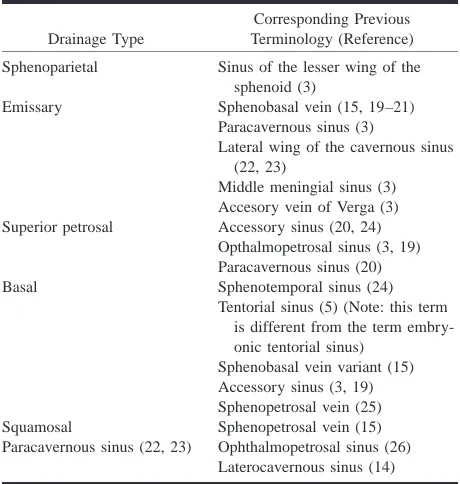
Table 2: Comparison of classification systems for variants of the superficial middle cerebral vein
Drainage Type
Corresponding Previous Terminology (Reference) Sphenoparietal Sinus of the lesser wing of the
sphenoid (3)
Emissary Sphenobasal vein (15, 19–21) Paracavernous sinus (3)
Lateral wing of the cavernous sinus (22, 23)
Middle meningial sinus (3) Accesory vein of Verga (3) Superior petrosal Accessory sinus (20, 24)
Opthalmopetrosal sinus (3, 19) Paracavernous sinus (20) Basal Sphenotemporal sinus (24)
Tentorial sinus (5) (Note: this term is different from the term embry-onic tentorial sinus)
Sphenobasal vein variant (15) Accessory sinus (3, 19) Sphenopetrosal vein (25) Squamosal Sphenopetrosal vein (15) Paracavernous sinus (22, 23) Ophthalmopetrosal sinus (26)
Laterocavernous sinus (14)
Previous classifications of the SMCV have been confusing and contradictory, without any basis in embryology. In some cases, several terms have been used to describe the same variant, and other times the same term has been used to describe dif-ferent variations. Table 2 lists the terms of our clas-sification system and the corresponding previous terminology. The confusion seems to have derived from a poor knowledge of embryology, a limited number of cadaveric specimens, inferior (low spa-tial resolution) angiographic images, and a poor comprehension of recent literature.
Three-dimensional CT angiography allows a va-riety of possible manipulations of the images from only one examination, high spatial resolution, and simultaneous visualization and evaluation of ves-sels and bone. However, in some cases, the sphen-oparietal, cavernous, and emissary types of drain-age were difficult to distinguish (16–18). This problem may be solved by examining images ob-tained in multiple directions and by limiting the images to the cavernous area by making recon-structions from thin-slice volume data. The supe-rior petrosal, basal, and squamosal types of drain-age were clearly differentiated. In addition, 3D CT angiography delineated the course and locality of veins that were previously obscure. In particular, we found that the emissary type flows out through the foramen ovale and the basal type runs laterally to the foramen ovale from anterior to posterior.
For neurosurgeons, information about the loca-tion of the SMCV along the floor of the middle cranial fossa, the outflow vein, and its junction is important for planning less invasive surgical ap-proaches via the pterional, anterior temporal,
sub-temporal, or other routes. Hacker’s classification (15) based on angiographic studies or examination of cadavers cannot provide accurate information about individual differences. In contrast, our clas-sification, based on embryologic development and 3D CT angiographic evaluation, offers precise and practical information. Preoperative 3D CT angiog-raphy, by displaying multiple axial and oblique views, is likely to be useful for showing the vessels and their anatomic relationship to the bone struc-ture as well as variations of the cranial venous system.
Conclusion
The classification of SMCV variants into seven types based on the changes seen during embryo-logic development combined with 3D CT angio-graphic evaluation can help identify and categorize the SMCV and its anatomic relationship to the mid-dle cranial fossa bone structure in individual patients.
References
1. DiChiro G. Angiographic patterns of cerebral convexity veins
and superficial dural sinuses. AJR Am J Roentgenol 1962;87:
308–321
2. Jones FW, ed. Buchanan’s Manual of Anatomy. London: Bailli-ere, Tindall and Cox; 1950:262
3. Wolf BS, Huang YP, Newton CM. The superficial sylvian venous
drainage system. AJR Am J Roentgenol 1963;89:398–410
4. Knosp E, Muller G, Perneczky A. Anatomical remarks on the
fetal cavernous sinus and the veins of the middle cranial fossa.
In: Dolenc VV, ed. The Cavernous Sinus: A Multidisciplinary
Ap-proach to Vascular and Tumorous Lesions. New York: Springer;
1987:104–116
5. Padget DH. The cranial venous system in man in reference to
development, adult configuration, and relation to the arteries.
Am J Anat 1956;98:307–355
6. Zuckerkandle E. Beitrag zur Anatomie des Schlafenbeins.
Mon-atsschr Ohrenheilk 1873;9:101–108
7. Bailey P. Intracranial Tumors. Springfield, Ill: Charles C Thomas; 1933
8. Padget DH. Development of cranial venous system in man, from
view point of comparative anatomy (Carnegie Institution of Washington publication 611). Contrib Embryol 1957;36:79–140
9. Bisaria KK. The superficial sylvian vein in humans: with
spe-cial reference to its termination. Anat Rec 1958;212:319–325
10. Bonneville JF, Cattin F, Racle A, et al. Dynamic CT of the
la-terosellar extradural venous spaces. AJNR Am J Neuroradiol
1989;10:535–542
11. Mercier R, Patouillard P, Vanneuville G, Aussilhou A.
Contri-bution a l’etude du sinus caverneux par l’utilisation simultanee de plusieurs modes d’investigation. C R Assoc Anat 1970;149:
877–890
12. Pauret G. Traite d’Anatomie Humaine. Paris: Masson; 1958;745– 752, tome III, fasc 2
13. Umansky F, Nathan H. The lateral wall of the cavernous sinus
with special reference to the nerves related to it. J Neurosurg
1982;56:228–234
14. San Millan Ruiz D, Gailloud P, de Miquel Miquel MA, et al.
Laterocavernous sinus. Acta Rec 1999;254:7–12
15. Hacker H. Abflusswege der Sylvischen Venengruppe. Radiologe 1968;8:383–387
16. Aoki S, Sasaki Y, Machida T, Ohkubo T, Minami M, Sasaki Y.
Cerebral aneurysms: detection and delineation using 3-D-CT angiography. AJNR Am J Neuroradiol 1992;13:1115–1120
17. Harbaugh RE, Schlusselberg DS, Jeffery R, Hayden S, Cromwell LD, Pluta D. Three-dimensional computerized tomography
an-giography in the diagnosis of cerebrovascular disease. J
AJNR: 21, May 2000 938 SUZUKI
18. Takahana Y, Uno E, Wakamatsu K, Okada Y, Kaneko T, Tsuchiya Y. Three-dimensional computerized tomography angiography
in a persistent primitive hypoglossal artery: a case report [in Japanese]. Jpn J Neurosurg 1998;7:125–128
19. Ito J. Supratentorial venous system. In: Maki Y, Kuru H, eds.
Neuroradiology I. Tokyo: Asakura Shoten;1986:443–484
20. Lang J. Tentorial sinus and transverse sinus. In: Lang L, ed.
Clinical Anatomy of the Head. Berlin: Springer;1983;
324–325
21. Oka K, Photon AL Jr, Barry M, Rodriguez R. Microsurgical
anatomy of the superficial veins of the cerebrum. Neurosurgery
1985;17:711–748
22. Padget DH. The development of the cranial arteries in the
hu-man embryo (Carnegie Institution of Washington publication 575). Contrib Embryol 1948;32:205–261
23. Padget DH. Designation of the embryonic intersegmental
ar-teries in reference to the vertebral arar-teries and subclavian stem. Anat Rec 1954;119:349–356
24. Knott JF. On the cerebral sinus and their variations. J Anat
Physiol (London) 1881;16:27–42
25. Yamakami I, Hirai S, Yamaura A, Ono J. Venous system playing
a key role in transpetrosal approach [in Japanese]. No Shinkei
Geka 1998;26:699–707