Blaise V. Jones, John C. Egelhoff, and Richard J. Patterson
Summary: We present five cases of hypertensive encephalopa-thy in children, three with MR imaging findings and two with CT findings alone. One of the five patients had MR perfusion imag-ing, which showed perfusion abnormalities that support the con-cept of vasodilatation as the major contributor to the syndrome. Hypertensive encephalopathy is rarely reported in children, and its true prevalence may be underestimated. Characteristic le-sions in the severely hypertensive child should be recognized as manifestations of hypertensive encephalopathy, and subsequent clinical management should focus on treatment of the hyperten-sion and/or its underlying causes.
Index terms: Brain, diseases; Children, diseases; Hypertension
Hypertensive encephalopathy is a condition characterized by varying degrees of headache, nausea, vomiting, visual disturbances, focal neurologic deficit, and seizures in the setting of severe systemic hypertension that is relatively acute in onset. The degree of hypertension var-ies, but systolic pressure greater than 250 mm Hg and diastolic pressure greater than 150 mm Hg are commonly encountered. It can be seen in patients with acute elevation of blood pres-sure related to nephritis or eclampsia; alterna-tively, it may be superimposed on chronic, un-treated, or inadequately treated essential hypertension (1, 2). Clinical findings typically resolve with adequate treatment of the hyper-tension; permanent deficits are seen in those cases complicated by frank infarction or hem-orrhage. We are aware of two previously re-ported cases of nonobstetric hypertensive en-cephalopathy in children (3, 4).
Case Reports Case 1
A 10-year-old boy had a generalized tonicoclonic sei-zure preceded by a severe headache. Blood pressure was
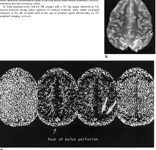
140/108 mm Hg. Findings at neurologic examination were normal. Findings on computed tomographic (CT) scans obtained at the time of presentation with and without in-travenous contrast material were also normal. Magnetic resonance (MR) imaging performed approximately 8 hours later showed regions of abnormal hyperintense sig-nal on T2-weighted images in the cortex and subcortical white matter of the high occipital lobes (Fig 1). There was minimal associated hypointense signal on T1-weighted images; no enhancement was seen after administration of contrast material. During a second MR examination per-formed 18 hours later, gradient-echo (24/15/1 [repetition time/echo time/excitations]) images with a 10° flip angle were obtained every 1.8 seconds at the level of the region of abnormal signal during bolus infusion of contrast mate-rial. This study showed mildly increased perfusion in the regions of abnormal signal. No cause of the hypertension was found, despite an extensive imaging and metabolic workup. The patient was treated medically, with good blood pressure control and no further seizure activity or neurologic symptoms.
Case 2
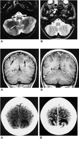
An 11-year-old girl had headache, mental status changes, and hypertension of 240/180 mm Hg. Abdomi-nal sonographic and CT studies showed a chronically shrunken kidney. MR examination of the brain showed regions of abnormal hyperintense signal throughout the occipital lobes on T2-weighted images. Additional foci of abnormal T2 prolongation were seen in the medulla ob-longata, pons, and cerebellar hemispheres (Fig 2A). There was no enhancement after administration of contrast ma-terial in any of the lesions. Embolization of the atrophic kidney was performed, with resolution of the systemic hypertension and neurologic symptoms. Follow-up MR imaging showed resolution of the lesions (Fig 2B).
Case 3
A 15-year-old boy with Addison disease was admit-ted with a new onset of generalized seizures and with blood pressure of 185/125 mm Hg. MR imaging at the
Received December 4, 1995; accepted after revision March 4, 1996.
Presented in part at the annual meeting of the American Society of Neuroradiology, Chicago, Ill, April 1995. From the Department Radiology, Children’s Hospital Medical Center, Cincinnati, Ohio.
Address reprint requests to Blaise V. Jones, MD, Department of Radiology, Room CG533B, M. S. Hershey Medical Center, PO Box 850, Hershey, PA 17033.
AJNR 18:101–106, Jan 1997 0195-6108/97/1801–0101©American Society of Neuroradiology
time of presentation showed patchy foci of hyperintense signal on T2-weighted images at the parietooccipital junction bilaterally. These lesions were hypointense on T1-weighted images, with local mass effect. There was mild gyral enhancement after administration of contrast material. Repeat contrast-enhanced MR imaging per-formed 1 week after treatment of the hypertension showed marked decrease in T1 and T2 signal abnor-malities and resolution of the abnormal enhancement (Fig 3).
Case 4
An 11-year-old girl had a seizure preceded by severe headache. She also had increasing abdominal girth over a period of several weeks. Blood pressure on admission was 165/117 mm Hg. A CT scan of the abdomen revealed a large primitive neuroectodermal tumor with abundant ma-lignant ascites. A contrast-enhanced CT scan of the head showed regions of abnormal decreased attenuation in the high occipital lobes, with some cortical enhancement after contrast administration (Fig 4). Neurologic findings re-Fig 1. Case 1: 10-year-old boy with idiopathic hypertension and seizures.
A, Axial T2-weighted (2500/110) MR image through the parietooccipital junction shows abnormal hyperintense signal in the subcortical white matter bilaterally (arrows), extending into the overlying cortex.
[image:2.587.51.549.106.584.2]Fig 2. Case 2: 11-year-old girl with ren-ovascular hypertension and headache.
A, Axial T2-weighted image (2500/90) through the posterior fossa shows abnormal hyperintense signal in the olives (black ar-rows) and the periphery of the cerebellar hemispheres (white arrow).
B, MR image obtained after treatment of hypertension shows resolution of the signal abnormalities.
Fig 3. Case 3: 15-year-old boy with hy-pertension caused by Addison disease.
A, Coronal postcontrast T1-weighted image (550/15) through the posterior pa-rietal lobes shows abnormal cortical en-hancement with mild associated mass ef-fect and surrounding low signal intensity (arrows).
B, MR image obtained after treatment of hypertension shows resolution of the ab-normal enhancement and mass effect and only minimal residual hypointense signal.
turned to normal with treatment of the hypertension; fol-low-up imaging was not performed.
Case 5
A 9-year-old boy with a recent renal transplant had seizures in the hospital associated with subacute graft re-jection and a blood pressure of 180/110 mm Hg. A con-trast-enhanced CT scan of the head showed bilateral and symmetric regions of decreased attenuation centered in the subcortical white matter of the parietooccipital junc-tions and the cerebellar hemispheres with extension into the overlying cortex. A less prominent region of decreased attenuation was seen in the right frontal lobe. There was no enhancement with contrast administration. Neurologic symptoms resolved with control of the hypertension, and follow-up imaging was not performed.
Discussion
The lesions of hypertensive encephalopathy were initially thought to be ischemic in nature. Autoregulatory vasoconstriction in the cerebral vasculature is seen in response to systemic hy-pertension. It has been theorized that in hyper-tensive encephalopathy this responsive vaso-constriction is severe enough to cause ischemia in the affected vascular territory, and that the symptoms and imaging findings are a reflection of the ischemia (5–7). This theory is supported by cases of angiographically demonstrated va-sospasm in patients with eclampsia and neuro-logic symptoms (7, 8). More recent investiga-tors have implicated vasodilatation rather than vasoconstriction as the major component of hy-pertensive encephalopathy. Animal studies have shown that severe and acute increases in cerebral blood pressure result in overdistention of arterioles, accompanied by hydrostatic edema and increased pinocytosis, resulting in extravasation of proteins and fluid into the in-terstitium (9 –11). Cerebral perfusion is actually increased in affected regions (12). These changes may be mediated by increased prosta-glandin synthesis (9). They are reversible up to a point; more severe and permanent endothelial damage occurs with higher pressures (9, 11). Arterioles situated a short distance from the cortical surface are most affected, and sympa-thetic nervous activity affords protection from these effects. The posterior circulation has sig-nificantly less sympathetic innervation than the carotid circulation (10, 13), which may explain why the majority of lesions in hypertensive en-cephalopathy are found in the vascular territory of the posterior circulation.
Previous authors have detailed the imaging findings in adults with hypertensive encepha-lopathy (4 – 6, 14, 15). The most commonly described abnormalities consist of foci of hyper-intense signal on T2-weighted MR images in the subcortical white matter of the occipital lobes. These lesions frequently have associated hy-pointensity on T1-weighted MR images and de-creased attenuation on CT scans. Focal or dif-fuse brain swelling and gray matter involvement are usually evident. Associated contrast en-hancement has been reported in a small num-ber of cases. Lesions are less often seen in other brain locations, including the cerebellum, brain stem, parietooccipital junction, basal ganglia, and frontal lobes. While hemorrhagic foci are commonly encountered in autopsy studies (16), they are infrequently seen at imaging, ex-cept in patients with chronic hypertension (4, 14, 15). The five cases reported here showed a characteristic distribution of imaging abnormal-ities, with the majority of lesions occurring in the vascular distribution of the posterior circulation. All cases had involvement of both subcortical white matter and adjacent gray matter, and all had lesions in the occipital lobes or at the pari-etooccipital junction. Focal swelling/edema was seen in all cases. Enhancement was seen in two cases, and two children had lesions in the cerebellar hemispheres. No lesions were seen in the basal ganglia, and only one patient had frontal lobe involvement. No hemorrhagic foci were seen. The child with the most lesions (case 2) had the greatest level of hypertension. All symptoms resolved in these five patients, and in all cases in which follow-up imaging was per-formed there was either resolution of or marked improvement in the abnormal findings.
previ-ously identified lesions on spin-echo images. The increased perfusion to these lesions seen on the gradient-echo images was mild in de-gree, but is consistent with the theory of vaso-dilatation with blood-brain barrier disturbance and extravasation of proteins as the primary event in hypertensive encephalopathy. These findings correspond well to those of Schwartz et al (15), who reported results of 99m Tc-hexa-methylpropyleneamine oxime (HMPAO) single-photon emission CT (SPECT) studies in two patients with hypertensive encephalopathy. In the one patient who was symptomatic during imaging, increased perfusion to regions of ab-normal signal was clearly demonstrated on MR examination. In their second patient, who was studied 1 day after resolution of acute signs and symptoms, only mild perfusion abnormalities were seen. Perfusion imaging may provide a greater insight into the mechanism of hyperten-sive encephalopathy, but this will require further investigation of additional cases.
It has been observed that previously normo-tensive persons develop hypernormo-tensive enceph-alopathy at lower blood pressures than do chronically hypertensive persons (1). In the adult, cerebral blood flow is maintained by au-toregulation over a range of systemic mean ar-terial pressures from 60 to 150 mm Hg. This range is shifted to higher pressures in untreated hypertensive patients. Normal systemic arterial pressures are lower in children than adults, with systolic measurements ranging from 105 mm Hg or less at 1 year of age to 135 mm Hg or less at age 18 (90th percentile) (18). It is reasonable to assume that the pressure range of cerebral blood flow autoregulation is accordingly lower in children as well. The five patients in this study had an average systolic pressure of 182 mm Hg and an average diastolic pressure of 128 mm Hg, somewhat lower than the values typically reported in adults. We postulate that children develop hypertensive encephalopathy at lower absolute pressures than adults owing to the rel-ative “left shift” of their range of cerebral blood flow autoregulation.
Hypertension is uncommon in children, and is often seen in conjunction with systemic dis-ease. Three of the five children in this report had systemic illnesses that directly contributed to their hypertension; a fourth had renovascular hypertension. Of two previously reported cases of hypertension in children, one occurred in a child with hypertension associated with Wilms
tumor and the other was in a teenager with renovascular hypertension. Given the infre-quency of hypertension in the pediatric popula-tion and the nonspecific and reversible nature of the findings in hypertensive encephalopathy, it is not surprising that the diagnosis has rarely been reported in children. The actual preva-lence may be underestimated.
More aggressive and effective treatment of hypertension in the obstetric population ap-pears to have decreased the frequency of hy-pertensive encephalopathy (1). The infrequent identification of infarct or hemorrhage in recent reports as compared with earlier imaging and autopsy studies suggests that these are compli-cations seen in the more severe cases of hyper-tensive encephalopathy. Such cases are ex-pected to be less common in children, as they are more likely to present with symptoms at lower absolute levels of systemic pressure, and they generally respond well to antihypertensive therapy. The characteristic intracranial lesions of hypertensive encephalopathy should be rec-ognized in the hypertensive child, and subse-quent clinical management should focus on treatment of the hypertension and/or its under-lying causes. It is our experience that in uncom-plicated cases follow-up imaging is not neces-sary.
References
1. Dinsdale HB. Hypertensive encephalopathy.Stroke1982;13:717– 719
2. Gifford RW. Management of hypertensive crises.JAMA1991;266: 829 – 835
3. Shanley DJ, Sisler CL. MR demonstration of reversible cerebral lesions in a child with hypertensive encephalopathy caused by Wilms’ tumor.AJR Am J Roentgenol1992;158:1161–1162 4. Weingarten K, Barbut D, Filippi C, Zimmerman RD. Acute
hyper-tensive encephalopathy: findings on spin-echo and gradient-echo MR imaging.AJR Am J Roentgenol1994;162:665– 670 5. Coughlin WF, McMurdo SK, Reeves T. MR imaging of postpartum
cortical blindness.J Comput Assist Tomogr1989;13:572–576 6. Waldron RL, Abbott DC, Vellody D. Computed tomography in
preeclampsia-eclampsia syndrome. AJNR Am J Neuroradiol
1985;6:442– 443
7. Will AD, Lewis KL, Hinshaw DB Jr, et al. Cerebral vasoconstriction in toxemia.Neurology1987;37:1555–1557
8. Trommer BL, Homer D, Mikhael MA. Cerebral vasospasm and eclampsia.Stroke1988;19:326 –329
9. Kontos HA, Wei EP, Deitrich D, et al. Mechanism of cerebral arteriolar abnormalities after acute hypertension. Am J Physiol
1981;240:511–527
Farrar JK. Effects of acutely induced hypertension in cats on pial arteriolar caliber, local cerebral blood flow, and the blood-brain barrier.Circ Res1976;39:33– 41
11. Nag S, Robertson DM, Dinsdale HB. Cerebral cortical changes in acute experimental hypertension.Lab Invest1977;36:150 –161 12. Skinhøj E, Strangaard S. Pathogenesis of hypertensive
encepha-lopathy.Lancet1973;1:461– 462
13. Beausang-Linder M, Bill A. Cerebral circulation in acute arterial hypertension: protective effects of sympathetic nervous activity.
Acta Physiol Scand1981;111:193–199
14. Sanders TG, Clayman DA, Sanchez-Ramos L, Vines FS, Russo L. Brain in eclampsia: MR imaging with clinical correlation. Radiol-ogy1991;80:475– 478
15. Schwartz RB, Jones KM, Kalina P, et al. Hypertensive encepha-lopathy: findings on CT, MR imaging, and SPECT imaging in 14 cases.AJR Am J Roentgenol1992;159:379 –383
16. Richards A, Graham D, Bullock R. Clinicopathological study of neurological complications due to hypertensive disorders of pregnancy. J Neurol Neurosurg Psychiatry 1988;51: 416 – 421
17. Tzika AA, Massoth RJ, Ball WS Jr, Majumdar S, Dunn RS, Kirks DR. Cerebral perfusion in children: detection with dynamic con-trast-enhanced T2*-weighted MR images.Radiology 1993;187:
449 – 458
18. Report of the Task Force on Blood Pressure in Children.Pediatrics