International Journal of Medical Science and Current Research (IJMSCR)
Available online at: www.ijmscr.com
Volume2, Issue 6,Page No: 315-320
November-December 2019
315
Medicine ID-101739732
Evaluation of Autonomic Nervous Dysfunction in Patients with Bronchial Asthma by Non
Invasive Tests and Its Comparison with Autonomic Nervous Dysfunction in Non
Asthmatic Controls
Dr. Dharmendra Jhavar1, Dr. Sarang Mahanaik2*, Dr. Archana Verma3, Dr. Dattaprasad Ganganpalli4, Dr. Umesh Kumar Chandra5, Dr. Sudhir Mourya6
1MD Medicine, Professor, 2,4PG Student, Junior Resident, 3DNB Neurology,
Associate Professor, 5MD Medicine, Senior Resident Department of Medicine, MGM Medical College, Indore, MP, India
*Corresponding Author:
Dr. Sarang Mahanaik
Junior Resident, Department of Medicine, MGM Medical College, Indore, MP, India
Type of Publication: Original Research Article Conflicts of Interest: Nil
ABSTRACT
Introduction
Airway and pulmonary vascular tone may be determined by a complex interplay between different components of the autonomic nervous system(ANS). There is now abundant evidence that neural control of the airways may be abnormal in airway disease and that neurogenic mechanisms may contribute to their pathophysiology. There is a complex interaction between inflammation and neural control of airways. ANS regulates many aspects of airway smooth muscle, airway secretions, blood flow, micro-vascular permeability and the migration and release of inflammatory cells. In this study we evaluated prevalence and degree of autonomic dysfunctions(AD) in bronchial asthma(BA) patients
Materials and Methods
This prospective observational study was conducted in 45 BA patients and 20 age and gender matched healthy controls in study duration of 1 year from March, 2004 to February, 2005 who were attending OPD and admitted in wards in Department of Medicine, MGM Medical College & MY Hospital, Indore, MP, India.
Results
Out of 45 patients of BA 33 had abnormal and out of 20 controls, no one had AD. Out of 45 patients, 12 had duration less than 5 years out of this 3 had abnormal tests whereas 33 patients had BA of more than 5 years out of which 30 had AD. 24 patients had severe BA out of which 22 had AD. Out of 21 mild to moderate degree BA patients 11 had AD and 10 had normal ANS. Indicating more severe the BA more the AD(P value <0.01). Parasympathetic dysfunctions were seen in most of the patients (46.67%). Out of 18 males, 14 had AD, and out of 27 females 19 had AD, which was statistically non significant (P =0.1).
Conclusions
BA patients display definitive dysfunction of autonomic nervous system as compared with age and gender matched controls. With chronicity more incidence of autonomic dysfunction is observed. As the severity of BA increases (as judged by symptoms, PEF variability, use of beta-2 agonist, and dose of inhaled steroids) it is observed that the incidence of autonomic dysfunction also increases.
Keywords: Autonomic dysfunctions, autonomic nervous system, bronchial asthma
INTRODUCTION
Bronchial asthma (BA) is a major chronic airway disorder which is a serious public health problem in countries throughout the world. In this inflammatory disease many cells play a role, particularly mast cells, eosinophils and T lympohocytes.1 In susceptible individuals this inflammation causes recurrent
Pag
e
316
Pag
e
316
Pag
e
316
Pag
e
316
Pag
e
316
Pag
e
316
Pag
e
316
Pag
e
316
Pag
e
316
Pag
e
316
Pag
e
316
Pag
e
316
Pag
e
316
Pag
e
316
Pag
e
316
Pag
e
316
Pag
e
316
Pag
e
316
Pag
e
316
Pag
e
316
Pag
e
316
associated increase in airway responsiveness to a variety of stimuli.
Three components of autonomic nervous system play a part in the control of airways and their secretions: the Parasympathetic via vagus nerve, the sympathetic system via its hormonal control of cAMP levels and the peptidergic or non-adrenergic non cholinergic (NANC) system. There are several studies showing the evidences that suggest the presence of autonomic dysfunction(AD) in the airways of BA patients.
Autonomic enervation of the lung is complex. In addition to classic cholinergic and adrenergic pathways, there are neural mechanisms that are
NANC, while non-adrenergic, non-cholinergic
mechanisms were originally envisaged as a separate neural system, it is now apparent that these effects are due to the release of co-transmitters from autonomic nerves. The various components of the autonomic nervous system(ANS) may interact with each other in a complex way both by affecting the
release of neurotransmitter (via pre-synaptic
receptors) at ganglia in the airway and by interaction at post-synaptic receptors.
In this study we evaluated AD in BA patients according to severity and chronicity of disease, and in healthy controls. We also tried to established correlation between gender and AD, in BA patients and healthy controls.
MATERIALS AND METHODS
The present study was conducted in 45 BA patients and, 20 age and gender matched healthy controls in study duration of 1 year from March, 2004 to February, 2005 who were attending OPD and admitted in wards in Department of Medicine, MGM Medical College & MY Hospital, Indore, MP, and India.
Inclusion criteria
BA patients
Exclusion criteria
Hypertension
Diabetes mellitus
Ischaemic heart disease
Alcoholics
Renal failure
Drugs causing autonomic dysfunction
Acute alcohol intoxication
Alcohol withdrawal
Delirium tremens
Hypoglycaemia at the time of performing test
And all other causes of dysautonomia.
The study protocol
Informed consent was obtained both from the patient and the control groups. The investigations were performed in a calm setting, after a rest of 10 minutes with considerable gap in between the test so as to minimise discomfort to the subjects as well as to enhance their enthusiastic participation and better results of the test. Normal sinus rhythm was confirmed in all the subjects and controls. All the tests were explained to the patients and controls. Detailed history and clinical examination was done of all the patients and controls. The subjects were divided in to three groups depending upon the severity of diseases.
Statistical analysis
Patients were divided in groups on the basis of clinical characteristics. Percentage analysis was used to describe demographic variables and presence of outcome events. The analysis was carried out using SPSS(Statistical Package for Social Science) for windows. ANOVA test was used for calculation of statistical significance. Mean and Standard Deviation was calculated with help of Microsoft Excel Office 2007 version. Probability values are two sided throughout and a P value of <0.05 was considered significant.
RESULTS
This study was conducted in 45 BA patients and 20 age and gender matched healthy controls. In this study we evaluated ANS dysfunctions in BA patients and its comparison with ANS dysfunction in healthy controls.
e
317
e
317
e317
e
317
e317
e
317
e317
e
317
e317
e
317
e
317
e
317
e
317
e317
e
317
e317
e
317
e317
e
317
e317
e
317
Table 1: Number of BA patients and controls with autonomic dysfunctions
ANS Controls(n=20) Percentage Patients(n=45) Percentage
Normal 20 100% 12 26.66%
Abnormal 0 0% 33 73.33%
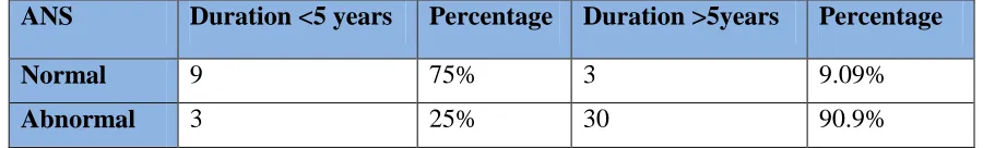
As shown in table 2, out of 45 patients, 12 patients had duration less than 5 years, out of this 3 had abnormal tests and 9 had normal tests, whereas 33 patients had BA of more than 5 years out of which 30 had AD and 3 had normal ANS.
Table 2: Comparison of duration of asthma with autonomic dysfunctions
ANS Duration <5 years Percentage Duration >5years Percentage
Normal 9 75% 3 9.09%
Abnormal 3 25% 30 90.9%
As shown in table 3, 24 patients had severe BA out of which 22 had AD and 2 had normal ANS. Out of 21, mild to moderate degree BA patients 11 had AD and 10 had normal ANS(P <0.01) indicating more severe the BA more the AD.
Table 3: Comparison between severity of bronchial asthma and number of patients with autonomic dysfunctions
ANS Mild to Moderate Percentage Severe Percentage
Normal 10 47.61 2 8.33
Abnormal 11 52.38 22 91.66
As shown in table 4, out of 18 males 14 had AD and out of 27 females, 19 had AD, and 8 had normal ANS (P< 0.1) which was statistically non significant.
Table 4: Number of males and females with autonomic dysfunction
ANS Male Percentage Female Percentage
Normal 4 22.22 8 29.62
Abnormal 14 77.77 19 70.37
Pag
e
318
Pag
e
318
Pag
e
318
Pag
e
318
Pag
e
318
Pag
e
318
Pag
e
318
Pag
e
318
Pag
e
318
Pag
e
318
Pag
e
318
Pag
e
318
Pag
e
318
Pag
e
318
Pag
e
318
Pag
e
318
Pag
e
318
Pag
e
318
Pag
e
318
Pag
e
318
Pag
e
318
Table 5: Other disorders which are common in bronchial asthma patients mainly with autonomic dysfunctions
ANS Anxiety
Disorder
Percent APD Percent GERD Peptic
Ulcer
Normal (n=12)
1 8.33 1 8.33% 100% 0%
Abnormal (n=33)
17 51.52 15 45.45% 80% 20%
Control (n=20)
1 5 0 0% 0% 0%
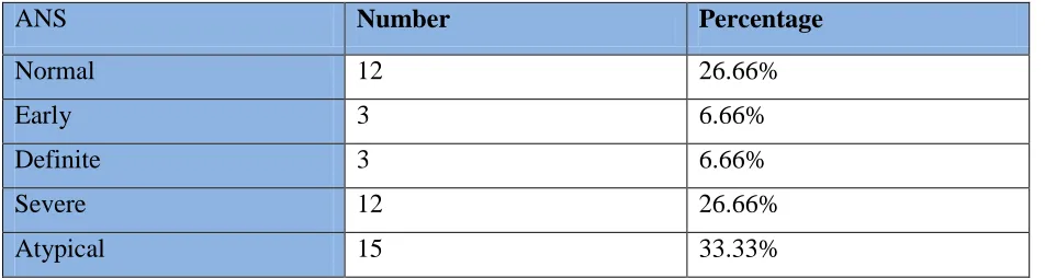
As shown in table 6, various category of AD and respective number of patients in each category.
Table 6: Categories into which autonomic dysfunction were divided
ANS Number Percentage
Normal 12 26.66%
Early 3 6.66%
Definite 3 6.66%
Severe 12 26.66%
Atypical 15 33.33%
Normal: All tests were normal or 1 borderline,
Early: 2 heart rate test border line or 1 heart rate test abnormal, BP tests normal
Definite: 2 heart rate test abnormal and BP test normal
Severe: 2 heart rate test abnormal and 2 BP test Borderline
Atypical: Any other combination of tests
DISCUSSION
The present study was conducted in 45 BA patients and, 20 age and gender matched healthy controls in study duration of 1 year from March, 2004 to February, 2005 who were attending OPD and admitted in wards in Department of Medicine, MGM Medical College & MY Hospital, Indore, MP, India.
In BA there is evidence of involvement of various types of cells including glands, blood vessels, mast cells, and smooth muscles. Recent evidence indicates an important role for the parasympathetic and
sympathetic system in regulation of airways smooth muscles in asthmatics.2 Bronchial smooth muscles constriction plays a major role in BA and recent studies implicate ANS. Parasympathetic system may affect the airway via reflexes involving bronchial smooth muscle or via increased mediator release. The
vagus nerve is important in normal
bronchoconstriction control in direct stimulation of
irritant receptors in airway epithelium (by
mechanical, chemical or pharmacological
stimulation) causing striking reflex
bronchoconstriction in vagal pathway. This reflex is exaggerated in asthma. Adrenergic innervation appears less potent. Beta adrenergic blockade has little or no effect in healthy subject but causes bronchoconstriction in asthmatic patients. Therefore bronchial hyperreactivity occurring in asthma apart from hyperplasia of smooth muscles may be due to abnormalities off parasympathetic or sympathetic nervous system. For cutaneous blood evaluation of beta adrenoreceptors and vagal activity. Most of the
e
319
e
319
e319
e
319
e319
e
319
e31
9
e
319
e319
e
319
e
319
e
319
e
319
e319
e
319
e319
e
319
e319
e
319
e319
e
319
administration of histamine or indirectly by studying the effects of beta blockers and beta-2 blockers. This study considered non–invasive test for beta receptor activity and parasympathetic activity. Inference made from studies varied from predominant alpha adrenergic hyperresponsiveness or parasympathetic vagal over activity or combination of both. The results were compared with the same studies carried out in normal healthy controls. The study considered of 45 patients of BA from 15-50 years of age. These patients were compared with 20 age and gender matched controls. The study population considered of 18 males and 27 female and 6 males and 14 females among control population. Then duration of asthma was noted in each for these patients. Duration was taken from the time of patient being diagnosed or having bronchial asthma till the date of performing the test. The duration varied from 8 months to 40 years.
It was found that out of 45 patients 33 patients had abnormal autonomic function tests and 12 had normal test. Most of our patients had more than one abnormal test. Control had all normal tests (P <0.001). This shows that BA patients have significant AD. Patients were further classified as those having early definite, severe or atypical involvement. Any patient who did not fit into the categories was considered as atypical. Most fit into atypical category i.e. 33%. A comparison was made between the relation between duration of BA and number of patients with AD. Arbitrarily duration was taken as <5 years and >5 years. 12 patients had duration less than 5 years out of this 3 patients with duration of BA less than 5 years had abnormal test, rest 9 had normal tests whereas 33 patients had BA for > 5 years out of these 30 had abnormal tests and only 3 patients had normal tests. P value<0.001 indicating that with chronicity the AD increases.
Comparing the severity of BA and AD, severity was defined on the basis of symptoms, number of hospitalization, number of exacerbation daily, use of beta-2 agonist, PEF variability etc. Out of 45 patients, 24 were classified as having severe BA and 21 as not severe. Out of these 24 patients, only 2 had normal autonomic function test rest 22 had AD, out of 21 patients classified as having mild to moderate BA only 11 had evidence of AD (P< 0.01), thus indicates that with severity the AD increases. Out of the 18 male patients, 14 had abnormal tests and 4 patients
did not have evidence of AD whereas 19 out of 27 females had AD and 8 had normal tests. P value <0.10 which implies that AD is observed irrespective of either sex. 21 of the study patients had only parasympathetic dysfunction i.e. 46.66%, 12 had both sympathetic and parasympathetic dysfunction i.e. 40% and only 6 had isolated sympathetic dysfunction i.e. 13.33%. This was in accordance with studies carried out by earlier workers who had stated that parasympathetic hyper responses are the main pathological event in causing AD in asthmatics.
Shah PK3 studied clinical dysautonomic in patients with BA. Their study comprises of 50 asthmatic patients and 20 healthy control subjects, carefully age and sex matched, were subjected to seven standardized tests to evaluate their autonomic status. Of the tests concerned with the parasympathetic system, the intravenous atropine test (p greater than 0.10) and heart rate response to standing (P greater than 0.01) which measured the basal parasympathetic tone, did not show a significant difference. Tests requiring stimulation of the parasympathetic system i.e. deep breathing test (p less than 0.01), valsalva maneuver (p less than 0.001) and carotid sinus massage (p less than 0.001) showed significantly heightened response. Postural fall of blood pressure (p greater than 0.10) and sustained hand grip test (p greater than 0.10) chiefly concerned with the sympathetic system, did not show a significant difference.
Kallenbach JM et al4 studied reflex rate control in asthma. They postulated that an abnormality in the autonomic control of airway calibre might be reflected by a parallel change in the reflex control of heart rate. They heart rate variations induced by deep breathing (respiratory examined the sinus arrhythmia ), the valsalva maneuver, and standing up from the recumbent position in asthmatic subjects and non asthmatic control subjects. The asthmatic patients had evidence of enhanced parasympathetic neural drive to the sinoatrial node, as manifested by a significantly greater magnitude of respiratory sinus arrhythmia, then the controls (p less than 0.0005) they were unable to induce a similar change in normal subjects by resistance breathing.
CONCLUSIONS
Pag
e
320
Pag
e
320
Pag
e
320
Pag
e
320
Pag
e
320
Pag
e
320
Pag
e
320
Pag
e
320
Pag
e
320
Pag
e
320
Pag
e
320
Pag
e
320
Pag
e
320
Pag
e
320
Pag
e
320
Pag
e
320
Pag
e
320
Pag
e
320
Pag
e
320
Pag
e
320
Pag
e
320
gender matched controls. With chronicity more incidence of autonomic dysfunction is observed. As the severity of BA increases (as judged by symptoms, PEF variability, use of beta-2 agonist, and dose of inhaled steroids) it is observed that the incidence of autonomic dysfunction also increases.
DISCLOSURES
Conflict of interest: Not declared
Funding: Nil
Ethical approval: Study was approved by institutional review board.
REFERENCES
1. P.J.Barnes, S.D.Shapiro, R.A.Pauwels.
Chronic obstructive pulmonary disease:
molecular and cellularmechanisms. European Respiratory Journal 2003; 22: 672-688.
2. Allison D. Fryer and David B. Jacoby. Muscarinic Receptors and Control of Airway Smooth Muscle. AJRCCM 1998; 158: 2.
3. Shah PK, Lakhotia M, Mehta S et al. Clinical dysautonomia in patients with bronchial asthma. Study with seven autonomic function tests. Chest 1990; 98:1408-13.