R E S E A R C H A R T I C L E
Open Access
Ancestral mesodermal reorganization and
evolution of the vertebrate head
Takayuki Onai
1*, Toshihiro Aramaki
2, Hidehiko Inomata
3, Tamami Hirai
1and Shigeru Kuratani
1Abstract
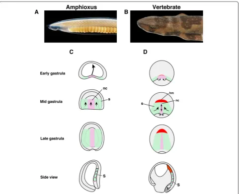
Introduction:The vertebrate head is characterized by unsegmented head mesoderm the evolutionary origin of which remains enigmatic. The head mesoderm is derived from the rostral part of the dorsal mesoderm, which is regionalized anteroposteriorly during gastrulation. The basal chordate amphioxus resembles vertebrates due to the presence of somites, but it lacks unsegmented head mesoderm. Gastrulation in amphioxus occurs by simple invagination with little mesodermal involution, whereas in vertebrates gastrulation is organized by massive cell movements, such as involution, convergence and extension, and cell migration.
Results:To identify key developmental events in the evolution of the vertebrate head mesoderm, we compared anterior/posterior (A/P) patterning mechanisms of the dorsal mesoderm in amphioxus and vertebrates. The dorsal mesodermal genesgsc,bra, anddeltaare expressed in similar patterns in early embryos of both animals, but later in development, these expression domains become anteroposteriorly segregated only in vertebrates. Suppression of mesodermal involution in vertebrate embryos by inhibition of convergence and extension recapitulates amphioxus-like dorsal mesoderm formation.
Conclusions:Reorganization of ancient mesoderm was likely involved in the evolution of the vertebrate head.
Keywords:Amphioxus, Head mesoderm, Vertebrate body plan, Somites
Introduction
How the highly complex vertebrate head—composed of brain, head muscles, and skull—evolved from non-vertebrate ancestors is a fundamental question in current evolutionary and developmental biology [1–3]. Recent comparative studies of the cephalochordate amphioxus and vertebrates suggest that a region hom-ologous to the vertebrate fore/mid/hind brain is also present in the rostral part of the central nervous system (CNS) of amphioxus [4–6]. Amphioxus is a basal chordate that has somites extending to the rostral end of the body, and is considered the best proxy for under-standing the origin of the vertebrate body plan [7].
The homology between amphioxus and the vertebrate CNS indicates that the unsegmented vertebrate head mesoderm evolved directly from the rostral somites of amphioxus [8]. Expression ofenin the rostral somites of
amphioxus (Branchiostoma floridae) anden2in the ven-tral part of the mandibular head mesoderm of shark (Scyliorhinus torazame) embryos supports this hypoth-esis [9, 10]. However, Bfpax3/7, a homologue of pax3 that serves as a somite marker in vertebrates, is expressed in the rostral somites, suggesting that the vertebrate head mesoderm did not evolve by simple modification of rostral somites of an amphioxus-like ancestor, but rather by fundamental reorganization that occurred in the dorsal mesoderm [2, 10].
During embryogenesis, the vertebrate head meso-derm derived from the rostral part of the dorsal mesoderm is regionalized along the A/P axis by a gradient of Wnt/β-catenin signalling [11, 12]. In the regionalization of the dorsal mesoderm, downstream regional marker genes of Wnt/β-catenin signalling are expressed in the progenitor domains; gsc is expressed
in the head mesoderm, whereas bra is expressed in
the presumptive notochord during the late-gastrula stage [13, 14]. Additionally, in the trunk mesoderm, delta expression is detected in the presumptive so-mite region [15, 16]. Previous functional studies have * Correspondence:[email protected]
1Kuratani Evolutionary Morphology Laboratory, RIKEN Center for
Developmental Biology, 2-2-3 Minatojima-minamimachi, Chuo-ku, Kobe 650-0047, Japan
Full list of author information is available at the end of the article
shown that overexpression of Xenopus laevis dkk1 (negative regulator of Wnt/β-catenin signalling) expands the gsc expression domain posteriorly in Xenopus em-bryos, whereasbra expression is activated by Wnt/β-ca-tenin signalling [11, 17, 18]. Additionally, delta has an essential role in somitogenesis, and is under the control of Wnt/β-catenin signalling [19].
In amphioxus, gsc and bra are co-expressed in the presumptive notochordal region at the gastrula stage
[20, 21]. The presumptive somite marker delta is
expressed in the first and second somites in the
late-gastrula stage [22]. Loss of gsc expression in the
notochord and gain of gsc expression in the head
mesoderm of vertebrates compared with amphioxus indicates that A/P re-arrangement of mesodermal gene expression occurred in the lineage of vertebrates. Excessive Wnt/β-catenin signalling in amphioxus
em-bryos induced by inhibition of GSK-3α/β does not
affect the expression of regional marker genes of the dorsal mesoderm, such as bra and fgf8/17/18, during the gastrula stage [23]. This suggests that, unlike in vertebrates, Wnt/β-catenin signalling does not play a role in dorsal mesoderm regionalization in amphioxus. If vertebrate embryos did evolve a rearrangement of gene expression in the dorsal mesoderm to generate the head mesodermal region, what was the key devel-opmental event in this process? We consider that re-arrangement of gene expression in the vertebrate dorsal mesoderm from an ancestral chordate evolved through a novel mesodermal cell movement present in vertebrates.
Amphioxus gastrulation occurs through simple inva-gination, with little mesodermal involution of the outer layer [24], whereas in vertebrates, an overt involution
takes place, as observed in Xenopus and lamprey
[25–27]. Thus, uniquely in amphioxus and distinct from the case in vertebrates, there is nearly no change in the relative positions of the ectoderm and mesoderm. However, it remains unclear how mesodermal involution affects A/P patterning of the dorsal mesoderm and how this change has led to two distinct types of ros-tral mesoderm in amphioxus and vertebrates.
To explore the molecular background of vertebrate head mesoderm evolution, we first investigated the de-velopmental stage at which the overall molecular topog-raphy of the dorsal mesoderm becomes distinctly different between amphioxus and vertebrates. We also examined whether mesodermal involution is important for dorsal mesoderm regionalization in vertebrate em-bryos, as a possible developmental factor that gave rise to vertebrate head mesoderm. Finally, we examined whether the genetic program for mesodermal involution is present in amphioxus. We hypothesize that vertebrate head mesoderm evolved from an amphioxus-like ancestral
mesoderm through anteroposterior reorganization of the genetic developmental architecture.
Materials and methods
Sources of amphioxus, lamprey, and shark embryos In the summer breeding season, adult amphioxus (B. floridae) were collected from Old Tampa Bay, FL, USA. The in vitro fertilization and culturing of the embryos were conducted as described [28]. Adult amphi-oxus (Branchiostoma japonicum) were collected from the ocean near Amakusa Island, Kumamoto, Japan. Adult lampreys (Lethenteron japonicum) were collected from the Miomote River in Niigata, Japan and the Shirubetu River in Hokkaido, Japan during the spring breeding season. In vitro fertilization was performed as described previously [29]. Adult sharks (S. torazame) were collected from Nakaminato Bay, Ibaraki, Japan in October and maintained in a seawater tank at 16 °C. Eggs were ob-tained from the adult females and mainob-tained in the seawater tank until they developed to the described stages [30].
In situ hybridization
Whole-mount in situ hybridization of amphioxus, lam-prey, shark, andXenopusembryos was performed as de-scribed in previous studies [10, 29, 31]. For section in situ hybridization of Xenopus embryos, larval-stage embryos were fixed using MEMFA for 2 h at room temperature then section in situ hybridization was per-formed as described [32]. For fluorescence in situ hybridization, the protocol used for amphioxus whole-mount in situ hybridization [23] was applied and an antibody (Anti-DIG-POD, Roche, Basel, Switzerland) and TSA system (PerkinElmer, Waltham, MA, USA) were used. Cellmask deep red (Life Technologies, Carlsbad, CA, USA) (1/1000) and DAPI (Invitrogen, Carlsbad, CA, USA) (1/1000) were used to stain the plasma membrane or nuclei. A Zeiss LSM 780 (Zeiss, Jena, Germany) was used to collect confocal images.
Plasmid construction and gene markers
The sequences of Bfdkk1/2/4 [21], Bfgsc [21], Bfbra [20], Bfwnt8 [21], Bfdelta [22], Stdkk1 (KF551566), Stgsc (KF564642), Stdelta(KF551567), Stbra (KF551568),
and Stwnt8 (KF551569) were amplified by PCR and
cloned into a TOPO cloning vector. Xldkk1-pCS2
(NM_001085592), Xlgsc-pCS2 (NM_001087809), Xldelta–
2-pCS2 (NM_001086082), Xlbra-pSP64 (M77243.1),
Xlwnt8-pCS2 (NM_001088168), and XlmyoD-pCS2
(NM_001085897) were gifts from Dr. Yoshiki Sasai
from RIKEN CDB, Japan. Xltbx1 (NM_001090445)
by PCR and cloned into a TOPO cloning vector. Xldd1-myc-pCS2, myc-XldshdelDEP-pCS2, and Bfrnd1 -pCS2 were linearized using NotI and transcribed using SP6 polymerase (mMessage mMachine, Ambion, Austin, TX, USA).
Xenopusexperiments
Embryos were staged according to the Normal Table published by Nieuwkoop and Faber [33]. The mRNA, in 1× modified Barth’s saline, was injected into the embryos using a fine glass capillary tube and a pressure injector (Narishige, Tokyo, Japan). The embryos were then trans-ferred into 0.1× Barth’s saline until further manipulation or harvest. For histological analysis, the embryos were fixed using Bouin’s fixative and then dehydrated and em-bedded in paraffin. Sections (6-μm-thick) were cut and stained with haematoxylin and eosin. The sequence of the Xlrnd1-morpholino antisense oligonucleotide (MO) has been published [34]. For the dorsal marginal zone assay, embryos were dissected at the early-gastrula stage (stage 10) and cultured in 1× low-calcium magnesium Riner’s supplemented with 0.2 % bovine serum albumin until stage 19.
Immunostaining of amphioxus andXenopusembryos Early-neurula-stage amphioxus embryos were fixed using 4 % paraformaldehyde in MOPS buffer at 4 °C overnight, and immunocytochemistry was performed as described [35], using a primary antibody against β-catenin (diluted 1:800 forXenopusembryos and 1:400 for amphioxus em-bryos; C-2206, Sigma, St. Louis, MO, USA), a secondary Alexa Fluor 488-conjugated antibody (1:400, Invitrogen), and DAPI (1:1000, Invitrogen). Whole embryos and histo-logical sections were imaged using a Zeiss LSM 710 con-focal microscope and a Zeiss Axio Zoom V16 microscope, respectively.
Phylogenetic tree analysis
Phylogenetic trees were constructed with MEGA5 soft-ware [36] using the maximum-likelihood method with 1000 bootstrap reiterations. All sequences were aligned using Clustal W (http://www.clustal.org/clustal2/).
Alignment analysis of Flrt3
For Flrt3 protein alignment analysis, we used the multiple alignment tool in Genetyx–Mac version 16.0.7 (http:// www.genetyx.co.jp/products/genetyx_mac_16/index.html).
Accession numbers
The sequences of the novel genes isolated were depos-ited in GenBank with following accession numbers. Ljgsc (KF551572), Ljdelta1 (KF564639), Ljwnt8 (KF551570), Stdkk1 (KF551566), Stgsc (KF564642), Stbrachyury (KF551568), Stdelta1(KF551567), and Stwnt8(KF551569).
Results and discussion
Genetic topography of the vertebrate dorsal mesoderm evolved through A/P expression domain shift of amphioxus mesodermal genes
In the dorsal mesoderm of amphioxus embryos, somites and notochord are regionalized during gastrulation, and an equivalent structure of the vertebrate unsegmented head mesoderm is thought to be absent in amphioxus [7] (Fig. 1). The dorsal mesoderm of vertebrates such as Xenopus is formed by massive mesodermal involution during the early-gastrula stage, which is not present in amphioxus embryos and is regionalized into head meso-derm, notochord, and somites [27, 37] (Fig. 1), suggest-ing that genetic programs govern differences between amphioxus and vertebrate mesodermal formation. We thus examined the molecular topography of the dorsal
mesoderm in amphioxus and Xenopus. We first
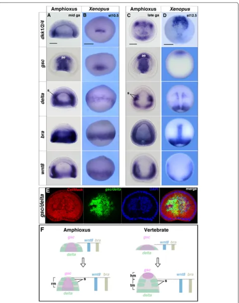
com-pared the mid-gastrula stage of amphioxus embryos and stage 10.5 Xenopus embryos because the orientation of dorsal mesodermal tissue is approximately parallel to the A/P body axis in both species, and thus comparable at these stages [33] (Additional file 1: Figure S1). The key regional genes include gsc (head mesoderm),bra (noto-chord), delta (somite), wnt8 (somite) and dkk1 (head mesoderm, somite).
By the mid-gastrula stage, all genes examined were expressed around the blastopore and showed similar pat-terns in amphioxus and vertebrates (Fig. 2a and b). However, by the late-gastrula stage of Xenopus, the ex-pression domains of the regional marker genes became separated anteroposteriorly, with the gsc expression do-main barely overlapping with those of delta, wnt8, and braas the head and trunk mesodermal identities became distinct (Fig. 2c–f ). These dynamic shifts were also ob-served in basal vertebrates, such as the lamprey (L. japo-nicum) and shark (S. torazame) (Additional file 1: Figures S2, S3 and Table S1). During the late-gastrula stage, the presomitic mesodermal region became distinct around the blastopore, and dkk1/2/4, delta, bra, and
wnt8 were co-expressed in Xenopus presomitic
meso-derm (Fig. 2d). In amphioxus, somites were found to form directly from the tail bud, and the expression of dkk1/2/4, delta, bra, and wnt8largely overlapped in the prospective tail bud region (Fig. 2c). These results sug-gest that during gastrula stages, the mesodermal genes segregate anteroposteriorly only in vertebrates, whereas in amphioxus, these genes overlap considerably (Fig. 2f ).
However, in amphioxus, the relative increase in mesoder-mal size was much lower than that in vertebrates (Fig. 3c). This suggests that, in vertebrates, the dynamic mesoder-mal gene shift is achieved by increasing the mesoderm, which is primarily dictated by mesodermal cell move-ments (e.g. involution, convergence and extension) (Fig. 3c). During gastrulation, mesodermal involution is controlled by convergence and extension of the dorsal axial mesoderm in vertebrates [38, 39]. To determine whether mesodermal involution is essential for the meso-dermal gene shift, we suppressed convergent extension by inhibiting the Wnt planar cell polarity (PCP) signalling pathway, (a key signal transduction pathway in conver-gence and extension) [40, 41]. For the loss of function
study of Wnt/PCP signalling, we injected Xldd1 (a
dominant-negative form of Xldsh; [42, 43]) mRNA into Xenopusembryos [26].
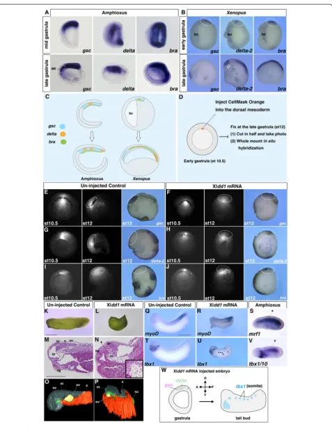
In the dye only-injected control embryos, labelled cells migrated anteriorly and expressedgsc but notdelta-2 or bra (Fig. 3d, e, g and i). In Xldd1 mRNA-injected em-bryos, however, labelled cells did not migrate anteriorly, but remained close to the blastopore, and gsc, delta-2, and bra were not separated anteroposteriorly as ob-served in the control embryos during the late-gastrula stage (Fig. 3f, h and j). Additionally, the size of the dorsal
mesoderm was much smaller in the Xldd1 mRNA–
injected embryos compared with control embryos (Fig. 3e–j), indicating that the developmental sequences of the dorsal mesoderm were somewhat similar to those in amphioxus. Microinjection of XldshdelDEP mRNA, a mutant dsh that specifically inhibits the Wnt/PCP sig-nalling pathway [44], indicated that the effect of Xldd1 injection resulted from suppression of the Wnt/PCP signalling pathway (Additional file 1: Figure S4A–F).
Overlapping head and trunk marker gene expression was also detected based on the results of the dorsal mar-ginal zone assay, suggesting that the effect of Xldd1 in-jection was not attributable to differences in the amount of yolk, but more likely to the loss of mesodermal cell movement (Additional file 1: Figure S4I–L).
These results are consistent with morphological changes observed at the tail bud stage in Xldd1-injectedXenopus embryos assimilated to an amphioxus-like condition. Specifically, the anterior-most somite, normally appearing just posterior to the anterior end of the notochord, was extended anteriorly into the prechordal domain (Fig. 3k–p). In these embryos, the somite marker
myoD was expressed normally, similar to mrf1 in
amphioxus embryos (Fig. 3q–s). Interestingly, ectopic expression of tbx1, a head mesoderm marker in verte-brates [10], was also detected in the Xldd1-injected Xenopus somites, similar to amphioxus tbx1/10, a so-mite marker in amphioxus [45], expression (Fig. 3t–w and Additional file 1: Figure S4G and H). These re-sults suggest that the vertebrate-specific mesodermal involution during gastrulation is likely responsible for the A/P distinction between head and trunk meso-derm, which does not occur in amphioxus.
A/P patterning in the dorsal mesoderm by different Wnt/
β-catenin-signalling input is a vertebrate novelty
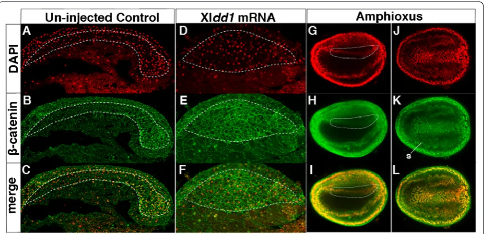
Previous functional studies in vertebrates have shown that the dorsal mesoderm is regionalized by a Wnt/β-ca-tenin-signalling gradient along the A/P axis during early embryogenesis [46]. The failure of A/P segregation of mesodermal regional gene expression in Xldd1 mRNA-injected embryos suggests that Wnt/β-catenin-signalling pathway control of these downstream genes is compro-mised in this context. We examined the nuclear localization of β-catenin, a downstream factor in the
Wnt/β-catenin signalling pathway, in Xldd1
mRNA-injected embryos. In the control embryos, nuclear localization of β-catenin was observed in the posterior dorsal mesodermal cells, but not in the anterior region
(Fig. 4a–c). In Xldd1 mRNA-injected embryos,
how-ever, β-catenin localized to the nucleus in some cells, but there was no clear A/P difference in the degree of nuclear localization (Fig. 4d–f ). The lack of obvious A/P difference in β-catenin nuclear localization was also ob-served in the amphioxus dorsal mesoderm (Fig. 4g–l).
Consistent with this result, a previous functional study showed that Wnt/β-catenin signalling had no role in the A/P patterning of the dorsal mesoderm during the gas-trula stages [23]. These findings suggest that anterior low and posterior high Wnt/β-catenin-signalling input is im-portant in the segregation of regional marker genes of the dorsal mesoderm along the A/P axis that evolved in vertebrates.
Evolution of head mesoderm in vertebrates
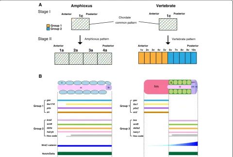
In this study, we propose that the vertebrate dorsal mesoderm evolved as an entirely novel pattern associ-ated with a new mechanism of mesoderm specifica-tion. As was first described by Ernst Haeckel [47], amphioxus gastrulation involves a simple invagination similar to that in cnidarians, with little change in the spatial relationships between the ectoderm and mesoen-doderm during development. Unlike in vertebrates, the dorsal mesoderm in amphioxus co-expresses both verte-brate head and trunk mesoderm marker genes (Fig. 5). Thus, the amphioxus dorsal mesoderm remains unspeci-fied along the A/P axis due to the absence of a vertebrate-specific developmental program (Fig. 5). Vertebrate meso-derm, however, is uniquely polarized along the A/P axis into the head and trunk mesoderm based on mesodermal patterning mediated by vertebrate-specific cell movement. By the end of gastrulation, the head and trunk mesoder-mal identities are specified by anteroposteriorly dislocated expression of regional marker genes. This patterning mechanism also controls A/P patterning of the overlying neuroectoderm. In vertebrates, the A/P regional identity of the CNS is organized by vertical signals from the under-lying dorsal mesoderm containing A/P pattern informa-tion [48]. In amphioxus, based on the topography of regional markers, the CNS is patterned into domains largely comparable to the fore-/mid-/hindbrain and the spinal cord in vertebrate embryos [5]. However, three major signalling centres (anterior neural ridge, zona limit-ans intrathalamica, and midbrain–hindbrain boundary) are absent in the amphioxus CNS [5, 49]. Given that our current study indicated that the A/P regional identity of the vertebrate dorsal mesoderm is fundamentally different from that of amphioxus (Fig. 5a), the three major signal-ling centres in the neuroectoderm of vertebrate embryos may have evolved through a reorganization of the dorsal mesoderm in an amphioxus-like chordate ancestor.
(See figure on previous page.)
Evolutionary reorganization of the entire dorsal meso-derm as described above would imply that individual amphioxus somites are not homologous to any specific region of the vertebrate head mesoderm. Our scenario instead favours the novel nature of the vertebrate mesoderm generated by modification in mesodermal patterning dynamics. From the perspective of vertebrate mesodermal specification, the segmented mesoderm in amphioxus appears as an intermediate between the head mesoderm and trunk somites, and vertebrate somites do not represent primitive traits, but rather derived traits established by removal of head mesoderm-like properties from ancestral somites. This scenario of vertebrate head evolution correlates with the observation that peripheral
nerves in amphioxus possess traits of both cranial and spinal nerves [50, 51]. The mesodermal developmental pattern is shared among chordates only in the early gastrulae, in which the mesoderm is not yet antero-posteriorly polarized, possibly representing a plesio-morphic state (Fig. 5). The A/P patterning of mesodermal identities through the different Wnt/β-catenin-signalling input takes place only in vertebrates later in the de-velopmental process; along with unique changes in cell movement, this can be considered a synapomorphic developmental trait for this animal subphylum. As pro-posed by Haeckel, this indicates that the vertebrate body plan is established by recapitulating an amphioxus-like an-cestral pattern (plesiomorphy) during the early-gastrula
(See figure on previous page.)
Fig. 3Inhibition of mesodermal involution in vertebrates recapitulates amphioxus A/P patterning. In situ hybridization ofgsc,deltaandbrain amphioxus (a) andXenopus(b) embryos. Thearrowheadsfordelta-2in the late gastrulaXenopusembryo indicate expression in somites.aeanterior endoderm,bcblastocoel.cSchematic diagram of relative size increase of the dorsal mesoderm during gastrulation in amphioxus and vertebrates.d Overview of the labelling study of the dorsal mesoderm inXenopus. CellMask Orange (fluorescent dye) was injected into the dorsal mesoderm at stage (st) 10.5 and the embryos were cultured until stage 12. Fixed embryos were cut in half and then subjected to in situ hybridization.e,g,iAt stage 12 in the control, labelled cells migrated anteriorly to form the head mesoderm (n= 21, 100 %) and expressedgsc(n= 2, 100 %) but notdelta-2(n= 2, 100 %) orbra(n= 3, 100 %).f,h,jAt stage 12 in the embryos that had been injected with 400 pg/cell Xldd1mRNA at the four-cell blastomere stage, the labelled cells stayed near the blastopore region (n= 40, 100 %), andgsc(n= 2, 100 %),delta-2(n= 6, 100 %), andbra (n= 3, 100 %) expression overlapped along the A/P axis.ststage. Thedotted circleindicates the cells labelled by CellMask Orange. External morphology (k,l), histological sections (m,n) and 3-D reconstruction (o,p) of control (n= 22, 100 %; left) and Xldd1mRNA–injected embryos (n= 35, 94 %; right).ncnotochord,ssomite,br brain,cgcement gland,ovotic vesicle, ntneural tube. Expression ofmyoD(muscle differentiation marker) (q;n= 15, 100 %,r;n= 14, 100 %) andtbx1(t;n= 12, 100 %,u;n= 20, 90 %) in control and Xldd1mRNA–injectedXenopus embryos, respectively.smrf1and (v)tbx1/10(pharynx and somite marker) expression in amphioxus embryos.wSchematic diagram of amphioxus-like developmental patterns in Xldd1mRNA–injectedXenopusembryos.Scale bars, 1 mm (k,q) and 500μm (m)
stage and engaging a novel pattern (synapomorphy) dur-ing the later stages. The comparison of mesodermal gene expression of vertebrates, amphioxus, and hemichordates (as an out-group taxon) suggests that the amphioxus mesoderm has an intermediate nature, possibly represent-ing a plesiomorphic state for deuterostomes (http:// www.ibiology.org/ibioseminars/evolution-ecology/marc-w-kirschner-part-3.html). This characterization of the evolu-tionary sequence of developmental dynamics in chordates also provides insight into the potential origins of mesoderm and mesodermal segments in bilaterians.
Possible mechanism of mesodermal involution unique to vertebrates
In this study, we showed that inhibition of mesodermal involution in vertebrate embryos recapitulated amphi-oxus development (Fig. 3). The lack of mesodermal
involution and likely convergent extension in amphioxus gastrulation indicates that the developmental program for mesodermal involution in vertebrates is absent in amphioxus. Disruption of cadherin-mediated cell–cell adhesion is essential during mesodermal involution and convergent extension in vertebrates, and fibronectin leucine-rich-repeat transmembrane 3 (Flrt3) and a small GTPase (Rnd1) control C-cadherin degradation [26, 34, 52, 53]. A BLAST search revealed a homologue of rnd1in amphioxus, but not of flrt3 (Additional file 1:
Figure S5A and B). In amphioxus, rnd1 expression
was observed around the blastopore (Additional file 1:
Figure S6A–H). However, overexpression of Bfrnd1
mRNA could not rescue the loss of endogenous rnd1
in Xenopus (Additional file 1: Figure S6I–M). These findings suggest that involvement of the cadherin degradation system in mesodermal involution as well
as convergence and extension may have emerged in the vertebrate lineage.
Conclusions
Our findings indicate that the A/P patterning of the vertebrate dorsal mesoderm evolved from an amphioxus-like ancestral mesoderm through A/P polarization of mesodermal specification to divide into the unsegmented head mesoderm anteriorly and the segmented trunk somites posteriorly. Vertebrate head mesoderm is thus an evolutionary novelty.
Additional file
Additional file 1: Figure S1.Dorsal mesoderm formation in chordates. Figure S2.Phylogenetic trees of dorsal mesoderm genes.Figure S3. Dorsal mesodermal gene expression in lamprey (L. japonicum) and shark (S. torazame) embryos.Figure S4.Suppression of the Wnt/PCP-signaling pathway inXenopusembryos.Figure S5.Flrt3 evolved in the vertebrate lineage.Figure S6.Flrt3-Rnd1 system is essential for mesoderm forma-tion in vertebrates.Table S1.Summary of expression pattern of genes in Figure S2.(DOCX 8727 kb)
Competing interests
The authors declare they have no competing interests.
Authors’contributions
TO and SK wrote the paper. TO and TH performed the experiments. TO, TA and HI designed the experiments. All authors read and approved the final manuscript.
Acknowledgements
We thank Linda Holland and Nick Holland of the University of California, San Diego, CA, USA for help with the collection of amphioxus and for discussion. We also thank Yasuhisa Henmi of Kumamoto University, Japan and Kinya Yasui of Hiroshima University, Japan for helping with amphioxus collection and Shigehiro Kuraku of RIKEN, Japan for preparing the phylogenetic trees. This research was supported by a KAKENHI Grant-in-Aid for Young Scientists (B) from the Japan Society for the Promotion of Science (grant number 24770222).
Author details 1
Kuratani Evolutionary Morphology Laboratory, RIKEN Center for Developmental Biology, 2-2-3 Minatojima-minamimachi, Chuo-ku, Kobe 650-0047, Japan.2Pattern Formation Group, Graduate School of Frontier
Biosciences, Osaka University, 1-3 Yamadaoka, Suita, Osaka 565-0871, Japan.
3
Laboratory for Axial Pattern Dynamics, RIKEN Center for Developmental Biology, 2-2-3 Minatojima-minamimachi, Chuo-ku, Kobe 650-0047, Japan.
Received: 9 June 2015 Accepted: 22 September 2015
References
1. Gans C, Northcutt RG. Neural crest and the origin of vertebrates: a new head. Science. 1983;220:268–74.
2. Onai T, Irie N, Kuratani S. The evolutionary origin of the vertebrate body plan: the problem of head segmentation. Annu Rev Genom Hum Genet. 2014;15:443–59.
3. Romer AS. The vertebrate as a dual animal–somatic and visceral. Evol Biol. 1972;6:121–56.
4. Holland LZ. Chordate roots of the vertebrate nervous system: expanding the molecular toolkit. Nat Rev Neurosci. 2009;10(10):736–46.
5. Holland LZ, Carvalho JE, Escriva H, Laudet V, Schubert M, Shimeld SM, et al. Evolution of bilaterian central nervous systems: a single origin? EvoDevo. 2013;4(1):27.
6. Wicht H, Lacalli TC. The nervous system of amphioxus: structure, development, and evolutionary significance. Can J Zool. 2005;83:122–50. 7. Holland LZ, Onai T. Early development of cephalochordate (amphioxus).
WIREs Dev Biol. 2011;1:167–83.
8. Holland LZ, Holland ND, Gilland E. Amphioxus and the evolution of head segmentation. Integr Comp Biol. 2008;48(5):630–46.
9. Holland LZ, Kene M, Williams NA, Holland ND. Sequence and embryonic expression of the amphioxus engrailed gene (AmphiEn): the metameric pattern of transcription resembles that of its segment-polarity homolog in Drosophila. Development. 1997;124(9):1723–32.
10. Adachi N, Takechi M, Hirai T, Kuratani S. Development of the head and trunk mesoderm in the dogfish, Scyliorhinus torazame: II. Comparison of gene expression between the head mesoderm and somites with reference to the origin of the vertebrate head. Evol Dev. 2012;14(3):257–76. 11. Kazanskaya O, Glinka A, Niehrs C. The role of Xenopus dickkopf1 in
prechordal plate specification and neural patterning. Development. 2000;127(22):4981–92.
12. Niehrs C. Regionally specific induction by the Spemann-Mangold organizer. Nat Rev Genet. 2004;5(6):425–34.
13. Cho KW, Blumberg B, Steinbeisser H, De Robertis EM. Molecular nature of Spemann’s organizer: the role of the Xenopus homeobox gene goosecoid. Cell. 1991;67(6):1111–20.
14. Smith JC, Price BM, Green JB, Weigel D, Herrmann BG. Expression of a Xenopus homolog of Brachyury (T) is an immediate-early response to mesoderm induction. Cell. 1991;67(1):79–87.
15. Peres JN, McNulty CL, Durston AJ. Interaction between X-Delta-2 and Hox genes regulates segmentation and patterning of the anteroposterior axis. Mech Develop. 2006;123(4):321–33.
16. Pourquie O. Vertebrate segmentation: from cyclic gene networks to scoliosis. Cell. 2011;145(5):650–63.
17. Martin BL, Kimelman D. Regulation of canonical Wnt signaling by Brachyury is essential for posterior mesoderm formation. Dev Cell. 2008;15(1):121–33.
18. Morley RH, Lachani K, Keefe D, Gilchrist MJ, Flicek P, Smith JC, et al. A gene regulatory network directed by zebrafish No tail accounts for its roles in mesoderm formation. Proc Natl Acad Sci U S A. 2009;106(10):3829–34. 19. Aulehla A, Wiegraebe W, Baubet V, Wahl MB, Deng C, Taketo M, et al. A
beta-catenin gradient links the clock and wavefront systems in mouse embryo segmentation. Nature Cell Biol. 2008;10(2):186–93.
20. Holland PW, Koschorz B, Holland LZ, Herrmann BG. Conservation of Brachyury (T) genes in amphioxus and vertebrates: developmental and evolutionary implications. Development. 1995;121(12):4283–91.
21. Yu JK, Satou Y, Holland ND, Shin IT, Kohara Y, Satoh N, et al. Axial patterning in cephalochordates and the evolution of the organizer. Nature.
2007;445(7128):613–7.
22. Rasmussen SL, Holland LZ, Schubert M, Beaster-Jones L, Holland ND. Amphioxus AmphiDelta: evolution of Delta protein structure, segmentation, and neurogenesis. Genesis. 2007;45(3):113–22.
23. Onai T, Takai A, Setiamarga DH, Holland LZ. Essential role of Dkk3 for head formation by inhibiting Wnt/beta-catenin and Nodal/Vg1 signaling pathways in the basal chordate amphioxus. Evol Dev. 2012;14(4):338–50. 24. Zhang S, Holland ND, Holland LZ. Topographic changes in nascent and
early mesoderm in amphioxus embryos studied by DiI labeling and by in situ hybridization for a Brachyury gene. Dev Genes Evol. 1997;206:532–5. 25. Damas H. Recherches sur le dévellopement de Lampetra fluviatilis L.
Contribution à l’étude de la Céphalogenèse des vertébrés. Archives de Biologie, Liége et Paris. 1944;55:1–248.
26. Hammerschmidt M, Wedlich D. Regulated adhesion as a driving force of gastrulation movements. Development. 2008;135(22):3625–41. 27. Winklbauer R, Damm EW. Internalizing the vegetal cell mass before and
during amphibian gastrulation: vegetal rotation and related movements. WIREs Interdiscip Rev Dev Biol. 2012;1:301–6.
28. Holland LZ, Onai T. Analyses of gene function in amphioxus embryos by microinjection of mRNAs and morpholino oligonucleotides. Methods Mol Biol. 2011;770:423–38.
29. Takio Y, Kuraku S, Murakami Y, Pasqualetti M, Rijli FM, Narita Y, et al. Hox gene expression patterns in Lethenteron japonicum embryos–insights into the evolution of the vertebrate Hox code. Dev Biol. 2007;308(2):606–20. 30. Ballard WW, Mellinger J, Lechenaud H. A series of normal stages for
31. Onai T, Matsuo-Takasaki M, Inomata H, Aramaki T, Matsumura M, Yakura R, et al. XTsh3 is an essential enhancing factor of canonical Wnt signaling in Xenopus axial determination. EMBO J. 2007;26(9):2350–60.
32. Butler K, Zorn AM, Gurdon JB. Nonradioactive in situ hybridization to xenopus tissue sections. Methods. 2001;23(4):303–12.
33. Nieuwkoop PD, Faber J. Normal table of Xenopus laevis. Amsterdam, Netherlands: North-Holland Publishing Co; 1956.
34. Ogata S, Morokuma J, Hayata T, Kolle G, Niehrs C, Ueno N, et al. TGF-beta signaling-mediated morphogenesis: modulation of cell adhesion via cadherin endocytosis. Genes Dev. 2007;21(14):1817–31.
35. Lu TM, Luo YJ, Yu JK. BMP and Delta/Notch signaling control the development of amphioxus epidermal sensory neurons: insights into the evolution of the peripheral sensory system. Development.
2012;139(11):2020–30.
36. Tamura K, Peterson D, Peterson N, Stecher G, Nei M, Kumar S. MEGA5: molecular evolutionary genetics analysis using maximum likelihood, evolutionary distance, and maximum parsimony methods. Mol Biol Evol. 2011;28(10):2731–9.
37. Keller R. Mechanisms of elongation in embryogenesis. Development. 2006;133(12):2291–302.
38. Shi J, Keller R. Gastulation in Xenopus laevis: involution–a current view. Semin Cell Dev Biol. 1994;5:85–90.
39. Keller R, Davidson L, Edlund A, Elul T, Ezin M, Shook D, et al. Mechanisms of convergence and extension by cell intercalation. Philos Trans R Soc Lond B Biol Sci. 2000;355(1399):897–922.
40. Habas R, Kato Y, He X. Wnt/Frizzled activation of Rho regulates vertebrate gastrulation and requires a novel Formin homology protein Daam1. Cell. 2001;107(7):843–54.
41. Solnica-Krezel L, Sepich DS. Gastrulation: making and shaping germ layers. Annu Rev Cell Dev Biol. 2012;28:687–717.
42. Sokol SY. Analysis of Dishevelled signalling pathways during Xenopus development. Curr Biol. 1996;6(11):1456–67.
43. Tanegashima K, Zhao H, Dawid IB. WGEF activates Rho in the Wnt-PCP pathway and controls convergent extension in Xenopus gastrulation. EMBO J. 2008;27(4):606–17.
44. Dzamba BJ, Jakab KR, Marsden M, Schwartz MA, DeSimone DW. Cadherin adhesion, tissue tension, and noncanonical Wnt signaling regulate fibronectin matrix organization. Dev Cell. 2009;16(3):421–32.
45. Mahadevan NR, Horton AC, Gibson-Brown JJ. Developmental expression of the amphioxus Tbx1/10 gene illuminates the evolution of vertebrate branchial arches and sclerotome. Dev Genes Evol. 2004;214(11):559–66. 46. De Robertis EM. Evo-devo: variations on ancestral themes. Cell.
2008;132(2):185–95.
47. Haeckel E. The evolution of man [English translation of third edition of Anthropogenie]. 3rd ed. New York: Fowle; 1876
48. Sharpe CR, Gurdon JB. The induction of anterior and posterior neural genes in Xenopus laevis. Development. 1990;109(4):765–74.
49. Pani AM, Mullarkey EE, Aronowicz J, Assimacopoulos S, Grove EA, Lowe CJ. Ancient deuterostome origins of vertebrate brain signalling centres. Nature. 2012;483(7389):289–94.
50. Fritzsch B, Northcutt RG. Cranial and spinal nerve organization in amphioxus and lampreys: evidence for an ancestral craniate pattern. Acta Anat (Basel). 1993;148(2–3):96–109.
51. Kuratani S. Spatial distribution of postotic crest cells defines the head/trunk interface of the vertebrate body: embryological interpretation of peripheral nerve morphology and evolution of the vertebrate head. Anat Embryol (Berl). 1997;195(1):1–13.
52. Egea J, Erlacher C, Montanez E, Burtscher I, Yamagishi S, Hess M, et al. Genetic ablation of FLRT3 reveals a novel morphogenetic function for the anterior visceral endoderm in suppressing mesoderm differentiation. Genes Dev. 2008;22(23):3349–62.
53. Fodor E, Zsigmond A, Horvath B, Molnar J, Nagy I, Toth G, et al. Full transcriptome analysis of early dorsoventral patterning in zebrafish. PLoS One. 2013;8(7):e70053.
Submit your next manuscript to BioMed Central and take full advantage of:
• Convenient online submission
• Thorough peer review
• No space constraints or color figure charges
• Immediate publication on acceptance
• Inclusion in PubMed, CAS, Scopus and Google Scholar
• Research which is freely available for redistribution