* To whom all correspondence should be addressed. E-mail: [email protected]
De-Noising Electroencephalogram (EEG)
Signal Using Iterative Clipping Algorithm
Padmesh Tripathi* and Abul Hasan Siddiqi
Department of Mathematics, School of Engineering and Technology, Sharda University, 32-34, Knowledge Park- III, Greater Noida - 201306, India.
http://dx.doi.org/10.13005/bbra/2470
(Received: 21 December 2016; accepted: 05 January 2017)
Signal de-noising has been a topic of great interest for a long period. EEG is used to detect the neurological diseases. In the process of EEG recording, signal is contaminated due to several factors. Hence, for analysis of EEG signal in order to detect the diseases, it is necessary that signal must be de-noised first. Here, de-noising of signal is expressed as an inverse problem with total variation. This is an optimization problem. The solution of this optimization problem is obtained by using the iterative clipping algorithm. In this article, iterative clipping algorithm is used for de-noising EEG signal. To measure the performance of method, signal to noise ratio(SNR) and root mean square error(RMSE) have been calculated. It has been observed that the approach used here, works well in de-noising the EEG signal.
Keywords: EEG; inverse problems; de-noising; total variation; iterative clipping algorithm.
Brain is one of the most important organs of human body which controls the coordination of muscles and nerves. Electroencephalogram (EEG) signal is a non-stationary biological signal and provides a lot of important information about different activities of the brain. It is the recording of electrical activity along the scalp with several electrodes placed on it over a short period of time. EEG is used to detect several neurological disorders1,2. There are many factors which affect
the recording and contaminate the EEG. Electrooculographic (EOG) is the most common physiological noise source that generates the EEG artefacts3. Therefore, denoising plays a vital role
in analysis of the EEG signal.
The problem of de-noising a signal is simply the noise removal from that signal. During
last few decades, many techniques have been used to denoise (remove artefacts) the EEG signals. Kalman filters4, adaptive filters5, 6), blind source
separation (BSS) method7, independent component
analysis (ICA) have been applied8, 9, 10, 11 in artifact
removing. Lagerlund et al12 explored the
applications of principal component analysis (PCA) in removing artefacts from EEG signals. Wavelet and its several variants have been widely used in artefact removing from EEG13, 14). Empirical Mode
Decomposition 15, 16) and ensemble empirical mode
decomposition (EEMD) have been used in17 and18
to remove different artefacts present in EEG. Another method proposed by Rudin et al. [19], total variation denoising is used in signal and image processing. Rodriguez & Wohlberg20,
Hu & Jacob21 and Bredies et al22 have applied it in
many methods are used to solve this problem. Selesnick and Bayram23 have developed an
algorithm based on24 which has been used in this
article.
Inverse problems deal with determining an input that produces an observed output, or determining an input that produces a desired output, often in presence of noise. Mathematically, let X and Y be spaces having appropriate structures such as Banach space, Hilbert space. Let A : X ’!Y be an operator which describes the
relationship between the data y and the model parameter x. Direct problem: given the input x, find the output y; inverse problem: given an observed output y, find an input x that produces it. The
solution minimizing
1 2
2 Lx
z
Ax− +λ
is known as total variation regularized solution. Here L is a smoothing and ë is
regularization parameter. Regularization parameter
ë plays important role in solving an inverse
problem25, 26.
In this article, iterative clipping algorithms proposed in23 have been used for de-noising EEG
signal. Two performance measuring parameters, Signal to Noise Ratios (SNR) and Root Mean Square Errors (RMSE) have been calculated for different values of the regularization parameter ë.
It has been observed that iterative clipping algorithm works well for de-noising the EEG signal.
MATERIAL AND METHODS Material
Data used here is taken from research project of Kocaeli University, Turkey, taken by Dr. Huya K. Sevindir on applications of wavelets methods to EEG data collected at the Hospital of Kocaeli University27.
Total Variation (TV) de-noising
The total variation of an N - point signal
( )
x n , 0 n ≤ ≤ N-1is defined as N 1
1 n 1
T V(x) | x(n) x(n 1) | Dx
−
=
=
∑
− − =where D is an N-1 x N matrix.
Let
y n
( )
be the measured (noisy) dataof the form
( )
( ) ( )
y n = x n + ε n , n= 0, 1, 2,..., N-1
...(1) where x(n) is (approximately) piecewise constant signal andå(n) is white Gaussian noise.
Then the inverse problem is to estimate x(n) given the noisy data y(n). TV de-noising estimates the signal x(n) by solving the optimization problem
{
2}
2 1
arg min ( ) n
x R∈ J x = y−x +λ Dx
... (2) The regularization parameter ë controls
the degree of smoothing18.
Algorithm for TV de-noising
Problem (2) is solved by replacing D by A and formulating the dual by using an auxiliary vector z5.
The iterative clipping algorithm for TV de-noising is given by:
( 1) ( )
( 1) ( ) ( 1)
2 2
,1
i t i
i i i
x y A z
z clip z Ax
λ αλ + + + = − ⎛ ⎞ = ⎜ + ⎟ ⎝ ⎠ ...(3)
where
max
(
t).
eig AA
α
≥
Numerical implementation and performance analysis
Total variation de-noising based on (3) is implemented by the MATLAB program described in23. Here á = 4 is set for de-noising. In the
MATLAB program, D is implemented with the diff
command. Number of iterations is kept fixed at 100 in algorithm.
To evaluate the performance, Signal to Noise Ratio (SNR) and Root Mean Square Error (RMSE) have been calculated for different values of regularization parameter ë in table1.
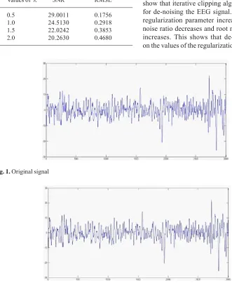
Figure1 shows the original data while Figure2, Figure3, Figure4 and Figure5 show the de-noised data.
RESULTS AND DISCUSSION
Table1. Calculation of SNR and RMSE
Values of λ SNR RMSE
0.5 29.0011 0.1756
1.0 24.5130 0.2918
1.5 22.0242 0.3853
2.0 20.2630 0.4680
Fig. 1. Original signal
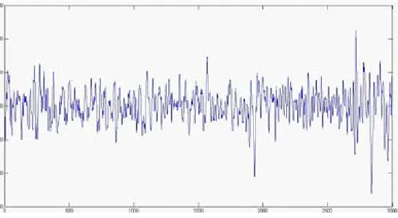
Fig. 2. Total variation de-noising using iterative clipping algorithm for regularization parameter λ = 0.5 (SNR = 29.0011, RMSE = 0.1756)
It is minimized using the iterative clipping algorithm. It has been used for de-noising the EEG signal. More SNR implies more de-noising and on the contrary, more RMSE implies lesser de-noising. Table1 shows the values of SNR and RMSE for different values of ë. From the result obtained, we observe that iterative clipping algorithm works well for de-noising the EEG signal. We also observe that as ë increases, SNR decreases and RMSE increases. Figure1 represents the original data.
Figure2 represents the de-noised data with SNR = 29.0011& RMSE = 0.1756for ë = 0.5, Figure3 represents the de-noised data with SNR = 24.5130& RMSE = 0.2918 for ë = 1.0, Figure4 represents the de-noised data with SNR = 22.0242 & RMSE = 0.3853 for ë = 1.5 and Figure5 represents the de-noised data with SNR = 20.2630 & RMSE = 0.4680 for ë = 2.0. Thus, through de-noising, the quality of EEG signal is enhanced.
CONCLUSIONS
Fig. 3. Total variation de-noising using iterative clipping algorithm for regularization parameter λ = 1(SNR = 24.5130, RMSE = 0.2918)
Fig. 4. Total variation de-noising using iterative clipping algorithm for regularization parameter λ = 1.5 (SNR = 22.0242, RMSE = 0.3853)
is reflected from table1. The calculated SNR and RMSE for different values of regularization parameter show a satisfactory result.
REFERENCES
1. Binder DK, Haut SR. Towards new paradigms of seizure detection. Epilepsy Behaviours. 2013;
26(3): 247-523.
2. Boutros NN, Struve F. Electrophysiological assessment of neuro psychiatric disorders. Seminars in Clinical Neuropsychiatry. 2002; 7(1): 30-41.
3. Croft RJ, Barry RJ. Removal of ocular artefact from the EEG: A review. Clinical Neurophysiology. 2000; 30(1): 5-19.
4. Sameni R, Shamsollahi M, Jutten C, Clifford G. A nonlinear Bayesian filtering framework for ECG denoising. IEEE Transactions on Biomedical Engineering. 2007; 54(12): 2172– 2185.
5. He P, Wilson G, Russel C. Removal of ocular artefacts from electroencephalogram by adaptive filtering. Medical and Biological Engineering and Computing. 2004; 42(3): 407-412.
6. Sweeney KT,AyazH, Ward TE, Izzetoglu M, McLoone SF, OnaralB. A methodology for validating artefact removal techniques for physiological signals. IEEE Transactions on Information Technology in Biomedicine. 2012; 16(5): 918-926.
7. Jung T-P, Makeig S, Humphries C, Lee TW, McKeown MJ, Iragui V. Removing electroencephalographic artefacts by blind source separation. Psychophysilogy. 2000; 37(2): 163–178.
8. Ille N, Beucker R, Scherg M. Spatially Constrained independent component analysis for artefact correction in EEG and MEG. NeuroImage. 2001; 13(6): 159-159.
9. Flexer A, Bauer H, Priplf J, Dorffner G. Using ICA for removal of ocular artefacts in EEG recorded from blind subjects. Neural Networks. 2005; 18(7): 998-1005.
10. Krishnaveni V, Jayaraman S, Kumar PMM, Shivakumar K, Ramadoss K. Comparison of independent component analysis algorithms for removal of ocular artefacts from electroencephalogram. Measurement Science Review. 2005; 5(2): 67-78.
11. Winkler I, Haufe S, Tangermann M. Automatic classification of artefactual ICA components for artefact removal in EEG signals. Behavioural and Brain Functions. 2011; 7(30): 1-15. 12. Lagerlund TD, Sharbrough FW, Busacker NE.
(1997). Spatial filtering of multichannel electroencephalographic recordings through principal component analysis by singular value decomposition. Journal of Clinical Neurophysiology. 1997; 14(1): 73–82. 13. Krishnaveni V, Jayaraman S, Aravind S,
Hariharasudhan V, Ramadoss K. Automatic identification and removal of ocular artefacts from EEG using Wavelet transform. Measurement Science Review Journal– Measurement in Biomedicine. 2006; 6(4): 45-57.
14. Akhtar MA, Mitsuhashi W, James C. Employing spatially constrained ICA and wavelet denoising for automatic removal of artefacts from multichannel EEG data. Journal of Signal Processing. 2012; 92(2): 401–416.
15. Raghavendra BS, Dutt DN. Correction of ocular artefacts in EEG recordings using empirical mode decomposition. Psychophysiology. 2010;
47(5): 955-960.
16. Lindsen J, Bhattachatya J. Correction of blink artefacts using independent component analysis and empirical mode decomposition. Psychophysiology. 2010; 47(5): 955–960. 17. Sweeney K, Ward T, McLoone S. Artefact
removal in physiological signals—Practices and possibilities. IEEE Transanctions on Information Technology in Biomedicine. 2012; 16(3): 488– 500.
18. Mijovi´c B, DeVos M, Gligorijevic I, Taelman J, Huffel SV. Source separation from single-channel recordings by combining empirical-mode decomposition and independent component analysis. IEEE Transactions on Biomedical Engineering. 2010; 57(9): 2188–2196. 19. Rudin L, Osher S, Fatemi E. Nonlinear total
variation based noise removal algorithms. Physica D. 1992; 60: 259-268.
20. Rodriguez P, Wohlberg B. Efficient minimization method for a generalized total variation functional. IEEE Transactions on Image Processing. 2009; 18(2): 322-332.
21. Hu Y, Jacob M. Higher degree total variation (hdtv) regularization for image recovery. IEEE Transactions on Image Processing. 2012; 21(5): 2559-2571.
22. Bredies K, Kunisch K, Pock T. Total generalized variation. SIAM Journal on Imaging Science. 2010; 3(3): 492-526.
23. Selesnick I, Bayram I. http://cnx.org/contents/ 8NLpidGL@1/total-Variation- Filtering, Accessed November 25, 2016.
Pattern Recognition, Lecture Notes in Computer Sciences, Springer, 3757 2000, pp. 136-152. 25. Solo V. Selection of regularisation parameters
for total variation de-noising, in Proc. IEEE international conference on Acoustics, Speech, Signal Processing, ICASSP, 1999, pp.1653-1655.
26. Wen Y W, Chan R H. Parameter selection for
total-variation-based image restoration using discrepancy principle, IEEE Transactions on Image Processing. 2012; 21(4): 1770-1781. 27. Siddiqi AH, Sevindir HK, Aslan Z, Yazici C.