Hamza WS et al. Int J Community Med Public Health. 2018 Aug;5(8):3212-3219
http://www.ijcmph.com pISSN 2394-6032 | eISSN 2394-6040
Original Research Article
Epidemiological analysis of Elizabethkingia meningoseptica infection
cluster among mechanically ventilated pediatric intensive care patients
Wafaa Seddik Hamza
1,2*, Samar Saeed Morsi
3,4, Ebtehal Saleh Al Roomi
5,
Vincent Olubunmi Rotimi
6INTRODUCTION
Elizabethkingia meningoseptica, formerly known as
Flavobacterium meningosepticum and Chryseobacterium meningosepticum or CDC II-a, is a motile, non-fastidious, catalase-and oxidase-positive, aerobic glucose
non-fermenting Gram-negative bacillus first described by King in 1959.1,2 It is commonly found in the environment worldwide and has been detected in soil, river water and reservoirs. However, it rarely causes human infections although it has been reported as causative agent of meningitis in newborn babies and meningitis or
ABSTRACT
Background: Elizabethkingia meningoseptica is frequently found in hospital environments and usually associated with healthcare-associated infections (HAIs), particularly in patients in the intensive care units (ICU). The current study report an outbreak of E. meningoseptica infection/colonization in the pediatric intensive care unit, highlighted the infection control methods used to stem the spread.
Methods: During a period of 7 months, May-November 2015, 4 patients were infected/ colonized by E. meningoseptica. Infection control measures were re-emphasized after each case and environmental swabs were cultured to detect possible source. Follow up for 25 months to ensure eradication of the pathogen.
Results: Four patients were colonized/ infected with E. meningoseptica, their mean age 22 months. The average time patients spent in ICU between admission and isolation of E. meningoseptica was 27.5±19.2 days. All patients were mechanically ventilated. 25% E. meningoseptica isolated from blood causing healthcare associated Central Line Associated Blood Stream Infection (CLABSI) while it was isolated from endotracheal tube (ETT) secretion in 75% as healthcare associated colonization. The 4 isolates confirmed as identical using pulsed field gel electrophoresis (PFGE).
Conclusions: Intensive infection control measures including healthcare workers education, emphasizing hand hygiene, comprehensive cleaning and disinfection of equipment and the environment are important to eradicate the bacterium.
Keywords: Elizabethkingia meningoseptica, Healthcare associated infections, Mechanical ventilation, Pediatric intensive care unit
1
Infection Control Office, 5Department of Microbiology, Chest Diseases Hospital, Kuwait
2
Department of Public Health and Community Medicine, Faculty of Medicine, Assiut University, Egypt
3
Infection Control Office, Al-Razi Hospital, Kuwait
4
Department of Microbiology and Immunology, Faculty of Medicine, Zagazig University, Egypt
6
Department of Microbiology, Faculty of Medicine, Kuwait University, Kuwait
Received: 03 June 2018
Revised: 02 July 2018
Accepted: 04 July 2018
*Correspondence:
Dr. Wafaa Seddik Hamza, E-mail: [email protected]
Copyright: © the author(s), publisher and licensee Medip Academy. This is an open-access article distributed under the terms of the Creative Commons Attribution Non-Commercial License, which permits unrestricted non-commercial use, distribution, and reproduction in any medium, provided the original work is properly cited.
bloodstream and respiratory infections in people with weakened immune systems.3
E. meningoseptica is usually isolated from any aquatic environment in the hospital; sinks, taps, fluids for preparing disinfectants, flushing saline used for medical devices, including feeding tubes, arterial catheters, and respirators. It has been shown that this organism can survive in chlorine-treated water deliveries, often colonizing sink basins and taps, intubation tubes, humidifiers and newborns ‘incubators, and has become a probable reservoir for hospital infections.4-6
E. meningoseptica causes potentially risky infections in patients on admission to critical care areas because of its multidrug-resistant (MDR) character and its ability to get used to different environments.7 It is organisms of low virulence as only a small percentage of colonized patients develop sepsis while others remain asymptomatic.8 However, it is associated with high mortality rates (23-52%), partly due to being an MDR organism.9,10 The prevalence of nosocomial infection by E. meningoseptica
has increased, predominantly in patients with severe underlying diseases, prolonged hospitalization, treatment with invasive procedures, prior use of broad-spectrum antimicrobials and concomitant infections. These factors have impacted survival rates.11
Several cases of E. meningoseptica infections have been recognized in outbreaks related to contamination of hospital tap-water, saline, disinfectants antibiotic solutions, lipid solution, sink drains and respiratory equipment, evolving it to be potentially important cause of hospital infections.6,12,13 The objectives of this study are to, report unusual clustering of E. meningoseptica
infections/colonization over a short period and highlight the infection control measures taken to control the infection.
METHODS
Study design
A prospective cohort study with ongoing daily epidemiological and microbiological surveillance for a cluster of Elizabethkingia meningoseptica that occurred in the Pediatric Intensive Care Unit (PICU) was carried out.
Setting
The PICU is a 7-bed unit (6-bed bay with 2 hand washing stations in addition to 1 cubicle isolation room with clinical sink). Infrared taps are used in all sinks. Infection control team provides support with daily rounds. Nurse-to-patient ratio is one-to-one. The PICU is located in the Chest Diseases Hospital, which is a 358-bed tertiary care hospital that is specialized in management of cardiac and cardiothoracic cases in Kuwait.
Study population
All pediatric patients admitted to the PICU in the study hospital with different cardiac and cardiothoracic diseases and stayed to receive medical management or postoperative care after open heart/thoracic surgeries.
Sample size
Investigation of the cluster of Elizabethkingia meningoseptica cases among all patients residing in PICU from May 2015 till December 2017.
Research tools
Surveillance form
Included; socio-demographic data of patients, hospital file number, location, and bed number. Date of admission, date of surgery, type and date of insertion and removal of invasive devices. Diagnosis on admission, history in details, underlying condition, clinical, laboratory and radiological evidence. Type of laboratory samples, collection date and report result. Medications name and dosage, daily progress of the condition and patient outcome.
Outbreak notification form
Included the following; type of outbreak, Incubation period, etiological agent, mode of transmission, date of outbreak was detected /reported to infection control, outbreak location. Healthcare facility source, Index case identified or not, if healthcare facility source was the index case; if it was a HCW or not. Date of onset of the first ill person, date of outbreak commenced, total number of affected cases; number of laboratory confirmed cases, number of patients still hospitalized, number of deceased patients. Outbreak status; ongoing or controlled, date of onset of last till person resolved, the date of outbreak completed.
Outbreak case list
This form is the summary information of the previous two forms.
Data collection
All cases' information was collected by infection control team from medical files, laboratory reports, nursing notes and environmental screening reports using the previous forms through their daily visits to the study location.
Data for the current research was retrieved from the surveillance form, outbreak notification form and outbreak case list for the diagnosed nosocomial
Laboratory work
Bacterial identification and antibiotic susceptibility testing
The isolates had been identified at the microbiology laboratory of the hospital using the API 20 NE system (bioMerieux, Marcy-l Étoile, France) and VITEK 2 ID System (bioMerieux). Antibiotic susceptibility testing against amikacin, ceftazidime, ciprofloxacin, colistin, gentamycin, imipenem, meropenem, piperacillin, piperacillin-tazobactam, trimethoprim-sulfamethoxazole, rifampicin and vancomycin was performed using the E-test (bioMérieux, Marcy-l Étoile, France) method according to the manufacturer's protocol. E. coli ATCC 25922 was included in each run for quality control. Results were interpreted according to the recommendations of the Clinical Laboratory Standard Institute.14
Genomic fingerprinting
Four E. meningoseptica isolates obtained from blood and endotracheal tube (ETT) secretions were sent to the anaerobic and hospital infection reference laboratory, to be fingerprinted by pulsed-field gel electrophoresis (PFGE) typing of the extracted whole-cell genomic DNA embedded in 1% agarose plugs and digested with Xba1. The Xba1-digested genomic DNA was electrophoresed in a 1% certified agarose gel with a voltage gradient of 6 V/h at 14°C at an angle of 120° using the CHEF-MAPPER XI System (Bio-Rad Laboratories, Hercules, CA, USA). Banding patterns were analyzed using FP Quest TM software (Bio-Rad Laboratories) and strains defined as having PFGE profiles of >94% similarity. A lambda ladder (New England Biolabs, Beverly, MA, USA) was included in each gel run. DNA relatedness was estimated using the criteria of Tenover and his colleagues in 1995.15
Environment screening
Water from all clinical taps in the critical care unit was sampled for bacterial colonization. A total of 100 mL of water was collected from each tap, filtered by using a 0.45-μ filter membrane, and incubated on MacConkey agar in air at 37°C for 48 hours. Oxidase-positive non– lactose-fermenting colonies were sub-cultured onto nutrient agar and a 10-μg meropenem disk placed on the inoculum. Organisms displaying meropenem resistance were further identified by the API 20 NE system (bioMérieux) and VITEK 2 ID System (bioMérieux).
RESULTS
Epidemiological analysis of the cluster of cases
Healthcare associated colonization/infections with E. meningoseptica were identified in 4 female patients. Their mean age was 22 months (range, 1 day – 7 years).
They were admitted on the PICU during a period of 7 months (May – November, 2015 The average time spent in the intensive care unit between admission and isolation of E. meningoseptica was 27.5±19.2 days (range 5-44 days).
Table 1: Frequency of possible risk factors in the study cluster.
Intervention Percentage (%)
PICU admission 100
Mechanical ventilation 100
Colistin use 25
Cefazolin 75
Central line 100
Arterial line 75
Parenteral nutrition 25
Bronchoscopy 25
Urinary catheter 100
Nasogastric tube 25
Tracheostomy care 25
All patients were mechanically ventilated for a period ranged 14-65 days, and had other invasive devices (central lines and urinary catheters). Three patients had open heart surgery. Cefazolin was the prophylactic antibiotic used for all cases before surgery and continued for 2-5 days post-operative. In 1 patient, E
meningoseptica was isolated from blood associated with healthcare-associated central line associated blood stream infection (CLABSI) while it was isolated from endotracheal tube (ETT) secretion in 3 patients classified as healthcare-associated colonization. Three of the cases had other clinically diagnosed healthcare-associated infections, such as catheter-associated urinary tract infection (CAUTI), ventilator-associated pneumonia (VAP), peritonitis, and CLABSI with Stenotrophomonas maltophilia, Pseudomonas aeruginosa, Klebseilla pneumoniae, Staphylococcus epidermis and
Staphylococcus hominies (Table 1 and 2). Three of the 4 patients expired (75% mortality rate) and the remaining 1 patient was boarded to United Kingdom to facilitate extubation and possible rehabilitations.
Infection control measures and interventions
Urgent infection control committee meeting discussed the situation and agreed up on the following measures:
Healthcare workers education
Table 2: Description of Elizabethkingia meningoseptica cluster of cases in pediatric intensive care unit.
Cases Age Gender Underlying condition E. meningoseptica
Detected
Mechanical ventilation
Duration of
ventilation Isolated from Other HAIs Outcome
1 1 day F Congenital heart HAIs Yes 21 days Blood CLABSI Expired
2 2 month F Congenital heart Trisomy 21
colonization
Yes 65 days ETT CLABSI
Peritonitis Expired
3 2 month F Congenital heart colonization Yes 14 days ETT VAP CAUTI Expired
4 7 year F
Post cardiac arrest VF and VT
Chronic Myocarditis
colonization Yes 30 days ETT Did not meet criteria
for any HAIs Boarded to UK
vf = ventricular fibrillation; vt = ventricular tachycardia; hais = healthcare-associated infections; ett = endotracheal tube; clabsi = central line-associated blood stream infection; vap = ventilator-associated pneumonia; cauti= catheter-ventilator-associated urinary tract infection.
Table 3: The antimicrobial susceptibility of the isolates.
Antimicrobial substance MIC interpretive criteria- Susceptibility %
S I R
Amikacin ≤16 32 ≥64 100
Piperacillin-Tazobactam ≤16 32-64 ≥128 100
Trimethoprim/Sulfa ≤2 - ≥4 100
Ciprofloxacin ≤1 2 ≥4 100
Levofloxacin ≤2 4 ≥8 100
Gentamicin ≤4 8 ≥16 100
Rifampicin ≤10 - ≥25 75
Amoxicillin-clavulanic acid ≤8 16 ≥32 0
Ceftazidime ≤8 16 ≥32 0
Ceftriaxone ≤8 16-32 ≥64 0
Cefuroxime ≤8 16 ≥32 0
Imipenem ≤2 4 ≥8 0
Meropenem ≤2 4 ≥8 0
Colistin ≤2 4 ≥8 0
HH compliance was measured to evaluate the impact of training for PICU team; doctors’ HH compliance was 51.6% but improved to 67.9% after the training. While nurses’ HH compliance were 85.6% and increased to 92.9% after education.
Screening of environmental samples
Environmental samples were collected from (ventilators, humidifiers, incubators, bed rails, sinks, treatment trolley, bedside tables, cleaning solutions open one and new one as well as water sample. Two samples (sink swab and a water sample) of the 24 (8.3%) environmental specimens obtained were identified positive for E. meningoseptica.
Patients and healthcare workers measures
Cohorting of patients as well as healthcare workers were done. Evacuation of the patients from the PICU was carried out to allow extensive cleaning and disinfection.
Environmental cleaning and disinfection
These were achieved by cleaning and disinfecting all equipment (e.g. ventilators) as per manufacturer instructions. All open containers of cleaning and disinfection solutions were discarded. Direct observation was carried out to ensure meticulous cleaning and disinfection of all environmental surfaces in PICU including beds, floors, and cleaning equipment (e.g. mops and buckets). In addition, removal and laundry of all curtains, cleaning and disinfection of the ceiling and air condition ducts, filters, outlets and inlets and hyper- chlorination of water supply to the unit, were all carried out.
Figure 1: Timeline of Elizabethkingia meningoseptica cluster of cases with 25 months of follow-up.
Follow up
Active daily surveillance of infections and follow-up for any possible colonization in patients who were in PICU
risk was performed by infection control team. There were no further cases of infection/colonization with E.
meningoseptica from November 2015 till December 2017 (Figure 1).
Antimicrobial susceptibility of the isolates
All the 4 isolates were susceptible to the aminoglycosides, fluoroquinolones, Piperacillin-Tazobactam and trimethoprim/sulfamethoxazole (Table 3).
Genomic typing of isolates
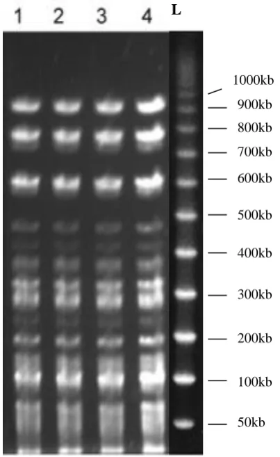
All the 4 isolates were identical (clonally related) on the pulsed-field gel electrophoresis (PFGE) as shown in Figure 2. The isolate from the index case was a blood isolate shown in lane 1 while the subsequent isolates from the ETT secretions of the 3 other patients are demonstrated in lanes 2, 3 and 4. Unfortunately, the 2 environmental isolates were lost in transit to the Reference Laboratory that performed the genomic typing.
Figure 2: Pulsed-field gel electrophoretic pattern of the four isolates of Elizabethkingia meningoseptica from blood stream infection and endotracheal tube (ETT) secretions of patients on the pediatric intensive
care unit.
Note: lanes 1 is the blood isolate, lanes 2, 3 and 4 are isolates from the ett secretions, and lane l is the ladder.
L
900kb
DISCUSSION
E.meningoseptica has a strong predilection for extremes of age with attendant high mortality rates. In this study, our patients were aged between 1 day and 7 years with a mortality rate of 75%. The organism is also well known to cause hospital-acquired infections in premature newborns and infants and associated with high mortality rate of approximately 43% in some studies although lower mortality rate of 9.1% has been reported for community-acquired infections.16
The predisposing factors for infection/colonization, in our study, were presence of comorbidity, congenital heart disease, open heart surgery and mechanical ventilation. These findings are supported by previous reports elsewhere which showed that admission to the ICU, the underlying severe illness, shock at presentation, tachycardia, use of life support devices, and prolonged antibiotic treatment were the main predisposing factors.9,16-18 Also being immuno-compromized and elderly may represent important host factors that predict susceptibility to E meningoseptica infection.9 Host susceptibility factors are critical determinants of risk of E. meningoseptica infection.4
It is also apparent from our study that the time interval between the admission of the patients to the hospital and development of infection/colonization with E.
meningoseptica vary. In this series, this interval appeared to have occurred relatively earlier, 5-44 days (mean 27.5±19.2 days), compared to other study conducted in 2011; where late infection was the common experience as it developed 50-70 days after hospital admission.11
The clinical spectrum of disease due to E. meningoseptica may range from simple colonization to symptomatic acute infection and furthermore to infection-related sequelae.19 Some hosts may be colonized by E. meningoseptica organism in the absence of signs and symptoms of active infection and thus may act as a source of an outbreak.20 It is reasonable to assume that contact to exogenous sources can lead to colonization with E. meningoseptica at mucous membranes as the respiratory tract and non-intact skin sites and that colonized patients may act as sources of infection for other susceptible individuals. Therefore, timely identification of patients colonized or infected with E. meningoseptica is of paramount importance in preventing further dissemination of the bacterium during an outbreak. 7
Meningoseptica infection is very challenging to both clinicians and microbiologists, as the organism is intrinsically resistant to multiple antibiotics, such as the β-lactams, aminoglycosides, tetracycline, tigecycline, colistin, chloramphenicol and carbapenems.4 However, it is susceptible to the agents used to treat Gram-positive bacterial infections such as rifampicin, ciprofloxacin, vancomycin and trimethoprim– sulfamethoxazole. Yet
adequate treatment for this organism has not been properly outlined. Although, vancomycin alone or when it is combined with rifampicin had been used efficiently, recent studies raised many inquiries about its efficacy.20
In our study, the 4 isolates were resistant to all β-lactam antibiotics, tigecycline, and colistin but susceptible to the other classes of antibiotics. Some studies have shown that susceptibility of E. meningoseptica was relatively high (>50%) to piperacillin, piperacillin-tazobactam, cotrimoxazole, ciprofloxacin, moxifloxacin, levofloxacin, tigecycline, vancomycin and showed multidrug resistance to ampicillin-sulbactam, ticarcillin, ceftazidime, ceftriaxone, cefepime, cefoperazone-sulbactam, cefepime-tazobactam, tetracycline, chloramphenicol, imipenem, meropenem, amikacin, gentamicin, tobramycin, and colistin.10, 21 Our study showed that the
E. meningoseptica isolates were fully susceptible to amikacin, piperacillin-tazobactam, trimethoprim/sulfa, ciprofloxacin, levofloxacin, and gentamicin.
All 4 clinical isolates were clonally related. Moreover, 2 samples from the environment revealed the growth of E. meningosepticas howling the same sensitivity pattern suggesting a point-source spread either from the index case or the environment. It is conceivable that the environment must have played a part in maintaining the organism within the PICU for the 7-month period. Although not proven, the hands of the healthcare workers in the unit would have been responsible for the spread. Outbreaks of meningitis due to E. meningoseptica have been reported on neonatal wards and ICUs in the past.5,17,22,23 Some of these outbreaks were investigated in order to determine whether transmission of E. meningoseptica from the environment to patients might have occurred. Despite examination of large numbers of screening cultures no firm evidence was obtained.17,23 However, another investigation considered that the hands of healthcare workers were the likely route of transmission of E. meningoseptica between pediatric patients.5
Screening of the environment and patients for E.
meningoseptica and other infection control measures by contrast with outbreaks of other bacteria such as vancomycin-resistant enterococci, has been debatable for a while, So far, there is no concrete agreement about who to screen and when to initiate active screening during a suspected outbreak of E. meningoseptica infection.24 However, we believe that implementation of screening strategy to detect asymptomatic carriers should be considered in an outbreak setting (or where E.
these, infusion containers and sinks have been identified most often as likely sources of contamination, although in our study, sink and tap water were the contaminated sources; a finding which is partly supported by the study of Amer et al in which the organism was found as colonizer in tap water, tubing of ventilators and in sink basins of the hospital wards.7,25 Several cases of E.
meningoseptica infections have been reported as part of outbreaks and source was traced to contaminated hospital water supply, saline, disinfectants, antibiotic solutions, water sinks, and respirators.26
Studies have also shown that the organism can survive in chlorine-treated water supplies as sink basins and taps, and becoming a possible reservoir for hospital infections.4,27 Thus, outbreaks may be controlled with strong emphasis on infection control measures. Given the importance of environmental sources of the bacterium in the epidemiology of nosocomial outbreaks of E. meningoseptica infection, it is not surprising that some investigators have reported success in controlling outbreaks using enhanced cleaning protocols, cite control of a cluster of infection involving 13 pediatric patients.5 Although an environmental source was not identified, the outbreak was terminated following introduction of two disinfectants (hypochlorite solution, and isopropanol spray) daily cleaning of the unit with particular emphasis on objects containing, or in contact with water.
Other measures that have successfully controlled E.
meningoseptica outbreaks in hospital settings include modification of empirical antimicrobial protocols, restriction of staff exchange and stoppage of new admissions, supplementation of hand hygiene regimens with chlorhexidine gluconate 4%, as well as actions relating to hospital water such as hyper chlorination, isolation of tanks from the common hospital feeder tanks and toileting of babies with sterile rather than tap water. It is satisfying to note that the same measures that have been used successfully by to eradicate E. meningoseptica
outbreaks in pediatric units including, among others, restriction of further admissions and thorough disinfection of the unit were effective to eradicate the cluster of E. meningoseptica cases in our study.6,12,28
CONCLUSION
Early recognition of patients colonized or infected with
E. meningoseptica assisted in preventing spread of the bacterium. Efficient investigations to identify and control the source of the microorganism and comprehensive cleaning of all equipment and environmental surfaces are necessary. Furthermore, the strengthening of standard infection control measures, included proactive contact isolation precautions, restricted patient movement and transfer, in addition to enhanced staff HH compliance and good antibiotic stewardship together were the actions that have eradicated E.meningoseptica cluster.
Recommendations
Development of robust interventions to contain outbreaks of this pathogen among critically ill patient is urgently needed to reduce the morbidity and mortality associated with this infection. Environmental sampling is required to identify the possible source.
Funding: No funding sources Conflict of interest: None declared Ethical approval: Not required
REFERENCES
1. King EO. Studies on a group of previously unclassified bacteria associated with meningitis in infants. Am J Clin Pathol. 1959;31:241-7.
2. Kim KK, Kim MK, Lim JH, Park HY, Lee ST. Transfer of Chryseobacterium meningosepticum and Chryseobacterium miricola to Elizabethkingia gen. nov. as Elizabethkingia meningoseptica comb. nov. and Elizabethkingia miricola comb. nov. Int J Syst Evol Microbiol. 2005;55:1287–93.
3. Center for Disease Control. Available athttps://www.cdc.gov/Elizabethkingia. Assessed at 16 June 2016.
4. Bloch KC, Nadarajah R, Jacobs R. Chryseobacterium meningosepticum: an emerging pathogen among immunocompromised adults. Report of 6 cases and literature review. Medicine. 1997;76(1):30-41.
5. Ceyhan M, Yildirim I, Tekeli A, Yurdakok M, Us E, Altun B, et al., Chryseobacterium meningosepticum outbreak observed in 3 clusters involving both neonatal and non-neonatal pediatric patients. Am J Infect Control. 2008;36(6):453-7.
6. Hoque SN, Graham J, Kaufmann ME, Tabaqchali S. Chryseobacterium (Flavobacterium) meningo-septicum outbreak associated with colonization of water taps in a neonatal intensive care unit. J Hosp Infect. 2001;47:188-92.
7. Jean SS, Lee WS, Chen FL, Ou TY, Hsueh PR. Elizabethkingia meningoseptica: an important emerging pathogen causing healthcare-associated infections. J Hosp Infect. 2014;86(4):244-9. 8. Swain B, Rout S, Otta S, Rakshit A. Elizabethkingia
meningoseptica: an unusual cause for septicaemia JMM Case Reports. 2014.
9. Lin YT, Chiu CH, Chan YJ, Lin ML, Yu KW, Wang FD, et al. Clinical and microbiological analysis of Elizabethkingia meningoseptica bacteremia in adult patients in Taiwan. Scand J Infect Dis. 2009;41:628-34.
11. Pereira Graziella H, de Oliveira GD, Saad AC, de Barros BVL, Lucas da SPS. Nosocomial infections caused by Elizabethkingia meningoseptica: an emergent pathogen. Braz J Infect Dis. 2013;17(5):606-9.
12. Güngör S, Ozen M, Akinci A, Durmaz R. A Chryseobacterium meningosepticum outbreak in a neonatal ward. Infect Control Hosp Epidemiol. 2003;24:613-7.
13. Steinberg JP, Burd EM. Other gram-negative and gram-variable bacilli. In Mandell GL, Bennett JE, Dolin R, editors. Principles and Practice of Infect Dis. Philadelphia: Churchill Livingstone Elsevier; 2010: 3015-3033.
14. Clinical and Laboratory Standards Institute (CLSI). Performance standards for antimicrobial susceptibility testing; 22nd International Supplement; Wayne, Pennsylvania. CLSI Document M100-S18. 2015.
15. Tenover FC, Arbeit RD, Goering RV, Mickelsen PA, Murray BE, Persing DH, et al. Interpreting chromosomal DNA restriction patterns produced by pulsed-field gel electrophoresis: Criteria for bacterial strain typing. J Clin Microbiol. 1995;33:2233-9.
16. Hung PP, Lin YH, Lin CF, Liu MF, Shi ZY. Chryseobacterium meningosepticum infection: antibiotic susceptibility and risk factors for mortality. J Microbiol Immunol Infect. 2008;41(2):137–44.
17. Weaver KN, Jones RC, Albright R, Thomas Y, Zambrano CH, Costello M, et al. Acute emergence of Elizabethkingia Meningoseptica infection among mechanically ventilated patients in a long-term acute care facility. Infect Control Hosp Epidemiol. 2010;31:54–8.
18. Jung SH, Lee B, Mirrakhimov AE, Hussain N. Septic shock caused by Elizabethkingia meningoseptica: a case report and review of literature. BMJ Case Reports. 2013: bcr2013009066.
19. Cabrera HA, Davis GH. Epidemic meningitis of the newborn caused by flavobacteria. I. Epidemiology and bacteriology. Am J Dis Child. 1961;201(3):289–95.
20. Ceyhan M, Celik M. Elizabethkingia meningosepticum (Chryseobacterium
meningo-septicum) infections in Children. Int J Pediatr 2011;2011:215237.
21. Tak V, Mathur P, Varghese P, Misra MC. Elizabethkingia meningoseptica: An emerging pathogen causing meningitis in a hospitalized adult trauma patient. Indian J Med Microbiol. 2013;31:293-5.
22. Tekerekoglu MS, Durmaz R, Ayan M, Cizmeci Z, Akinci A. Analysis of an outbreak due to Chryseobacterium meningosepticum in a neonatal intensive care unit. New Microbiol. 2003;26:57-63. 23. Maraki S, Scoulica E, Manoura A, Papageorgiou N,
Giannakopoulou C, Galanakis EA. Chryseobacterium meningosepticum colonization outbreak in a neonatal intensive care unit. Eur J Clin Microbiol Infect Dis. 2009;28:1415-9.
24. Johnston BL, Bryce E. Hospital infection control strategies for vancomycin-resistant Enterococcus, methicillin-resistant Staphylococcus aureus and Clostridium difficile. Can Med Assoc J. 2009;180:627-31.
25. Amer MZ, Bandey M, Bukhari A, Nemenqani D. Neonatal meningitis caused by Elizabethkingia Meningoseptica in Saudi Arabia. J Infect Dev Ctries. 2011;5:745–7.
26. Bhat KS, Priya R, Krishnan L, Kanungo R. Elizabethkingia meningoseptica bacteremia in a neonate: A case report and mini-review of the literature. J Curr Res Sci Med. 2016;2:42-5. 27. Chiu CH, Waddingdon M, Greenberg D,
Schreckenberger PC, Carnahan AM. Atypical Chryseobacterium meningosepticum and meningitis and sepsis in newborns and the immune-compromised, Taiwan,” Emerging Infectious Diseases. 2000;6(5):481–6.
28. Hazuka BT, Dajani AS, Talbot K, Keen BM. Two outbreaks of Flavobacterium meningosepticum type E in a neonatal intensive care unit. J Clin Microbiol. 1977;6(5):450–5.
Cite this article as: Hamza WS, Morsi SS, Roomi ESA, Rotimi VO.Epidemiological analysis of