Int. J. Nanosci. Nanotechnol., Vol. 14, No. 2, June. 2018, pp. 93-99
Synthesis of Cu-CuO and Cu-Cu2O
Nanoparticles via Electro-Explosion of Wire
Method
Reza Ahmadi1,*, Ahmad Razzaghian1, Zahra Eivazi2 and Kobra Shahidi1
1Department of Metallurgy and Materials Engineering, Faculty of Engineering, Imam
Khomeini International University (IKIU), P.O.Box 34148-96818, Qazvin, Iran. 2 Department of Materials Nano Materials, Faculty of Engineering,Tarbiat Modarres
University, P.O.Box 14115-111,Tehran, Iran.
(*) Corresponding author: [email protected] (Received: 09 February 2017 and Accepted: 05 March 2018)
Abstract
We report an investigation on the synthesis of Copper Oxide nanostructures via the Electro-Explosion of Wire method. Cu-CuO and Cu-Cu2O nanoparticles with mean particle size of 21.1 to 59.1 nm were
synthesized by changing the applied current density and amount of used Tetra Methyl Ammonium Hydroxide as the surfactant. The X-ray diffraction analysis, field emission scanning electron microscopy and Fourier transform infrared spectroscopy techniques have been used for characterization of the prepared samples. The results confirmed the influence of surfactant application on the size, morphology and chemical composition of the synthesized nanoparticles, while the applied current density has only had a significant effect on the mean particle size.
Keywords:Electro-explosion of wire, Copper oxide, Nanoparticle, Tetra methyl ammonium hydroxide, Field emission microscopy.
1. INRODUCTION
The synthesis, properties and application of Nanostructured materials have been studied vastly in recent years [1-5]. The unique mechanical, electric, magnetic, chemical and biological properties of these structures are mainly due to their large surface to volume ratio. These unique properties lead to different applications comparing to the traditional materials. Besides, various synthesis methods are employed for each application.
Among different techniques for synthesis of nanostructured materials such as coprecipitation [6-8], sol-gel [9-11], hydro-thermal [12, 13], hydro-thermal decomposition [14] and Electro-Explosion of Wire (EEW), the latter is one of the most promising methods especially for synthesizing nano-metric powders from metallic precursors. In this approach, very thin wires of ductile and conductive metals are exploded under electric discharge conditions originated
The effect of medium temperature on the mechanism of particles formation and also size and morphology of Copper Oxide nanoparticles has been studied recently. According to the published results, the mechanism of nucleation and growth of Cu-CuO particles changes from Ostwald Ripening (OR) to Oriented Attachment (OA) by increasing temperature and the mean particle size is maximum at low temperature regime of OR growth [15]. Cu and Cu-CuO nanoparticles have various electronic and optoelectronic applications due to their desired electronic properties. Some of these applications include field emission uses, electrode for Li ion batteries, solar cells and field effect transistors [15, 24]. Also, their implement-tation in antimicrobial applications [25], chemical sensors [26] and magnetic storage media [27] have been considered recently. The size, morphology and chemical composition of the Cu-CuO nanoparticles strongly affect their performance in the mentioned applications. Copper and Copper Oxide nanoparticles have been synthesized via EEW technique frequently. In one study, Cu/CuO/Cu2O nanoparticles have been prepared by explosion of Copper wire in DI water and characterized via HRTEM, XRD, XPS, FT-IR, VSM and Raman Spectroscopy techniques. The XRD results showed only the presence of Cu and Cu2O phases and the amount of the metallic phase was about twice of the others, but HRTEM, XPS, FT-IR and Raman Spectroscopy results verified the presence of trace amounts of CuO phase too [28]. The results of some other studies have revealed the presence of the Cu2O phase in the structure of the nanoparticles synthesized via EEW method [29, 30]. Cu2O might be oxidized to CuO due to the explosion conditions, but the very short time of the phenomenon can prevent the oxidation reaction and so the Cu2O phase will be the stable phase in the final product [29]. In the present study, Cu-CuO nanoparticles have been synthesized via EEW method in various experimental
current densities and surfactant amounts. The synthesized nanoparticles, have been studied using Field Emission Scanning Electron Microscopy (FESEM), X-ray Diffraction (XRD) and Fourier Transform Infrared (FT-IR) techniques. The results show that the size, morphology and chemical composition of particles are affected by the surfactant to particles ratio, but the amount of current density only affects the mean size of the particles. The results revealed that Cu-CuO and Cu-Cu2O nanoparticles with mean particle size between 21 and 59 nanometer have been produced via the EEW method.
2. MATERIALS AND METHODS 2.1. Sample Preparation
The experimental conditions for the synthesis of samples A1 to A7 are presented in table 1. All samples were synthesized via EEW method in various current densities between 3 and 16 kA/mm2. Samples A1 to A4 were synthesized via explosion of 0.2 mm copper wires in DI water without surfactant and samples A5 to A7 were synthesized in the presence of various amounts of Tetra Methyl Ammonium Hydroxide (TMAH) as surfactant. The synthesized samples were prepared as suspension and solid specimens for FESEM, XRD and FT-IR characterizations.
2.2. Characterization Methods
3. RESULTS AND DISCUSSION
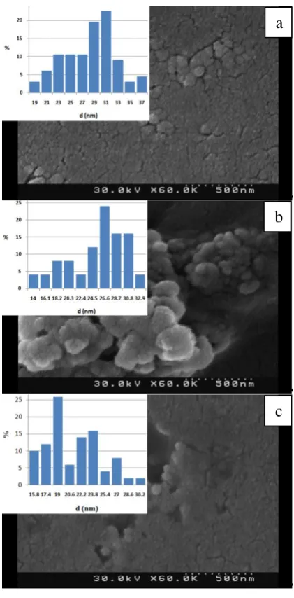
FESEM images of samples A1-A4 are presented in Figure 1a-d, respectively. As seen from the size distribution diagrams, the mean particle size decreases from 59.1 to 32 nanometers by increasing the current density from 3 to 16 kA/mm2. This is due to the fact that in high induced electric energies, small number of atoms form the initial embryo and reduces the final particle size. In other words, by increasing input electric power, the explosion temperature raises, so the cooling rate of initial Cu atoms increases and each Cu or CuO particle contains only a few number of Cu or O atoms. On the other hand, in the low levels of the applied energies and achieved temperatures, the total explosion and evaporation process could be prolonged and so the particle can grow to larger sizes. This result is in good coincidence with other
reports [17, 31-33]. Figure 1. FESEM images of samples a) A1,
b) A2, c) A3 and d) A4 according to table 1.
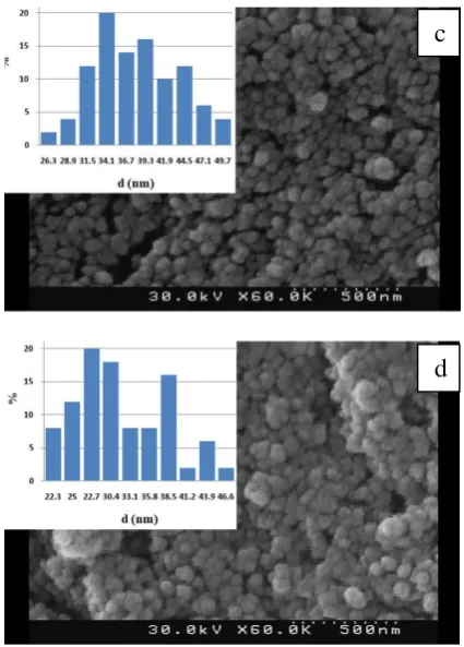
Figures 2a-c present the FESEM images of samples A5-A7, respectively. These samples were synthesized in the presence of TMAH. With increasing the molarity of the TMAH as surfactant, particle size decreases. TMAH molecules surrounded the initial embryos prevent from the fast growth of the particles.
The mean aspect ratios of the synthesized samples are listed in Table 1. This parameter is calculated by considering at least 50 particles for each sample. It seems that by increasing the amount of surfactant, the preferred growth planes are covered with TMAH molecules, so there is no significant difference between the growth rates of various crystallographic plans. In these conditions, spherical growth of particles is the dominate phenomenon and the mean aspect ratio decreases with the increasing of the surfactant amount. XRD patterns of samples A4 and A7 which synthesized with and without TMAH are shown in Figure 3.
a
b
c
Figure 2. FESEM images of samples a) A5, b) A6 and c) A7 according to the table 1.
Table 1. The experimental conditions for the synthesis of samples A1 to A7
It can be seen from marked peaks that Cu, CuO and Cu2O phases formed. Using Rietveld method, the amount of Oxide phase in sample A7 is about twofold of sample A4 (66% respect to 32%). This is
decomposes in high temperature of wire explosion and copper oxide compounds form duo to the formation of the free O atoms. This phenomenon is in contrast with the usual effect of surfactants in other synthesis methods in which the surfactant protects the surface from oxidation. Besides, the dominate Copper Oxide phases in A4 and A7 are Cu2O and CuO, respectively. The O:Cu ratio in sample A7 is more than sample A4 due to the extra Oxygen from the decomposition of TMAH in A7.
As seen in Figure 3, the XRD peaks are sharper in the sample A4 comparing to the sample A7 and the mean crystallite sizes are 31.1 and 20.5 nm for samples A4 and A7, respectively. These results are in good agreement with the FESEM findings (Figures 2 and 3). The recent values have been calculated using Scherrer formula, the useful equation for calculating crystallite size that has been described elsewhere [34].
Figure 3. XRD patterns of samples A4 (upper) and A7 (lower).
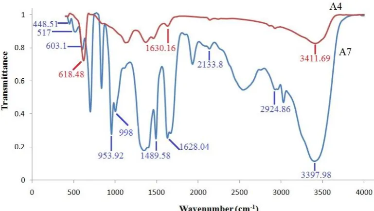
The FT-IR spectra of samples A4 and A7 are shown in Figure 4. The characteristic bands of sample A7 appearing at 448.51 cm -1, 517 cm-1, and 603.1 cm- 1 can be attributed
to the Au and Bu modes of CuO along the [101] direction [35, 36]. The peaks b
related to the asymmetric stretching vibration mode of the C-N band and the asymmetric methyl deformation mode from TMAH, respectively [37]. The broad peak at 3397.98 cm-1 is assigned to O-H band and the sharp peak at 1628.04 cm-1 is related to the absorbed water [38]. The peak at 2924.86 cm-1 is probably due to the ethanol contamination during to the FT-IR sample preparation [39]. The peak at 953.52 cm-1 could be assigned to the Cu-O-H bending mode. The peak at 2133.8 cm-1 probably is due to the NO2+ species from the decomposition of N(CH3)4OH [40].
The distinguishing peak seen at the FT-IR pattern of sample A4 is located at 618.48 cm-1 and is related to the C-O band of Cu2O [41]. No peak from Cu-O band at CuO is seen in this spectrum. This is in good agreement with the XRD results and verifies the presence of CuO and Cu2O phases in the samples A7 and A4, respectively. The peak at 1630.16 cm-1 is related to the absorbed water, similar to the sample A7.
Figure 4. FT-IR spectra of samples A4 and A7.
4. CONCLUSION
Cu-CuO and Cu-Cu2O nanoparticles have been synthesized via EEW method with the mean particle size between 21.1 and 59.1 nm and aspect ratio between 1.03 and 1.35. The Oxygen content of the products was increased by increasing of TMAH due to the surfactant decomposition. XRD and FT-IR results verified the formation of Cu2O and CuO phases with and without TMAH,
respectively. According to the SEM findings, the particle size was decreased with increasing the amount of applied current density and TMAH (as the surfactant). The mean aspect ratio of particles was decreased with increasing TMAH which probably due to its effect on the growth of different crystallographic planes.
REFERENCES
1. Ahmadi R., Madaah Hosseini H. R., Masoudi A., Omid H., Namivandi-Zangeneh R., Ahmadi M., Ahmadi Z., Gu N., (2013).“Effect of concentration on hydrodynamic size of magnetite-based ferrofluid as a potential MRI contrast agent”, Colloids Surf. A Physicochem. Eng. Asp., 424: 113– 117.
3. Hedayati K., Azarakhsh S., Ghanbari D., (2016). “Synthesis and magnetic investigation of cobalt ferrite nanoparticles prepared via a simple chemical precipitation method”, J. Nanostruct., 6: 127-131.
4. Najafi F., Rajabi M., (2015). “Thermal gravity analysis for the study of stability of graphene oxide–glycine nanocomposites”, Int. Nano Lett., 5: 187-190.
5. Nasirian A., (2012). “Synthesis and characterization of Cu nanoparticles and studying of their catalytic properties”, Int. J. Nano Dimens., 2: 159-164.
6. Mansouri F., Amiri G., Fatemi M., (2016). “Synthesis and tissue distribution of CoFe2O4 nanoparticles coated with DMSA in rats liver”, Nanomed. J., 3: 196-201.
7. Durga Vijaykarthik D., Kirithika M., Prithivikumaran N., Jeyakumaran N., (2014). “Synthesis and characterization of Cadmium Oxide nanoparticles for antimicrobial activity”, Int. J. Nano Dimens., 5: 557-562. 8. Pallab Chandra S., Abdus Subhan M., Nizam U., Prosenjit S., (2017). “Synthesis, Structure, Spectroscopy and
Photocatalytic Studies of Nano Multi-Metal Oxide MgO∙Al2O3∙ZnO and MgO∙Al2O3∙ZnO-Curcumin Composite”, Int. J. Nanosci. Nanotechnol., 13: 69-82.
9. Ahmadi P., Alamolhoda S., Mirkazemi S. M., (2017). “Phase Formation, Microstructure and Magnetic Properties of BiFeO3 Synthesized by Sol-Gel Auto Combustion Method Using Different Solvents”, Int. J. Nanosci. Nanotechnol., 13: 195-201.
10.Janitabar Darzi S., Mahjoub A., Bayat A., (2015). “Sulfur modified ZnO nanorod as a high performance photocatalyst for degradation of Congoredazo dye”, Int. J. Nano Dimens., 6: 425-431.
11.Aghabeygi S., Zare-Dehnavi M., Farhadyar A., Farhadyar N., (2015). “Preparation and Characterization of ZrO2/ZnO Nanocomposite under Ultrasonic Irradiation via Sol-gel Route”, Int. J. Bio-Inorg. Hybrid Nanomater., 4: 35-38.
12.Ahmadi R., Masoudi A., Madaah Hosseini H. R., Gu N., (2013). “Kinetics of magnetite nanoparticles formation in a one step low temperature hydrothermal process”, Ceram. Int., 39: 4999-5005.
13.Alaei Z., Mohammad Nejad S., Yousefi M. H., (2016). “The Effects of Different Seed Layers and Growth Time on the Quality of ZnO NRs Arrays”, Int. J. Nanosci. Nanotechnol., 12: 119-130.
14.Waseem S., Ali Z., Bibi M., Usmen Z., (2016).“Magnetic nanobeads: Synthesis and application in biomedicine”, Nanomed. J., 3: 147-154.
15.Krishnan S., Haseeb A., Johan M. R., (2014). “Low dimensional CuO nanocomposites synthesis by pulsed wire explosion and their crystal growth mechanism”, Ceram. Int., 40: 9907-9916.
16.Kotov Y. A., (2003). “Electric explosion of wires as a method for preparation of nanopowders”, J. Nanopart. Res., 5: 539-550.
17.Debalina B., Kamaraj M., Murthy B. S., Chakravarthi S. R., Sarathi R., (2012). “Generation and characterization of nano-tungsten carbide particles by wireexplosion process”, J. Alloys. Compd., 496: 122– 128.
18.Sarathi R., Sindhu T. K., Chakravarthy S. R., Sharma A., Nagesh K. V., (2009). “Generation and characterization of nano-tungsten particles formed by wire explosion process”, J. Alloys. Compd., 475: 658– 663.
19.Sarathi R., Sindhu T. K., Chakravarthy S. R., (2007). “Generation of nanoaluminium powder through wire explosion process and its characterization”, Mater. Charact., 58: 148-155.
20.Yao W. T., Yu S. H., Zhou Y., Jiang J., Wu Q. S., Zhang L., Jiang J., (2005).“Formation of uniform CuO nanorods by spontaneous aggregation: selective synthesis of CuO, Cu2O, and Cu nanoparticles by a solid−liquid phase arc discharge process”, J. Phys. Chem. B, 109: 14011- 14016.
21.Navendu G., Pen S., (2017). “Water-driven stabilization of cadmium sulphide nanoparticles”, Appl. Surf. Sci., 425: 576-584.
22.Navendu G., Pen S., (2004). “Water-induced stabilization of ZnS nanoparticles”, Solid State Commun., 132: 791-794.
23.Navendu G., Pen S., (2014). “Water driven stabilization of ZnS nanoparticles prepared by exploding wire technique”, Mater. Res. Express, 1: 1-14.
24.Ghosh S., Avasthi D. K., Shah P., Ganesan V., Gupta A., Sarangi D., Bhattacharya R., Assmann W., (2000). “Deposition of thin films of different oxides of copper by RF reactive sputtering and their characterization”, VACUUM, 57: 377-385.
25.Barua S., Das G., Aidew L., Buragohain A. K., Karak N., (2013). “Copper-copper oxide coated nanofibrillar cellulose: a promising biomaterial”, RSC Adv., 3: 14997-15004.
26.Chowdhuri A., Sharma P., Gupta V., Sreenivas K., Rao K. V., (2002). “H2S gas sensing mechanism of SnO2 films with ultrathin CuO dotted islands”, J. Appl. Phys., 92: 2172-2180.
27.Darezereshki E., Bakhtiari F., (2011). “A novel technique to synthesis of tenorite (CuO) nanoparticles from low concentration CuSO4 solution”, J. Min. Metall. Sect. B-Metall., 47: 73-78.
29.Mosa A. O., Akomolafe T., Carter M. J., (1998). “Production of cuprous oxide, a solar cell material,by thermal oxidation and a study of its physical and electrical properties”, Sol. Energy Mater. Sol. Cells, 51: 305-316. 30.Park E., Park H. W., Lee J., (2015). “Synthesis of hierarchical copper oxide composites prepared via electrical
explosion of the wire in liquids method”, Colloids. Surf. A Physicochem. Eng. Asp., 482: 710-717.
31.Kotov Y. A., Samatov O. M., (1999).“Production of nanometer-sized AlN powders by the exploding wire method”, Nanostruct. Mater., 12: 119-122.
32.Kwon Y. S., Jung Y. H., Yavorovsky N. A., Illyn A. P., Kim J. S., (2001).“Ultra-fine powder by wire explotion method”, Scripta Mater., 44: 2247–2251.
33.Poonam W., Sharma P. K., Jha A. K., (2013). “Synthesis of Copper Nanoparticles through Wire Explosion Route”, Int. J. Eng. Res. App., 3: 1664-1669.
34.Ahmadi R., Sadrnezhad S. K., Namivandi Zangene R., Oghabian M. A., (2016). “Kinetics of oxygen adsorption on ZnS nanoparticles synthesized by precipitation process”, Mater. Sci-Poland, 34: 260-265.
35.Ethiraj A. S., Kang D. J., (2012). “Synthesis and characterization of CuO nanowires by a simple wet chemical method”, Nanoscale Res. Lett., 7: 70-74.
36.Kliche G., Popovic Z. V., (1990). “Far-infrared spectroscopic investigations on CuO”, Phys. Rev. B, 42: 10060-10066.
37.Lim Y. F, Choi J. J., Hanrath T., (2012). “Facile Synthesis of Colloidal CuO Nanocrystals for Light-Harvesting Applications”, J. Nanomater., 4: 1-6.
38.Lin B. C., Chen S. Y., Shen P., (2012). “(Zn,H)-codoped copper oxide nanoparticles via pulsed laser ablation on Cu-Zn alloy in water”, Nanoscale Res. Lett., 7: 272-280.
39.Korzhavyi P. A., Soroka I. L., Isaev E. I., Lilja C., Johansson B., (2012). “Exploring monovalent copper compounds with oxygen and hydrogen”, Proc. Natl. Acad. Sci. USA, 109: 686-689.
40.Hoost T. E., Laframboise K. A., Otto K., (1995). “NO adsorption on Cu-ZSM-5: assignment of IR band at 2133 cm−1”, Catal. Lett.,33: 105-116.