INTRODUCTION
Methylphenidate hydrochloride is a medicine which is used in attention-deficit hyperactivity disorder1. The chemical structure and phisico-chemical properties of the drug are tabulated in Table 12. Methylphenidate hydrochloride is used as part of a program for the treatment of people who have attention-deficit hyperactivity disorder. Methylphenidate hydrochloride works by affecting certain chemicals in the brain, which may help to reduce some of the
Hollow-Fiber liquid-phase Microextraction Followed by
High Performance Liquid Chromatography for the
Determination of Trace Amounts of Methylphenidate
Hydrochloride in Biological Fluids
SEYEDEH NEDA MIRAEE
1, MAHNAZ QOMI*,
FOROUGH SHAMSHIRI
2and PARVIZ RAOUFI
3Department of Medicinal Chemistry (Pharmaceutical Sciences Research Center) Pharmaceutical Sciences Branch, Islamic Azad University, Tehran, Iran
http://dx.doi.org/10.13005/bpj/546
(Received: August 10, 2014; accepted: November 05, 2014) ABSTRACT
Methylphenidate hydrochloride is used fortreatment ofhyperactivity disorder. In the current work, for the first time a microextraction technique was introduced to detection and quantification of methylphenidate hydrochloride in urine and plasma samples. Hollow ûber based liquid phase microextraction (HF-LPME) followed by high performance liquid chromatography (HPLC) coupled with ultraviolet (UV) detection was used for extraction of methylphenidate hydrochloride. The organic membrane solventconsists of 1-Octanol immobilized in the pores of a hollow fiber. A pH gradient was driving force to migrate analyte from sample solution, through the organic liquid membrane into an acidic acceptor solution which was located inside the lumen of hollow fiber.Extraction recoveries upper than 80% were obtainedin different biological matrices which resulted inpreconcentration factors upper than 112 and acceptable repeatability (2.4< RSD% <4.8). The method offers good linearity with estimation of coefficient higher than 0.9990. Finally, it was applied to the determination and quantification of methylphenidate hydrochloride in biological samples.
Key words: Methylphenidate hydrochloride; High performance liquid chromatography; Hollow fiber based liquid phase microextraction; Microextraction.
Severalmethodshave been presented in order to detection and quantification ofMethylphenidate hydrochloride up to now. There are some methods for detection and quantification of Methylphenidate hydrochlorideconcentration in biological samples includingreverse phase high performance liquid chromatography (RP-HPLC)7, 8, liquid chromatography-mass spectroscopy9, 10, Liquid chromatography– tandem mass spectrometry11, 12,HPLC with chemiluminescence detection13, gas chromatography-mass spectroscopy14, 15 and gas chromatography with electron capture detection16, 17.
Sample preparation steps should be used for determination of the drug in biological samples in all of these methods. It is difficult to obtain low detection limits without sample preparation steps. We believe that microextraction technique has not been reported for extraction and preconcentration of Methylphenidate hydrochloride from body fluids. In this work, for the first time, three phasehollow ûber based liquid phase microextraction (HF-LPME) followed by HPLC with ultraviolet (UV) detection was used and validated for detection of Methylphenidate hydrochloride in biological samples.
HF-LPME as one of LPME was introduced for the first time by Pedersen-Bjergaard[18, 19]. In HF-LPME technique, polypropylene porous hollow fiber membrane is used as the organic solvent carrier, in which target analyts transfer across organic liquid membrane from sample solution to acceptor phase. HF-LPME can provide better stability and sample clean-up ability than other LPME methods. HF-LPME divided into two-phase HF-LPME and three-phase HF-LPME. The extraction vial is filled with the sample solution. A Measured piece of a porous HF may be either a rod with a sealed bottom or a u-shape where both ends are connected to guiding tubes. Before extraction, the HF is first dipped in the organic solvent for a few times to immobilize solvent in the pores, and excess solvent is removed.
The acceptor solution fills the lumen of the HF. This acceptor solution can be an organic solvent in which the same as that used for the organic solvent in HF pores, resulting in a
two-phase LPME, or the acceptor solution may be an acidic or basic aqueous solution, resulting in a three-phase LPME. In the two-phase LPME, the target analytes are extracted from the aqueous sample and into the organic solvent (acceptor solution) present both in the porous wall and inside the lumen of the HF20-24. In three-phase LPME, the analytes are extracted from the aqueous sample, through the organic solvent in HF pores, and further into the aqueous acceptor solution present inside the lumen of the HF25-37.
In this work, the effects of various variables on HF-LPME efficiency were investigated and optimized. After optimization, the method followed by HPLC-UV was applied for extraction and determination of Methylphenidate hydrochloride in plasma and urine sample as biological samples.
EXPERIMENTAL Chemicals and materials
All chemicals were of analytical-reagent grades and used as received. Methylphenidate hydrochloridestandard were kindly donated byDrug and Food Administration (Tehran, Iran).1-Octanol, dodecane, n-decane, n-hexane, were purchased from Merck (Darmstadt, Germany).Acetonitrile and methanol of HPLC were obtained from Merck (Darmstadt, Germany). Sodium hydroxide and sodium chloride were obtained from Sigma– Aldrich (St. Louis, MO, USA). Distilled water was deionized by a Milli-Q water purification system from Millipore (Madrid, Spain).
The PPQ3/2 polypropylene hollow fiber (600 µm ID, 200 µm wall thickness and 0.2 µm pore size) was purchased from Membrana GmbH (Obernburg, Germany) and used as received. Stock solutions of analyte of about 1000 mg L-1 were precisely prepared in methanol. They were all stored in the darkness at 4 ÚC and working analyte mixtures were daily prepared by dilution with the appropriate volume of distilled water. Apparatus and software
mixing valve with a 10 µL sample loop, YL9101 vacuum degasser and a YL 9120 UV-Vis detector. Chromatography data were recorded and analyzed using Younglin Auto Chro 3000 software.The separations were performed on an ODS-3 column (250 mm × 4.6 mm, with particle size of 5 µm) from MZ-Analysentechnik (Mainz, Germany).The mobile phase consisted of 50 mMpotassium dihydrogen orthophosphate and methanol mixture (57:43), under isocratic condition. The flow rate of the mobile phase was set at 1.0 mL min-1 andtotal analysis time was 15 min.The injection volume was 10 µL for all of the samples and detection was performed at a wavelength of 210 nm.
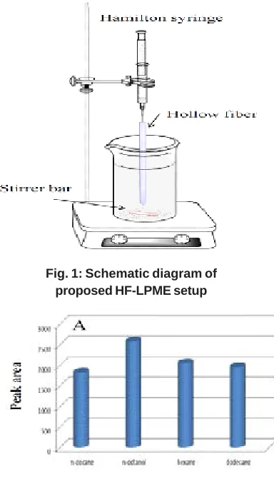
HF-LPME procedure
Ten mL of sample solution was ûlled into a 15 mL vial. Extraction process was shown in fig. 1.A 5.0 cm piece of fresh ûber was inserted into the needle tip of a 25 µL Hamilton syringe that was previously ûlled with acceptor phase. Subsequently, the fiber dipped for a 10 s period into the organic solvent. After filling hollow fiber wall pores with organic membrane solvent, excess amount of organic solvent washed with distilled water, and 10 µL of acceptor solution with pH=1.9 was injected into the lumen of hollow ûber with the Hamilton syringe, and the lower end of the hollow fiber was mechanically sealed by a piece of foil. Subsequently, the ûber was placed in the sample solution vial. Extraction vial was placed on a magnetic stirrer plate to provide effective stirring condition during the extractions. During extraction, the solution was stirred at 750 rpm. After extraction, the acceptor solution was collected into a micro-vial by Hamilton syringe.Finally, acceptor solution was injected for analysis into the HPLC instrument. Real sample analysis
Drug-free human plasma was kindly denoted by Iranian Blood Transfusion Organization (Tehran, Iran). Urine samples were obtainedfrom healthy young volunteer. The samples were stored at “4°C, thawed and shaken before extraction. Calculation of preconcentration factor, extraction recovery and relative recovery
The preconcentration factor (PF) was defined as the ratio of the final analyte concentration in the acceptor phase (Cf,a) and the initial concentration
of analyte (Ci,s) in the sample solution:
...(1)
where Cf,a was calculated from a calibration graph obtained by direct injection of analytes standard solutions (0.2-200 mg L-1) in 10 mMHCl. Extraction recovery (ER) was defined as the percentage of the number of moles of analyte which was extracted to the acceptor phase (nf,a) divided by the number of moles of analyte originally presented in the sample solution (ni,s).
...(2)
...(3)
whereVf,a and Vi,s are the volumes of acceptor phase and sample solution, respectively. Relative recovery (RR) was calculated by the following equation:
...(4)
whereCfound, Creal, and Cadded are the concentrations (µg L-1) of analyte after addition of known amount of standard into the real sample, the concentration of analyte in real sample, and the concentration of known amount of standard which was spiked into the real sample, respectively.
RESULTS AND DISCUSSION
In order to obtain the maximum extraction efficiency for preconcentration and determination of Methylphenidate hydrochloride in biological fluids, the major parameters on HF-LPME, including, organic membrane solvent, sample solution stirring rate, extraction time, pH in donor and acceptor phases, and temperature were investigated and optimized by one variable at the time method.. All optimizations steps were performed in ultra-pure water.
Organic membrane solvent
Table 1: Chemical structures, pKa and logP of methylphenidate hydrochloride.
Name Chemical structure IUPAC name pKa logP
Methylphenidate methyl 2-phenyl-2- 9.09 2.25
hydrochloride (piperidin-2-yl)acetate
Reference [2]
Table 2: Figures of merit of HF-LPME in drug-free distilled water sample
LOD (ngmL-1) LOQ (ngmL-1) Linearity (ngmL-1) R2 PFa RSD% b
Within day Between day
3.0 12.0 12.0-5000.0 0.9990 112 2.5 3.5
a Drugs were present at 500 ng mL-1.b Within day and between day RSDs% were obtained by four replications.
Table 3: Determination of methylphenidate hydrochloride in different urine and plasma samples Sample Creal (ngmL-1) C
added (ng mL
-1) C
found(ng mL
-1) RSD% (n = 3) RR%
Plasma 1 nda 0.2 0.82 2.4 82
Plasma 2 nd0.5 1.8 2.6 90
Plasma 3 nd1.0 4.0 3.2 80
Urine 1 nd0.2 0.17 3.2 85
Urine 2 nd0.5 0.84 3.6 84
Urine 3 nd1.0 1.6 4.3 80
Urine 4 nd2.0 4.3 4.8 86
a Not detected
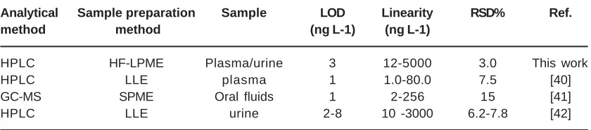
Table 4: Comparison of the HF-LPME with other analytical techniques for determination of methylphenidate hydrochloride
Analytical Sample preparation Sample LOD Linearity RSD% Ref.
method method (ng L-1) (ng L-1)
HPLC HF-LPME Plasma/urine 3 12-5000 3.0 This work
HPLC LLE plasma 1 1.0-80.0 7.5 [40]
GC-MS SPME Oral fluids 1 2-256 15 [41]
HPLC LLE urine 2-8 10 -3000 6.2-7.8 [42]
in HF-LPME, selection of an organic membrane solvent is necessary. The organic solvent forms a thin layer within the wall of the hollow fiber. The
Fig. 2: Optimization of (A) organic membrane solvent, (B) donor sample solution pH, (C) acceptor phase pH and (D) stirring rate for extraction of methylphenidate hydrochloride
Fig. 1: Schematic diagram of proposed HF-LPME setup
organic membrane solvent should not be miscible in sample and acceptor solution and should have good affinity for the target analyte in order to extraction target analyte from sample solution to
acceptor phase38. Therefore,1-Octanol, n-dodecane, n-decane, and n-hexane were investigated as organic membrane solvent. As shown in Fig. 2A,1-octanol showed the higher extraction efficiency than the others for Methylphenidate hydrochloride. Therefore, 1-octanol was selected as optimal organic membrane solvent.
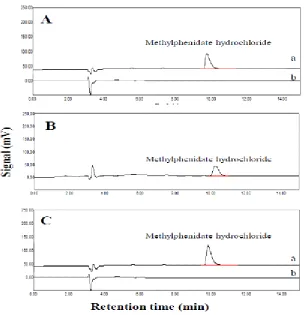
Fig. 4: Chromatograms obtained after HF-LPME extraction of (A) urinesample, (B) patient that used methylphenidate hydrochloride tablet and (C) plasma sample ((a) spiked sample and (b) non-spiked sample at a concentration level of 1.0 mg L-1)
hydrochloride being around 9.78 values. For this purpose, donor and acceptor phase pH was investigated in pH from 9 to 11.7 and 1.5 to 2.2, respectively. As shown in fig. 2B and 2C, the highest extraction efficiency was obtained using pH=11.2 and pH=1.9for Methylphenidate hydrochloride. Therefore, pH=11.2 for donor phase and pH=1.9 for acceptor phasewere used for following experiments.
Effect of stirring speed
Stirringof sample solution have greet effect to the analyte diffusion, which can be acceleratedextraction process and shorten the time of extraction to reach the extraction equilibrium.Stirring of sample solution facilitates analyte diffusion from donor phase into the acceptor phase[38].In the present work, the effect of the stirring speed from 100 to 1000 rpm onthe extraction efficiency was studied. As shown in fig. 2D, when the stirring speed was increased from 250 to 750 rpm, the extraction efficiency oftarget analytewas increased. However, increasing in stirring rate over 750 rpm, decrease extraction efficiency due to bobble formation around the hollow fiber in extraction process. Hence,thestirring rate of 750 rpm was chosen as the optimal stirring speed for following experiment.
Salting-out effect
The salting-out influence is widely used to increase the extraction recovery of uncharged target compounds from aqueous sample. It consists in decreasing the solubility of uncharged compounds in donor phase by increasing the ionic strength. In present work, the effect of various concentrations of NaCl from 0 to 40 percent to extraction of target analytewas tested. Theextraction efficiency enhanced when the NaCl concentration was increased from 0 to 30% (w/v). Over 30% salt addition decreased extraction efficiency. As shown in fig. 3A, 30% salt addition showed high extraction efficiency, and used in the future experiments. Extraction time effect
The extraction time is amajor parameter in HF-LPME technique.HF-LPME like other miniaturized sample preparation techniques such as SPME; several minutes to hours take to reach an equilibrium that ensures optimal extraction
efficiency. In this work, efficiency of methylphenidate hydrochloride extraction was studied in the range of 20–60 min. Fig. 3Billustratethe result ofdifferent extraction time on extraction efficiency. The extraction efficiency increased by the increasing of the extraction time up to 50 min. Future increasing in the extraction time to 60 min, decreasing the extraction efficiency due to instability in organic membrane solvent. Therefore, 50 min was selected as the optimal extraction time in the future experiments.
Influence of extraction temperature
The temperature of extraction has to be investigated because this parameter cans influence the partition coefficient of the analyte between the various phases. To study the effect of the temperature, the extraction vial was placed in an oil bath to heat the donor phase from 5 ÚC to 60 ÚC. The results, presented in Fig. 3C show that a temperature enhancement increased transfer of target analyte to the acceptor phase until 25 °C. A strong decrease of the extraction efficiency was observed by more increasing extraction temperature over 25 °C. This can be due to increasing the miscibility of 1-octanol in water at high temperature or to the partial evaporation of this organic solvent. In conclusion, a temperature of 25 °C was chosen as optimal extraction temperature.
As a consequence, the optimal conditions were attained by using 11.2 and 1.9 as donor and acceptor phases’pH, respectively, and using 750rpm as stirring speed for 50 min. In addition, the organic membrane composition was 1-Octanol. Thirtypercent salt addition and of 25 °C assample temperature was selected as best condition for Methylphenidate hydrochloride extraction.
Method performance
range of 12–5000 µg L-1 with coefûcient of determination (r 2)more than 0.9990. The relative standard deviations (RSD %) for extraction of the analytewere less than 2.5% and 3.5% for intraday and interday experiment, respectively. LODs less than 3.0 µg L-1 wasviewed for target analyte. PF values higher than 112-fold were obtained for the extraction of methylphenidate hydrochlorideby comparison slope of calibration curve before and after extraction process.
Analysis of real sample
HF-LPME is a powerful method for isolation and cleanup of target analyte from untreated biological fluids. Thus, the optimal conditions of HF-LPME were used for extraction of the target analytefrom human plasma and urine samples. To reduce matrix effects calibration curves were plotted in drug free urine and plasma samples.
Extraction from human urine sample
Drug-free human urine was spiked with proper amount of the target drug and extraction was accomplished after dilution of urine samples (1:3) and addition of proper amount of NaOH solution to achieve pH 11.2. The results are summarized in Table3. RSD% values less than 4.8% confirm the acceptable precision of proposed HF-LPME method. To evaluate the applicability of HF-LPME for human urine, four urine samples were analyzed with the proposed method. Since no methylphenidate hydrochloride was found in samples, all urine samples were spiked with the target drug at a different concentration level. Chromatograms are shown in Fig. 4A. To investigate the capability and accuracy of the proposed HF-LPME method, a urine sample was collected from a volunteer used 10 mg methylphenidate hydrochloride tablet, after 12 h of the last use. Figs. 4B show the typical chromatograms of real urine sample that collected from a patient that used methylphenidate hydrochloride tablet.
Extraction from human plasma sample
Plasma sample was diluted with water (1:3) and adjusted to pH 11.2 by addition of proper amount of NaOH solution. The drug was spiked into the human plasma and their quantitative analysis was evaluated under optimized conditions. Precision of the method was determined by three-replicate extraction of the drugs from samples at different concentration level. The RSD% was found less than 3.2% for Methylphenidate hydrochloride. To evaluate the applicability of HF-LPME for human plasma, three plasma samples were analyzed with the proposed method. Since no methylphenidate hydrochloride was found in samples, all plasma samples were spiked with the target drug at a different concentration level that showed in Table 3. Chromatograms are shown in Fig. 4C.
Comparison of the proposed method with other techniques
The present method was compared with the other methods in terms of validation and precision. As can be seen, the method is quite comparable to those mentioned in Table 4.
CONCLUSIONS
The present study exhibited an excellent performance of the HF-LPME technique for the extraction of methylphenidate hydrochloride drug from biological fluids. Up to 112-fold enrichment factor and effective sample clean-up were obtained. Accordingly, it is concluded that HF-LPME is an effective method to preconcentration of methylphenidate hydrochloride drug from the biological samples prior to HPLC analysis. The results indicated that hollow ûber microextraction method has an excellent cleanup, high enrichment, factor and can be served as a simple and sensitive method for monitoring of methylphenidate hydrochloride drug in the biological samples.
REFERENCES
1. J.K. Dunnick, J.R. Hailey, Experimental studies on the long-term effects of methylphenidate hydrochloride, Toxicology,
103 (1995) 77-84.
U.S. Department of Health and Human Services, 2001.
3. J.J. Panos, J.P. O’Callaghan, D.B. Miller, S.A. Ferguson, Effects of developmental methylphenidate (MPH) treatment on monoamine neurochemistry of male and female rats, Neurotoxicology and Teratology, 45 (2014) 70-74.
4. N. Easton, F.H. Marshall, C.A. Marsden, K.C.F. Fone, Mapping the central effects of methylphenidate in the rat using pharmacological MRI BOLD contrast, Neuropharmacology, 57 (2009) 653-664. 5. R. Ferreira, G.S. Bassi, A. Cabral, M.J. Nobre,
Withdrawal from methylphenidate increases neural reactivity of dorsal midbrain, Neuroscience Research, 68 (2010) 290-300.
6. S.M. Morris, V.N. Dobrovolsky, J.G. Shaddock, R.A. Mittelstaedt, M.E. Bishop, M.G. Manjanatha, S.D. Shelton, D.R. Doerge, N.C. Twaddle, J.J. Chen, C.-J. Lin, M.G. Paule, W. Slikker Jr, C.E. Hotchkiss, D. Petibone, J.D. Tucker, D.R. Mattison, The genetic toxicology of methylphenidate hydrochloride in non-human primates, Mutation Research/ Genetic Toxicology and Environmental Mutagenesis, 673 (2009) 59-66.
7. S. Pokkula, S. Thota, V. Raj Kumar, V.K. Nagabandi, Development and validation of RP-HPLC method for the determination of methylphenidate hydrochloride in API, Inter national Jour nal of Phar mTech Research, 6 (2014) 462-467.
8. M. Lalande, D.L. Wilson, I.J. McGilveray, HPLC determination of methylphenidate in human plasma, Journal of Liquid Chromatography, 10 (1987) 2257-2264. 9. C.C. Combs, E.L. Hankins, C.L. Copeland,
S.D. Brown, B.B. Pond, Quantitative determination of d- and l-threo enantiomers of methylphenidate in brain tissue by liquid chromatography-mass spectrometr y, Biomedical Chromatography, 27 (2013) 1587-1589.
10. S.K. Bushby, N. Thomas, P.A. Priemel, C.V. Coulter, T. Rades, J.A. Kieser, Determination of methylphenidate in Calliphorid larvae by liquid-liquid extraction and liquid chromatography mass spectrometry
-Forensic entomotoxicology using an in vivo rat brain model, Journal of Pharmaceutical and Biomedical Analysis, 70 (2012) 456-461.
11. K. De Cássia Mariotti, G. Rübensam, F. Barreto, V.C. Bica, L.Z. Meneghini, R.S. Ortiz, P.E. Froehlich, R.P. Limberger, Simultaneous determination of fenproporex, diethylpropione and methylphenidate in oral fluid by LC-MS/MS, Chromatographia, 77 (2014) 83-90.
12. S.M. Paterson, G.A. Moore, C.M. Florkowski, P.M. George, Deter mination of methylphenidate and its metabolite ritalinic acid in urine by liquid chromatography/ tandem mass spectrometry, Journal of Chromatography B: Analytical Technologies in the Biomedical and Life Sciences, 881-882 (2012) 20-26.
13. M. Wada, K. Abe, R. Ikeda, R. Kikura-Hanajiri, N. Kuroda, K. Nakashima, HPLC determination of methylphenidate and its metabolite, ritalinic acid, by high-performance liquid chromatography with peroxyoxalate chemiluminescence detection, Analytical and Bioanalytical Chemistry, 400 (2011) 387-393.
14. H.J. Leis, G. Fauler, G. Raspotnig, W. Windischhofer, Negative ion chemical ionization for the determination of methylphenidate in human plasma by stable isotope dilution gas chromatography/mass spectrometry, Journal of Mass Spectrometry, 35 (2000) 1100-1104.
15. F.T. Delbeke, M. Debackere, Isolation and detection of methylphenidate, phacetoperane and some other sympathomimetic central nervous stimulants with special reference to doping. I. Gas chromatographic detection procedure with electron capture detection for some secondary amines, Journal of Chromatography, 106 (1975) 412-417. 16. R. Huffman, J.W. Blake, R. Ray, J. Noonan,
P.W. Murdick, Methylphenidate blood plasma levels in the horse determined by derivative gas liquid chromatography electron capture, Journal of Chromatographic Science, 12 (1974) 382-384.
Tharp, Detection of methylphenidate and methamphetamine in equine body fluids by gas chromatographic analysis of an electron-capturing derivative, American Journal of Veterinary Research, 33 (1972) 27-31.
18. S. Pedersen-Bjergaard, K.E. Rasmussen, Liquid”Liquid”Liquid Microextraction for Sample Preparation of Biological Fluids Prior to Capillary Electrophoresis, Analytical Chemistry, 71 (1999) 2650-2656.
19. B. Ebrahimpour, Y. Yamini, A. Esrafili, Emulsification liquid phase microextraction followed by on-line phase separation coupled to high performance liquid chromatography, Analytica Chimica Acta, 751 (2012) 79-85.
20. O. Sha, X. Zhu, Y. Feng, W. Ma, Aqueous two-phase based on ionic liquid liquid–liquid microextraction for simultaneous determination of five synthetic food colourants in different food samples by high-performance liquid chromatography, Food Chemistry, 174 (2015) 380-386.
21. J. Zhou, Q. Zhang, J.B. Sun, X.L. Sun, P. Zeng, Two-phase hollow fiber liquid phase microextraction based on magnetofluid for simultaneous determination of Echinacoside, Tubuloside B, Acteoside and Isoacteoside in rat plasma after oral administration of Cistanche salsa extract by high performance liquid chromatography, Journal of Pharmaceutical and Biomedical Analysis, 94 (2014) 30-35.
22. S. Yu, Q. Xiao, B. Zhu, X. Zhong, Y. Xu, G. Su, M. Chen, Gas chromatography–mass spectrometry determination of earthy–musty odorous compounds in waters by two phase hollow-fiber liquid-phase microextraction using polyvinylidene fluoride fibers, Journal of Chromatography A, 1329 (2014) 45-51. 23. J. Zhou, P. Zeng, J.B. Sun, F.Q. Wang, Q.
Zhang, Application of two-phase hollow fiber liquid phase microextraction coupled with high-performance liquid chromatography for the study of the echinacoside pharmacokinetics in Parkinson’s disease rat plasma, Journal of Pharmaceutical and Biomedical Analysis, 81–82 (2013) 27-33. 24. M. Arvand, E. Bozorgzadeh, S. Shariati,
Two-phase hollow fiber liquid Two-phase microextraction for preconcentration of pyrethroid pesticides residues in some fruits and vegetable juices prior to gas chromatography/mass spectrometr y, Journal of Food Composition and Analysis, 31 (2013) 275-283.
25. Y.-Y. Chao, Y.-M. Tu, Z.-X. Jian, H.-W. Wang, Y.-L. Huang, Direct deter mination of chlorophenols in water samples through ultrasound-assisted hollow fiber liquid– liquid–liquid microextraction on-line coupled with high-performance liquid chromatography, Jour nal of Chromatography A, 1271 (2013) 41-49. 26. T.S. Ho, J.L. Egge Reubsaet, H.S. Anthonsen,
S. Pedersen-Bjergaard, K.E. Rasmussen, Liquid-phase microextraction based on carrier mediated transport combined with liquid chromatography–mass spectrometry: New concept for the determination of polar drugs in a single drop of human plasma, Journal of Chromatography A, 1072 (2005) 29-36.
27. L. Hou, G. Shen, H.K. Lee, Automated hollow fiber-protected dynamic liquid-phase microextraction of pesticides for gas chromatography–mass spectrometric analysis, Journal of Chromatography A, 985 (2003) 107-116.
28. A. Rodríguez, S. Pedersen-Bjergaard, K.E. Rasmussen, C. Nerín, Selective three-phase liquid three-phase microextraction of acidic compounds from foodstuff simulants, Journal of Chromatography A, 1198–1199 (2008) 38-44.
29. Y. Wu, B. Hu, Simultaneous determination of several phytohormones in natural coconut juice by hollow fiber-based liquid–liquid– liquid microextraction-high performance liquid chromatography, Jour nal of Chromatography A, 1216 (2009) 7657-7663. 30. L. Xia, B. Hu, Y. Wu, Hollow fiber-based liquid–liquid–liquid microextraction combined with high-performance liquid chromatography for the speciation of organomercury, Journal of Chromatography A, 1173 (2007) 44-51.
liquid–liquid microextraction for the determination of organosulfur pesticides in environmental and beverage samples by gas chromatography with flame photometric detection, Journal of Chromatography A, 1193 (2008) 7-18. 32. J. Zhang, T. Su, H.K. Lee, Development and
application of microporous hollow fiber protected liquid-phase microextraction via gaseous diffusion to the determination of phenols in water, Journal of Chromatography A, 1121 (2006) 10-15.
33. M. Villar-Navarro, M. Ramos-Payán, R. Fernández-Torres, M. Callejón-Mochón, M.Á. Bello-López, A novel application of three phase hollow fiber based liquid phase microextraction (HF-LPME) for the HPLC determination of two endocrine disrupting compounds (EDCs), octylphenol and n-nonylphenol, in environmental waters, Science of The Total Environment, 443 (2013) 1-6.
34. X.-W. Tan, Y.-X. Song, R.-P. Wei, G.-Y. Yi, Determination of Trace Bisphenol A in Water Using Three-phase Hollow Fiber Liquid-phase Microextraction Coupled with High Perfor mance Liquid Chromatography, Chinese Journal of Analytical Chemistry, 40 (2012) 1409-1414.
35. L. Guo, H.K. Lee, Ionic liquid based three-phase liquid–liquid–liquid solvent bar microextraction for the determination of phenols in seawater samples, Journal of Chromatography A, 1218 (2011) 4299-4306. 36. M. Ghambarian, Y. Yamini, A. Esrafili, Three-phase hollow fiber liquid-Three-phase microextraction based on two immiscible organic solvents for determination of tramadol in urine and plasma samples, Journal of Pharmaceutical and Biomedical Analysis, 56 (2011) 1041-1045.
37. S. Shariati, Y. Yamini, M. Darabi, M. Amini, Three phase liquid phase microextraction of phenylacetic acid and phenylpropionic acid from biological fluids, Journal of Chromatography B, 855 (2007) 228-235. 38. M. Mirzaei, H. Dinpanah, Three phases
hollow fiber LPME combined with HPLC-UV for extraction, preconcentration and determination of valerenic acid in Valeriana officinalis, Journal of Chromatography B, 879 (2011) 1870-1874.
39. C. Desoubr ies, F. Chapuis-Hugon, A. Bossée, V. Pichon, Three-phase hollow fiber liquid-phase microextraction of organophosphorous nerve agent degradation products from complex samples, Journal of Chromatography B, 900 (2012) 48-58.
40. H.-J. Zhu, J.-S. Wang, K.S. Patrick, J.L. Donovan, C.L. DeVane, J.S. Markowitz, A novel HPLC fluorescence method for the quantification of methylphenidate in human plasma, Journal of Chromatography B, 858 (2007) 91-95.
41. D.Z. Souza, P.O. Boehl, E. Comiran, K.C. Mariotti, F. Pechansky, P.C.A.V. Duarte, R. De Boni, P.E. Froehlich, R.P. Limberger, Determination of amphetamine-type stimulants in oral fluid by solid-phase microextraction and gas chromatography– mass spectrometry, Analytica Chimica Acta, 696 (2011) 67-76.