89 0 0 1 3 0 0 8 2
Liposomes
as a
Gene Delivery System
A th e sis p resen te d by
Yvonne P errie, M RPharmS
in p a rtial fulfilm ent of th e req u irem en ts for th e degree of D octor of Philosophy, U niversity of London
ernrr-f
All rights reserved
INFORMATION TO ALL USERS
The quality of this reproduction is dependent upon the quality of the copy submitted.
In the unlikely event that the author did not send a complete manuscript and there are missing pages, these will be noted. Also, if material had to be removed,
a note will indicate the deletion.
uest.
ProQuest 10104705
Published by ProQuest LLC(2016). Copyright of the Dissertation is held by the Author.
All rights reserved.
This work is protected against unauthorized copying under Title 17, United States Code. Microform Edition © ProQuest LLC.
ProQuest LLC
789 East Eisenhower Parkway P.O. Box 1346
To
m y Mum, D ad
hypothesis by an ugly fact."
First and foremost I would wish to express my thanks and sincere gratitude to my supervisor, Professor Gregory Gregoriadis, whose much valued advice and guidance has been invaluable throughout this work.
I would also like to thank my friends and colleagues from the Centre for Drug Delivery Research with whom I had the pleasure of working. To Brahim, Brenda, Christina, Janny, Jean-Christophe, Kent, Malini, Mia, Parinya, Sakthi, Sevtap, Sherry, Steve, and Sudax I extend my thanks, especially for succeeding to decipher my “foreign” accent (unfortunately Jean-Christophe never really got the hang of it!, C ’est la Vie).
My thanks also go to Dave, Donna and Steve from the animal unit for their generous and invaluable assistance with the animal work performed. Thanks for making potentially monotonous experimental work much less of a drudgery.
Annie and Graham from the computer unit have also provided lots assistance throughout my PhD for which I am grateful.
To my good friend Alison who, whilst working in The Division of Molecular Genetics, Institute of Biomedical and Life Sciences, University of Glasgow introduced me to the joys of plasmid purification I would like to say “cheers!”.
I would like to thank my sisters Loma and Vicki for all their support during my thesis. To my mum Maijorie my many thanks for all her support throughout my studies without which this would not have been possible and also thanks to my sadly departed dad Thomas who I know would be proud to see his daughter achieve her PhD.
Lastly, a special thanks go to Craig for all his help and support throughout this work. A big thank you also for cooking dinner every night for about the last six months - I greatly appreciated (and enjoyed) your cooking, long may it continue!.
Several non-viral gene delivery systems are currently being investigated, including
liposomes. The present studies attempt to characterise and assess the applicability of
liposomes as DNA delivery systems in gene therapy and genetic immunisation.
The dehydration-rehydration technique was used to entrap plasmid DNA into liposomes
(dehydration-rehydration vesicles; DRV) of varying compositions. Using this procedure, it
was shown that plasmid DNA can be efficiently entrapped in DRV in the absence of any
detrimental effect on the DNA. The use of cationic lipids as components of liposomes was
beneficial but not essential, with vesicles devoid of cationic lipids or incorporating anionic
lipids instead, exhibiting significant DNA entrapment values. Entrapped DNA was resistant
to nuclease degradation to an extent which was dependent on the DRV lipid composition.
The zeta potential and the z-average diameter of DRV vesicles was also dependent on the
lipid composition of DRV and, in the case of cationic DRV, on the ratio of cationic lipid to
anionic DNA. Microelectrophoresis studies of cationic DRV revealed DNA entrapment in-
between the bilayers of DRV (similarly to when no electrostatic attractions are present) as
well as binding of some of the DNA to the surface of DRV. Although systemic delivery of
DNA-containing DRV did not produce detectable gene expression, the formulation
employed was shown to offer significant protection to the incorporated plasmid DNA in the
presence of plasma at 37°C. In terms of the ability of DNA-containing DRV to induce
immune responses to antigens encoded by the DNA, results indicated that DRV with
entrapped DNA induce IgG antibody responses against the antigens that are significantly
higher than those of equivalent amounts obtained with naked DNA and that variations in the
vesicle lipid composition influence the magnitude of such responses. These results suggest
that DRV liposomes can act as a vehicle for the delivery of gene vaccines in genetic
Page No.
CONTENTS
Chapter 1 In tro d u ctio n ... 1
1.1: Gene delivery system s...2
1.1.1: Requirements of a gene delivery system ...4
1.1.2: Gene gun delivery ... 6
1.1.3: Polycations... 6
1.1.4: Liposomes ... 7
1.2: Basic aspects of liposomes... 7
1.2.1: Techniques employed for encapsulation of D N A ... 9
1.2.1.1: Lipid h ydration ... 9
1.2.1.2: Sonication procedures...9
1.2.1.3: Reverse-phase evaporation...10
1.2.1.4: Ca^^-EDTA chelation... 10
1.2.1.5: Ether injection...10
1.2.2: Cationic lipid based DNA-complexes... 11
1.2.2.1: Structural characteristics of cationic liposome-DNA complexes . 13 1.2.2.2: Cationic liposome-DNA complexes: Potential disadvantages . . . . 14
1.2.3: Incorporation of DNA into dehydration-rehydration liposomes ...15
1.2.3.1 : Transfection with liposome incorporated DNA in v it r o... 16
1.2.3.2: Transfection with liposome incorporated DNA in vivo...17
1.3: Genetic vaccination... 17
1.3.1.1: Live attenuated vaccines ...18
1.3.1.2: Non-live vaccines ... 18
1.3.2: Application of DNA in vaccine development... 19
1.3.2.1: The plasmid vaccine construct... 21
1.3.2.2: Modes of genetic immunisation... 22
1.3.2.3: Possible mechanism of genetic immunisation ... 23
1.3.2.4: Possible advantages of genetic immunisation... 26
1.3.3: DNA vaccination mediated by delivery system s... 27
1.3.4: Liposome-mediated DNA vaccination ...29
1.3.4.1: Mechanism of liposome-mediated vaccination... 30
1.4: Sum m ary...31
Chapter 2 Materials and M ethods... 34
2.1: Materials ...35
2.1.1: Chemicals...35
2.1.2: DNA plasmids ... 36
2.1.3: L ip id s...36
2.2: M ethods...36
2.2.1 : Extraction and purification of plasmid D N A ...36
2.2.1.1: Amplification of plasm ids...36
2.2.1.2: Purification of plasm ids...37
2.2.2: Spectrophotometric assay of D N A ... 38
2.2.4: Radiolabelling of plasmid D N A ...39
2.2.5: Preparation of liposomes ... 41
2.2.5.1: Preparation of multilamellar vesicles (M L V s)... 41
2.2.5.2: Preparation of small unilamellar vesicles (SUVs) ... 42
2.2.5.S: Preparation of dehydration-rehydration vesicles (D R V s)... 42
2.2.6: Separation of incorporated from non-incorporated plasmid D N A ... 43
2.2.7: Monitoring the extent of plasmid DNA incorporation... 43
2.2.8: Plasmid DNA extraction from liposom es... 44
2.2.9: Determination of liposomal DNA protection from nucleases ...44
2.2.10: Sizing of vesicles by photon correlation spectroscopy ... 45
2.2.11 : Sizing of vesicles by laser diffraction... 45
2.2.12: Characterisation of charged liposomes by microelectrophoresis...45
2.2.13: Experimental anim als... 46
2.2.14: Immunisation schedules... 46
2.2.15: Enzyme-linked immunosorbent assay... 47
2.2.16: Statistical analysis ... 48
2.2.17: Luciferase a ssay ...49
2.2.17.1: Construction of a luciferase standard c u r v e ... 49
2.2.18: Intravenous injection of DRV incorporating plasmid DNA encoding luciferase ... 49
2.2.18.1: Assay of luciferase activity in tissue extracts... 50
2.2.19: Radioiodonation of luciferase by the chloramine-T m e th o d ... 50
2.2.20.1: Assay of luciferase activity and radioactivity in tissues
extracts ...51
2.2.21: Luciferase stability in tissue extracts ... 52
2.2.22: Determination of protein by bicinochoninic acid assay... 52
Chapter 3 Entrapment of plasmid DNA in DRV: Initial characterisation stu d ies... 53
3.1: Characterisation studies ...54
3.2: Purification and characterisation of plasmid D N A ... 54
3.2.1: Plasmid purification... 54
3.2.2: Spectrophotometric assay of DNA concentration and p u rity ... 56
3.2.3: Characterisation of plasmids using restriction enzyme digestion...57
3.3: Radiolabelling of plasmid D N A ... 59
3.4: Incorporation of plasmid DNA into liposom es... 61
3.4.1: Confirmation of DNA integrity during and after the DRV process...63
3.4.2: Co-entrapment of plasmid DNA with other plasmids in DRV vesicles . . . . 65
3.5: Nuclease degradation of DNA: The extent of protection provided by DRV entrapment...67
3.6: The effect of using DOPE as a “helper lipid” in DRV formulations... 70
3.7: Entrapment of DNA in DRV formulations using non-ionic surfactants...75
Chapter 4 An investigation into DRV formulations incorporating charged
lip id s ... 78
4.1: The use of cationic lipids in DNA delivery system s... 79
4.2: The initial stages of the DRV process: Characterisation of SUV-DNA complexes ... 79
4.2.1 : The z-average diameter of SUV before and after mixing with plasmid D N A ... 80
4.2.2: The zeta potential of cationic SUV-DNA com plexes... 83
4.3: Physicochemical studies of DRV formulations supplemented with charged lipids...86
4.3.1: DRV formulations incorporating cationic lipid DOTAP: DNA incorporation stu d ie s... 86
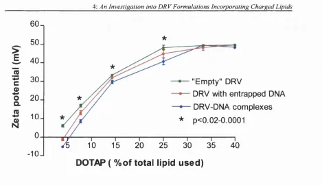
4.3.2: Zeta potential studies: Cationic DRV formulations...94
4.3.3: Zeta potential studies: Anionic DRV formulations...97
4.3.4: z-Average diameter of DRV vesicles: The effect of charged lip id s...99
4.4: Conclusions...108
Chapter 5 Liposome-mediated DNA immunisation: The effect of vesicle composition ... 120
5.1: Genetic immunisation... I l l 5.1.1: The effect of DOPE presence in liposome-mediated DNA immunisation . 113 5.1.1.1: Immunisation protocol ... 114
5.3: Induction of neonatal tolerance in mice ... 130
5.3.1: DRVfDNA) immunisation of neonatal mice ... 131
5.3.1.1: Immunisation protocol ... 131
5.3.1.2: Effect of immunisation with “empty” D R V ...132
5.4: Conclusions...135
Chapter 6 Liposome-mediated intravenous gene delivery ... 139
6.1: Gene th era p y ... 140
6.1.1: Lipid based plasmid DNA delivery systems... 140
6.1.1.1: The use of reporter genes in the study of gene expression . 143 6.1.1.2: Quantification of luciferase content... 144
6.1.1.3: Luciferase stability during extraction from tissues... 146
6.1.1.4: Luciferase activity after extraction from tissues of mice injected with the enzyme...147
6.2: Characterisation of DRV(DNA) after exposure to plasma ... 151
6.3: Intravenous injection of DRVfDNA)...154
6.4: Conclusions... 155
Chapter 7 Final discussion and future w o r k ... 157
7.1: Final discussion... 158
7.2: Future w o rk ... 162
R eferen ces... 163
LIST OF FIGURES Page No.
Chapter 1
1.1: Luciferase reporter plasmid circle m a p ...4
1.2: A schematic representation of gene transfer into a eukaryotic cell using a
non-viral delivery sy stem ...5
1.3: A schematic representation of a multilamellar liposom e...8
1.4: Examples of some lipids used to prepare liposom es... 11
1.5: A schematic representation of proposed mechanisms of DNA immunisation . . . 25
Chapter 3
3.1: Agarose gel electrophoresis of samples taken during the pGL2 plasmid
purification process ... 55
3.2: Spectrophotometric assay of pGL2 purified by the QIAGEN procedure...56
3.3: Agarose gel electrophoresis pGL2 before and after restriction enzyme
digestion... 58
3.4: Agarose gel electrophoresis pRc/CMV HBS before and after restriction
enzyme d ig estio n ... 58
3.5: Elution profile of ^^S-labelled pGL2 plasmid D N A ... 60
3.6: Agarose gel electrophoresis of radiolabelled pGL2 separated fi"om
non-incorporated ^^S-ATP by gel chromatography... 60
3.7: Agarose gel electrophoresis of DNA extracted fi*om neutral and cationic DRV . . 64
3.8: Agarose gel electrophoresis of DRV entrapped plasmid DNA ranging in size
3.9: Agarose gel electrophoresis of co-entrapped DNA extracted from D R V ...66
3.10: Gel electrophoresis of pRSVhGH plasmid DNA after exposure to D N a s e 70 3.11: The schematic representation of the inverted hexagonal phase formed by DOPE ...74
Chapter 4 4.1: A schematic representation of Dehydration-Rehydration Vesicles ... 79
4.2: z-Average diameter of SUV before and after mixing with D N A ...82
4.3: SUV mixed with D N A ... 82
4.4: The zeta potential of SUV-DNA...85
4.5 - 4.7: Gel electrophoresis of DRV(DNA) and DRV-DNA incorporating DOTAP . 91 4.8: Zeta potential of DRV liposom es... 95
4.9: Zeta potential of DRV(DNA) incorporating DOPE or cholesterol in the formulation...95
4.10: Zeta potential and entrapment efficiency of anionic D R V ...98
4.11: z-Average diameter of cationic “empty” DRV and DRV(DNA)...101
4.12: z-Average diameter of anionic “empty” DRV, DRV(DNA) and DRV(polylysine) ... 102
Chapter 5
5.1: Comparison of IgGi immune responses in mice 20 days after the final injection 115
5.2: Comparison of IgGi immune responses in mice 31 days after the final injection 116
5.4: The effect of DOTAP content in DRV(DNA) on IgGi, IgG2a and IgG2b immune
responses in mice 3 days after the final in jectio n... 124
5.5: The effect of DOTAP content in DRV(DNA) on IgGi, IgG2& and IgG2b immune responses in mice 25 days after the final in jectio n ... 125
5.6: The effect of DOTAP content in DRV(DNA) on IgGi, IgG2a and IgG2b immune responses in mice 34 days after the final in jectio n ... 126
5.7: The effect of DOTAP content in DRV(DNA) on IgGi, IgG2a and IgG2b immune responses in mice 41 days after the final in jectio n ...127
5.8: The effect of DOTAP content in DRV(DNA) on IgGi, IgGia and IgG2b immune responses in mice 56 days after the final in jectio n ...128
5.9: Neonatal tolerance studies: IgG immune responses ...133
5.10: Neonatal tolerance studies: IgGi immune responses... 133
Chapter 6 6.1: Luciferase standard curve... 145
6.2: Plasma clearance of luciferase after IV injection ...148
6.3: Detection of luciferase in the liver after IV injection ...148
6.4: Activity of luciferase after incubation under various conditions... 149
LIST OF TABLES Page No.
Chapter 3
3.1: Incorporation of plasmid DNA into various DRV liposome formulations... 62
3.2: Incorporation of plasmid DNA in DRV and its retention after exposure to deoxyribonucleases ... 70
3.3: Incorporation of DNA and zeta potential of DRV form ulations...72
Chapter 4 4.1 : Theoretical +/- charge ratio of cationic lipid to D N A ... 83
4.2: Incorporation of plasmid DNA into cationic D R V ... 88
4.3: Z-average diameter (nm) of cholesterol containing D R V ... 104
4.4: Size of DRV entrapping increasing amounts of DNA or polylysine... 105
Chapter 6 6.1 : Recovery of luciferase after the extraction process... 147
Chapter One
1.1 GENE DELIVERY SYSTEMS
Gene therapy has been described (Behr, 1993) as one of medicine*s greatest dreams:
molecular surgery at the root of life. The aim of gene therapy is to introduce genetic material
into a patient’s cells to cause these cells to produce the therapeutic protein. In this respect,
gene therapy has the potential to be a new type of weapon in the fight against not only
hereditary diseases but also against acquired diseases resulting fi*om multigenic disorders
such as cancer, or those resulting fi"om foreign viral genes (Behr, 1993). However the
administration of genetic material for therapeutic purposes has been recognised for some
time. Some of the earliest experiments describing the transfer of DNA (deoxyribonucleic
acid) into the cells of living animals were reported by Stasmey et al. (1950), and Ito et al.
(1958), where crude preparations of DNA isolated fi*om tumours were injected into
experimental animals and consequently induced the formation of tumours. These results
were confirmed by Ortho et al. (1964) and Israel et al. (1979) using purified and
recombinant viral DNA respectively. More recently, Hepatitis B proteins (Will et al, 1982),
insulin (Nicolau et a l, 1983) and reporter molecules (Wolff et a l, 1990) were expressed in
vivo after injection of plasmid DNA encoding for the corresponding genes.
Since these early studies, more current research on gene therapy has focussed on the
development of viral approaches to deliver therapeutic genes to cells. However, non-viral
vehicles for genes have emerged as a potentially safe and effective approach to gene therapy
(Tomlinson and Rolland, 1996). The use of plasmid DNA (a closed circular form of DNA;
Fig. 1.1) in non-viral delivery systems, which transfects cells epichromosomally, means the
as retroviruses, which result in integration of the delivered gene into the chromosome, has
potentially drastic side effects if integration results in disruption of required genes or
activation of oncogenes. Adenoviruses do not integrate into the host genome, but may trigger
immune and inflammatory responses which could mean repeat administrations are not
feasible. Furthermore, even although viral delivery systems are designed to be unable to
replicate in the host, there is the potential risk of mutations >\iiich can result in an infectious,
replication competent virus, either during production or in use for gene therapy (Varmus,
1988). The use of non-viral plasmid DNA delivery systems means that, essentially the size
of the plasmid DNA that can be accommodated in such systems is unlimited, unlike viral
vectors which have packaging constraints (eg.7-13 kb for adenovirus) (Jolly et a l, 1987;
Varmus, 1982). With regard to the safety profile of plasmid DNA, high resolution techniques
such as polymerase chain reaction have shown that the chance of plasmid DNA causing
malignancy is less than 1 in 10^ patients, and it has been estimated that 100 kg of plasmid
DNA must be injected for the occurrence of one tumorigenic event (Moelling, 1997).
Plasmid DNA usually includes the gene encoding the required protein. Fig. 1.1 shows an
example, the pGL2 plasmid DNA which encodes for the reporter protein luciferase, under
the control of a promoter sequence (simian virus 40 (SV40) promoter) to drive the
transcription of the protein gene insert and an mRNA stability polyadenylation region at the
3' end of the insert to ensure translation. In addition, an origin of replication for the
amplification of the plasmid in bacteria and a gene for antibiotic resistance to select the
poly (A) signal (for/uc roporter)
E n h an cer
pGL2-Control Vector (6046bp)
poly(A) signal (for background reduction)
S rrw l 3 Kpn\ 12 Sk I 18 MIkjl 2 2 Nhet 2 8
7 Xhol 3 3
Bgt ■ 3 7 ‘ Prom oter
Hind III 239
2 2 Z 5 P m
855ECORI
Fig. 1.1. Luciferase reporter plasmid (pGL2-Control Vector; 6.046 kb) circle map. The simian virus 40 promoter and enhancer regions are shown in black and the luciferase coding region in magenta. Enzyme digestion sites are also marked. Amp" (shown in blue) denotes the ampicillin resistance coding region.
1.1.1 Requirements of a gene delivery system
Ideally, a gene delivery system should allow control over location and function of the
administered gene, be administered by a convenient and conventional route, ensure
controlled distribution, be non-toxic, efficient, and inexpensive. Both ex vivo and in vivo
gene delivery methods are under development for gene therapy, however direct in vivo
administration of genes to the patients to cause controlled production and distribution of
therapeutic proteins within the body would represent an ideal approach for clinical practice
(Tomlinson and Rolland, 1996).
Fig 1.2 shows a very basic schematic presentation of gene transfer into a eukaryotic cell
using a non-viral delivery system. Even before arrival at a cell, for transfection to occur, a
degradation in vivo allowing delivery of the intact plasmid to the target cell. Once it reaches
the target cell, the DNA must enter the nucleus of the cell to allow transfection to occur (Fig.
1.2). This may involve the plasmid DNA delivery system being taken up by endocytosis or
phagocytosis, the plasmid DNA escaping the endosome and entering the nucleus (Szoka et
al, 1996). For the synthesis of proteins to occur from a DNA template, the DNA must be
transcribed into messenger ribonucleic acid (mRNA) which then leaves the nucleus and
enters the cytoplasm. The mRNA becomes attached to a ribosome and tRNA (transfer RNA),
codons of three bases which correspond to a particular amino acid, link with the
Chromosomal DNA Target cell
tRNA
L oW effect Ribosome
Intracellular
effect
À
Endosome
Systemic effect Plasmid DNA delivery system
complementary sequence of bases on the mRNA. Peptide bonds form between adjacent
amino acids and the completed protein is released into the cytoplasm. The protein can then
act intracellularly, locally or systemically. Ideally, elements of the delivery system should
facilitate the passage of the plasmid DNA into the cell and into the nucleus, delivering the
plasmid DNA in a form which allows transcription and hence translation to occur. This may
involve properties of the delivery system that could enhance release of the plasmid DNA
from the endosome after endocytosis. Several types of synthetic non-viral delivery systems
have been developed to aid in the “running of the gauntlet" between the site of
administration of the plasmid DNA and eventual cell transfection in vivo. These include
particle bombardment or gene gun systems, polycations, as well as liposomes.
1.1.2 Gene gun delivery
This system employs a gene gun which uses an adjustable electric discharge to generate a
shock wave which accelerates DNA-coated gold or tungsten particles (1-3 pm) directly into
the cytosol of target tissue or skin cells. It is thought that during its transit in the tissue, DNA
detaches itself from the gold particles to end up intracellularly. The advantages of this are
that targeting is easy and is reported to be pain free, but on the negative side, responses are
variable (Schofield and Caskey, 1995).
1.13 Polycations
These carry a positive charge which complexes and condenses the negatively charged DNA.
Condensation of DNA protects against DNA degradation by deoxyribonucleases (DNases)
include protamine, polylysine and spermidine. Lipopolyamines have been developed (Behr,
1996; Behr et al., 1989) including DOGS (dioctadecylamidoglycylspermine) and DPPES
(dipalmitoyl phosphatidylethanolamido spermine). These compounds together will form
miceUar structures. DOGS is commercially available as the transfection agent "Transfectam"
(Promega, UK).
1.1.4 Liposomes
Liposomes were first observed and their semipermeable nature described by Bangham and
colleagues (Bangham et ah, 1965; Papahadjopoulos and Bangham, 1966) in the mid 1960's
and were originally studied to reveal insight into biomembrane properties (Chapman, 1994).
However their potential usage as drug delivery systems was soon realised (Gregoriadis et al.,
1971; Gregoriadis and Ryman, 1972) . Mixing dry phospholipids with water gives rise to
bilayers of phospholipid molecules which arrange themselves spontaneously to form closed
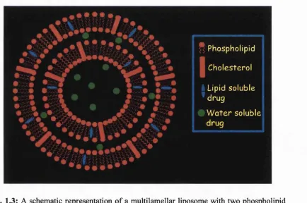
multilayered spherules known as liposomes (Gregoriadis, 1984). (Fig. 1.3). As they form,
liposomes entrap water and any water-soluble solutes (eg. drugs) that are present. Lipid-
soluble compounds, on the other hand, will be incorporated into the liposomal membrane
(Gregoriadis, 1984). The original procedure of making liposomes was modified in numerous
ways (Deamer and Uster, 1983; Gregoriadis, 1984) to give updated versions of the system
with high drug to lipid ratios.
1.2 BASIC ASPECTS OF LIPOSOMES
As already mentioned liposomes are closed vesicles, normally composed of phospholipids,
lamellae in which the phospholipids are organised in a bilayer configuration (Fig. 1.3).
Liposomes can range in size from 25 nm up to several micrometers. Water soluble
(hydrophilic) molecules, including DNA are located in the aqueous core, and in the case of
multilamellar vesicles (MLV) in the aqueous space between the bilayers. Liposomes are
particularly versatile as drug delivery systems as their lipid compositions and properties can
be widely varied. Liposomes, due to their sealed structure, can protect their entrapped
material from the external environment. Traditionally, liposomes are made of
phosphatidylcholine (Fig. 1.4 (a)), with or without cholesterol and supplemented with
anionic lipids such phosphatidlyglycerol or phosphatidylserine (Fig. 1.4 (b) and (c)
respectively) (Gregoriadis, 1984).
* Phosph olipid Cho e s t e r o I Lipid s o lu b le ' d r u g
W a t e r s o lu b le
With regard to the use of liposomes in DNA delivery, the possibility of employing liposomes
to introduce segments of genetic material to defective cells was noted as early as 1972
(Gregoriadis and Ryman, 1972). Liposomes were initially demonstrated to deliver
encapsulated DNA to bacterial cells (Fraley et al. , 1979) using liposomes produced by the
ether injection method (Deamer and Bangham, 1976) and composed of PC and PG.
Liposome-mediated transformation of fungal cells was reported by Radford era/. (1981) in
which chromosomal DNA was entrapped in liposomes composed of PS. Delivery of
liposome encapsulated DNA and RNA was shown by Dimitriadis (1978) in vitro, again
using anionic liposomes prepared by various methods. Transfection in all these experiments
was very low (Lurguin, 1984).
1.2.1 Techniques employed for encapsulation of DNA
1.2.1.1 Lipid hydration
Lipids dissolved in chloroform, form a dry film after evaporation of the chloroform. This
film can be rehydrated with water to form large MLV which are normally sonicated to
reduce their size. By hydrating the lipid film with DNA dissolved in water, DNA can be
entrapped in the aqueous core (Mukheijee et a l, 1978).
1.2.1.2 Sonication procedures
Many methods of liposome preparation involve a short sonication step to reduce the size of
the liposomes, however even low power sonication has been reported to cause significant
1.2.1.3 Reverse-phase evaporation
This phase involves first making an emulsion containing DNA, lipids dissolved in a nonpolar
solvent and a buffered aqueous solution. The nonpolar solvent is then removed under partial
vacuum to produce liposomes of 100 - 500 nm, with entrapment of DNA of up to 40 %
(Fraley et aL, 1980). This procedure often involves sonication of the liposome preparations.
1.2.1.4 - EDTA chelation
SUV composed of PS are firstly prepared and incubated with calcium to form large
multilamellar cochleates, DNA is then added to this solution and by the addition of EDTA
the cochleates are transformed into large unilamellar vesicles (LUV) with DNA incorporated
inside their lumen (Gould-Forgerite and Mannino, 1985; Itani et a i, 1987; Kondoro and
Duda, 1982; Mannino et a l, 1979; Szelei and Duda, 1989). The major disadvantage of this
is that the liposomes can only be formed with a negative charge (Gould-Forgerite et a l,
1989)
1.2.1.5 Ether injection
This technique was first proposed by Deamer and Bangham (1976). An ether solution of
phospholipid and steroid or other lipids is slowly injected into an aqueous DNA solution
heated at 60°C and the ether is evaporated off. This leads to the formation of liposomes
encapsulating DNA with a relatively low efficiency (0.95 - 15% of initial amount of DNA
1.2.2 Cationic Upid based DNA-complexes
The use of preformed positiveljjchargedliposomes which can form complexes with DNA as
novel gene delivery systems was first described by Feigner et a/. (1984) using a liposome
formulation known as “Lipofectin”. The authors described a DNA-transfection protocol that
made use of a synthetic cationic lipid A^-[l-(2,3-dioleyloxy)propyl]-A^,A^,iV-
trimethylammonium chloride (DOTMA). SUV liposomes containing only DOTMA or
combined with dioleoyl phosphatidylethanolamine (DOPE) (1:1 wt ratio) interacted
spontaneously with DNA to form lipid-DNA complexes with 100% complexation of DNA
(Feigner et a l, 1984). It was proposed that DOTMA facilitated fusion of the complex with
the plasma membrane of culture cells, resulting in both uptake and expression of DNA. This
provided a technique which was simple, highly reproducible and effective for both transient
and stable expression of transfected DNA in vitro (Feigner et a l, 1984). DOTMA is
characterised by one positively charged ammonium group linked to two oleoyl chains via
ester linkages, and this cationic charge interacts with the anionic phosphate groups of the
DNA by simple mixing. O
II R-C^
u —u r t
O
^ ^ (a) Phosphatidylcholine (PC) CH
H 2 C — O — P — O —C H 2 - C H 2 - N ( 0 ^ 3 ) 3
R - C - O ^ - ---
----R—
O—ÇH2 (b) Phosphatidylglycerol (PG)
J C H V
R'— C -C r " i^ c-O -P -O -C H z-C H -C H z-O H
o O- OH
' ^ ^ 0 —Ofc
Fig. 1.4 (continued)
CH2 o
C \ ,0—P —O — (CH2)2NH3*
O CHz
\ j
(d) dioleoyl phosphatidylethanolamine (DOPE)
(e) l,2-dioleoyl-3-trimethylammonium propane (DOTA?)
O CH2—NH (CH3)2
(f) l,2-dioleoyl-3-dimethylammonium propane (DODAP)
(CH3)2—NCH2—NH—C—O
(g) 3((N-(N’,N’)-dimethylaminoethane)-carbamoyl)cholesterol (DC-Chol)
A variety of synthetic cationic lipids have now been developed and some of those are shown
in Fig. 1.4. l,2-dioleoyl-3-trimethylammonium propane (DOTAP), a synthetic analogue of
DOTMA was synthesised by Browne/a/. (1989), as was l,2-dioleoyl-3-dimethylammonium
propane (DODAP) (Fig. 1.4 (e) and (f) respectively). The DOTMA/DOPE reagent is now
commercially available from Gibco, and DOTAP is marked as a transfection system by
Boeringer Mannheim. Other cationic lipids include cationic dérivâtes of cholesterol, for
instance 2 (N, (N’, N ’-dimethylamino-ethane)-carbamoyl) cholesterol (DC-chol) (Fig. 1.4
(g)) and cholesterol hemisuccinate choline ester (Brigham and Schreier, 1993; Friend et a l,
1990). Many quaternary ammonium-type compounds have also been synthesised, including
cetyldimethylethyl ammonium bromide (CDAB) and dimethyldioctadecyl ammonium
bromide (DDAB). All of these and other cationic lipids have been screened for transfection
efficiency in cell culture and shown variable success.
In many of the cationic liposome transfection systems DOPE (Fig. 1.4 (d)) is a key
component. DOPE is known to be a strong destabiliser of lipid bilayers (Litzinger and
Huang, 1992) due to its ability to enter the hexagonal phase (Connor and Huang, 1986;
Ellens et a l, 1984). The presence of DOPE in a liposomal formulation, in combination with
a cationic lipid, is thought to enhance the intrinsic fusogenic properties (Bennett et al.,
1996).
1.2.2.1 Structural characteristics o f cationic liposome-DNA complexes
Cationic liposome-DNA complexes have been reported to display a variety of polymorphic
of the two oppositely charged components (Gershon et al, 1993; Lasic et al., 1997, Radier
et a l, 1997); fibrillar structures, among them spaghetti-liketubules (Sternberg et a l, 1994)
and map-pinstructures (Sternberg et a l, 1998); and finally non-bilayer lipid arrangements
(Sternberg et a l, 1994 and Mok et a l, 1996). Such polymorphic structures have also been
reported (Sternberg et a l, 1998) to depend on the ratio of cationic lipid to anionic DNA.
1.2.2.2 Cationic liposome-DNA complexes: Potential disadvantages
The aforementioned cationic lipid based DNA delivery systems are dependent on cationic
lipids binding to DNA through electrostatic interactions, and the resultant structure having
a net positive charge. The net positive charge allows the particles to bind to the surface of
cells which have a net anionic charge, an event that is thought to be the first step of the
mechanism of transfection. Indeed in the first report of cationic liposome mediated
transfection (Feigner et a l,1987 ), it was described how the presence of serum inhibited the
ability of “Lipofectin” to promote transfection. It is known fi’om early studies (Black and
Gregoriadis, 1976) that the positive surface charge of cationic liposomes is masked by
plasma proteins which impose a net negative charge on the surface of vesicles. Such
interactions between liposome-DNA complexes and anionic molecules in the serum
neutralise the positive charge of the complex and decrease transfection efficiency (Li and
Huang, 1996). When studying transfection mediated by cationic liposomes, many groups
have observed little or no correlation between in vitro and in vivo transfection activity
(Szoka et a l, 1996). These vectors are also said to be unsuitable for intravenous
administration, and give poorly reproducible results (Li and Huang, 1996). Such poor results
DNA release from the complex and subsequent degradation (Li et al, 1998).
1.23 Incorporation of DNA into dehydration-rehydration liposomes
A procedure which was recently used to incorporate plasmid DNA into liposomes
(Gregoriadis et a l, 1996) is the dehydration-rehydration method. First described by Kirby
and Gregoriadis (1984), this novel method of liposome preparation is simple to use, employs
mild conditions and is capable of efficient entrapment of a wide range of materials including
small drugs (Kirby and Gregoriadis, 1984), drug-cyclodextrin complexes (McCormack and
Gregoriadis, 1996), peptides (Gregoriadis e ta l, 1993), interleukins (Gursel and Gregoriadis,
1997), antigens (Gregoriadis et a l, 1987) and other proteins (Skalko et a l, 1996). Using this
method to incorporate DNA (or any other solute) in dehydration-rehydration vesicles (DRV)
has the advantage that the DNA is not subjected to high temperatures, organic solvents
and/or sonication. Additionally, the DRV can be prepared with anionic, cationic or neutral
lipids. Briefly, to prepare DRV, a preparation of SUV of the desired lipid composition is
mixed with DNA and freeze-dried to produce a dehydrated powdery material in which the
flattened bilayers of liposomes are brought into intimate contact with the DNA. This
dehydrated powder is rehydrated under controlled conditions (Kirby and Gregoriadis, 1984)
to form multilamellar (Gregoriadis et a l, 1993; Skalko et al, 1998) DRV vesicles with
incorporated DNA (for a more detailed outline of this procedure see section 2.2.5 and Fig.
4.1 showing a schematic representation of the procedure), ranging in size from 600 - 900 nm
(Gregoriadis e/a/., 1996).
plasmid DNAs (Gregoriadis et a l, 1996; Gregoriadis et al., 1997; Morrison-Perrie and
Gregoriadis 1997; Gregoriadis et al, 1998; Ferrie and Gregoriadis, 1998), incorporation
values of plasmid DNA (based on ^^S-labelled plasmid DNA assay) were considerable with
values o f44 - 55% for neutral and anionic DRV and 57 - 95% (of the initial amount of DNA
used) for cationic DRV, with no apparent relationship between entrapment values and the
amount of DNA used (10 - 500 pg) (Gregoriadis et a l, 1998). Significant amounts of
incorporated DNA was shown (Gregoriadis et a l , 1996) to be resistant to deoxyribonuclease
degradation whether entrapped in cationic (49 - 93%), anionic (38 - 69%) or neutral (41 -
72% of initially incorporated DNA) liposomes. These results were confirmed by gel
electrophoresis studies (Gregoriadis et a l, 1996; Morrison-Perrie and Gregoriadis, 1997).
1.2.3.1 Transfection with liposome incorporated DNA in vitro
In vitro studies with DRV incorporating pGL2 plasmid DNA (expressing the luciferase
reporter gene; Fig. 1.1) revealed significant levels of luciferase activity with each of the
formulations tested (Gregoriadis et a l, 1996). However, DRV composed of
PC:DOPE:DOTMA (16:8:4 pmoles) and PC:DOPE:SA (16:8:4 pmoles) gave transfection
results which were approximately 10-fold higher than those seen with DRV composed of
PC:DOPE (16:8 pmoles) or PC:DOPE:PS (16:8:4 pmoles). In this study (Gregoriadis e ta l,
1996), DRV with entrapped DNA were also microfluidised to produce smaller vesicles (210
- 380 nm) which, with the exception of uncharged ones, retained substantial proportions of
the originally incorporated DNA, again in a form largely protected fi’om DNase degradation
(Gregoriadis et a l, 1996). The microfluidised DRV with incorporated DNA
formulations tested.
1.2.3.2 Transfection with liposome incorporated DNA in vivo
Plasmid DNA incorporated into DRV liposomes has also been shown to transfect cells in
vivo in immunisation experiments and is discussed in section 1.3.4.
1.3 GENETIC VACCINATION
The goal of any vaccination is to induce immunity to protect the host from disease. Vaccines
should generate long term protective immune responses which perform immune surveillance
against specific antigens (Wang et al, 1998). Vaccination against pathogenic
microorganisms represents one of the most important advances in the history of medicine
with vaccines including those against polio, measles, mumps, rubella, hepatitis A, hepatitis
B, pertussis and other diseases, having dramatically improved and protected more human
lives than any other avenue of modem medicine (Koprowski and Weiner, 1998). It is widely
believed that smallpox, which was responsible for more deaths in the twentieth century than
World Wars I and II combined has been completely eradicated as a result of the vaccine
against smallpox (Koprowski and Weiner, 1998).
1.3.1 Currently available vaccines
Traditionally, there are two categories of vaccines, ie. either live infectious material which
was manufactured in a weakened or attenuated state, so as to prevent the vaccine from
1.3.1.1 Live attenuated vaccines
Live attenuated vaccines (eg. polio and smallpox), generate immune responses as they
replicate in the host, however their ability to replicate in humans is limited so as to avoid
induction of the disease in the inoculated individual. Since these live attenuated viruses
replicate within the host cells, they can be customised by the host resulting in life-long
immunity. The viral proteins produced within the host are leaked or shed into the
extracellular space surrounding the infected cells and are then taken up and processed by
antigen presenting cells (APC) (including macrophages, dendritic cells and B cells). These
APC expand the immune response, a function which involves the recirculation of small
fragments of the antigen to their surface, where they are attached to major histocompatibility
complex (MHC) II antigens. The combination of MHC and the foreign peptide antigen forms
part of the signal with which APC trigger the action of a central population of immune cells,
the T helper lymphocyte cells (Koprowski and Weiner, 1998). APC also express co
stimulatory molecules which help to promote T cell expansion and activation. Activated T
cells secrete soluble molecules such as cytokines which further activate immune cells.
Production of viral proteins within cells also results in small fragments of viral proteins
being drawn to the infected cell surface, combined with the host MHC I antigens. These
complexes are recognised by killer cytotoxic T cells which, when combined with secondary
stimulation provided by professional APC and cytokine production (from stimulated helper
T cells), results in the destruction of infected cells.
1.3.1.2 Non-live vaccines
weaker immune responses and are unable to induce life-long immunity. Since they are not
produced within host cells and generally end up in the extracellular space after
administration, they can generate T helper and humoral immune responses but are usually
devoid of inducing significant T cytotoxic responses (Koprowski and Weiner, 1998).
However, from a practical view point they are generally easier to manipulate and produce
than live vaccines and also do not carry the danger of reversion to a more pathogenic form,
or the problem of inducing disease in persons with weak or compromised immune systems.
Recombinant subunit and synthetic peptide vaccines which can elicit specific immune
responses have also been developed. Unfortunately, subunit and peptide vaccines are weak
imunogens and are often unable to induce appropriate immune responses. A great variety of
experimental immunological adjuvants (Gregoriadis e ta l, 1993; Powel and Newton, 1995)
can render such vaccines stronger and more efficient. However, about seventy years since
the introduction of aluminium salts as an adjuvant, only one other adjuvant, liposomes
(Gregoriadis, 1990) has been approved for human use (Gregoriadis, 1990).
The benefits of the non live vaccines over live attenuated vaccines have resulted in the
former being the vaccine of choice, however as discussed they are far from ideal (Koprowski
and Weiner, 1998).
1.3.2 Application of DNA in vaccine development
A new side line of gene therapy, DNA immunisation, arose from the unexpected observation
cell mediated immunity against the DNA encoding a protein (Ulmer et al., 1993). Thereafter
came a succession of publications ft-om 1992 onwards showing the ability of plasmid DNA
to induce an immune (antibody) response to the encoded foreign protein (human growth
hormone) (Tang et al., 1992). Using DNA encoding influenza nucleoprotein, hnmunity was
found to be both humoural and cell-mediated and to protect mice challenged with the virus
(Fynan e ta l, 1993; Ulmer e ta l, 1993). Similarly, around the same time, humoural and cell
mediated immunity against human immunodeficiency virus type 1 (HTV 1) using plasmids
encoding the HTV revand envproteins was reported (Wang et a l, 1993) and a short time
later similar results were obtained with a gene for the hepatitis B surface antigen (HBsAg)
(Davis et a l, 1994). Cancer immunotherapy also employed DNA immunisation with
injections of plasmids encoding antigens resulting in the induction of immune responses
(Bright et al,1996; Corny et a l, 1995) which were protective in an animal model (Bright
et al,1996). Since these initial reports, an ever increasing number of plasmids encoding
immunogens form bacterial, viral and parasitic pathogens and a variety of tumour cells have
been adopted for studies in genetic immunisation. In a recent publication Gregoriadis (1998)
tabulated over 40 different plasmid DNA-encoded immunogens which are currently under
investigation in animal models ranging from mice (eg. Fu et al, 1997) to monkeys (eg.
Yasutomi et a l, 1996). In many of these studies, genetic immunisation led to protection of
animals fi*om infection (eg. Bright et al, 1996; Fooks et al, 1996; Fu et al, 1997; Gardner
et al, 1996; Lai et al, 1997; Luke et al, 1997; Ulmer et al, 1993; Xu and Liew, 1995) or
allergie reactions (eg. Hsu et al, 1996) (Gregoriadis, 1998). Progress in naked DNA
immunisation has been rapid and already clinical trials for therapy or prophylaxis against
1997). Since DNA vaccines are non-replicating and are produced within the host cells, they
can be constructed to function with the safety advantages of a subunit non-live vaccine and
yet mimic the immune potentiating aspects of a live attenuated vaccine (Koprowski and
Weiner, 1998).
1.3.2.1 The plasmid vaccine construct
Plasmid DNA used in vaccine or gene therapy must be supercoiled (Makickan et al, 1997).
Its components should consist of the gene encoding the antigen of interest (normally the
section of the target pathogen which elicits protective immunity), a promoter sequence
(usually derived for cytomegalovirus (CMV) or Rous sarcoma virus (RSV)) to drive the
transcription of the antigen gene insert, an mRNA stability polyadenylation region at the 3'
end o f the insert to ensure translation, the plasminogen activator gene which controls the
secretion of the recombinant product, and ancillary signals (Gregoriadis, 1998). In addition,
an origin of replication for the amplification of the plasmid in bacteria and a gene for
antibiotic resistance to select the transformed bacteria is required.
Originally, the extent of immune response elicited by the immunised host was thought to be
proportional to the amount of antigens produced by the plasmid (Chattergoon et al, 1997),
however it was observed (Sato et al, 1996) that plasmid vectors expressing large quantities
of encoded protein did not necessarily promote immune responses against the protein and
that the presence of short immunostimulatory DNA sequences (ISS) was required in the
plasmid construct. Thus, the more effective promoters were those that included a six residue
(Sato et al., 1996). It is known that bacterial DNA is much richer in unmethylated CpG
sequences than mammalian DNA, hence the immunostimulatory properties ofbacterial DNA
(Satoe/fl/., 1996).
In recent work (Chow et a l, 1996) it has been shown that HBsAg DNA vaccines co
expressing interlukin 2 (IL-2) were not only much more effective than identical vaccines
lacking the IL-2 gene but were also able to overcome MHC-linked nonresponsiveness to
HBsAg. This suggests that a variety of ancillary signals appended to DNA could contribute
further to a more effective vaccine including sequences encoding cytokines that could drive
immune responses towards the desired modes (Gregoriadis, 1998).
1«3.2«2 Modes o f genetic immunisation
Many genetic immunisation studies have opted for the intramuscular route and to a lesser
extent the intraepidermal route, however other routes (oral, vaginal, intravenous,
intraperitoneal and subcutaneous) have been used (Spier et a l, 1996). Other factors which
vary in the way vaccines are administered include the amount of plasmid per dose (ranging
between 1 - 200 pg), the number of doses (often 5 or more injections), the time intervals
between doses and the number of sites over which one dose is distributed (Gregoriadis,
1998). In most situations where immunisation with naked plasmid DNA worked, protocols
of three injections with three week intervals between injections have been applied (Spier et
1.3.2.3 Possible mechanisms o f genetic immunisation
For an immune response to occur, the DNA encoded antigen must be produced, thus naked
plasmid DNA must enter the cells and end up in the nucleus after injection. Hefeneider et
a l (1992) suggest that bacterial DNA enters cells by receptor mediated endocytosis.
Normally this would then result in DNA degradation in the lysosome however, since
transfection does occur, this cannot be the full story. It has been proposed (Gregoriadis,
1998) that some DNA may escape the lysosomorptropic pathway, either due to the
endosomes carrying the DNA breaking up either spontaneously or because of DNA presence,
releasing their contents in the cytosol, after which, possibly via internal receptors, can the
DNA enter the nucleus. This would explain how DNA enters the cells and ends up in the
nucleus. However, a further question which needs to be addressed refers to which cells are
involved in DNA uptake and transfection.
Myocytes have been shown (Davis et a l, 1993) to take up plasmid DNA to a small extent
and results (Chattergoon e ta l, 1997; Cohen and Steinman, 1997; Davis e ta l, 1993; Fynan
et a l, 1993; Manickan et a l, 1997; Moelling, 1997; Svensson et a l, 1996; Ulmer et a l,
1993) confirm that IM injection of DNA vaccines leads to such types of immunity as CTL.
This is surprising since antigen presentation requires the function of professional APC
(Matzinger, 1994) and myocytes are not professional APC (Gregoriadis, 1998). However,
myocytes carry MHC class I molecules and can present endogenously produced viral
peptides to the CD8^ cells to induce CTL, albeit inefficiently (Spier, 1996). Furthermore,
myocytes apparently lack expression of MHC class II or co-stimulatory molecules (Kumar
the transfer of antigenic material between the muscle cells and professional APC. Kumar and
Sercarz (1996) suggested the following mechanism: since muscle cells are unlikely to play
a major role in the initial priming of T cells, it is more likely that residual professional APC
such as macrophages or dendritic cells, capture the antigen released by myocytes and
process/present appropriate peptides in the context of MHC class I and II molecules,
resulting in the priming of both CD4^ and CD8^ cells upon their migration to the draining
lymph node cells. Stimulation of CD4^ and CD8^ T helper cells results in an antibody and
cytotoxic response. Gregoriadis (1998) further proposed that it could also be likely that
plasmid is taken up as such directly by APC, such as dendritic cells infiltrating the injection
site, which will express and present peptides to CD8^ cells following transport to the lymph
nodes or spleen. Manickan et a l (1997) recently reported that dendritic cells are the
essential APC involved in immune responses elicited by IM injection of DNA vaccines and
this may explain the high efficiency of DNA vaccination achieved after injection into the
dendritic cell-rich epidermis reported by Nakano et a l (1997) (Gregoriadis, 1998). There
are also several known examples where endogenous antigens enter presentation pathways
relevant to CD8^ T cell induction (Manickan et a l, 1997). Alternatively, it has been
proposed (Gregoriadis, 1998) that CD4^ cells may be activated by APC via MHC class II
presentation of antigen secreted by the myocytes (or released from them after their
destruction via a cytotoxic T cell response) and captured by the cells. Such an event would
lead to both cellular and humoral immunity. Fig. 1.5 shows a schematic representation of the
proposed (Gregoriadis, 1998; Kumar and Sercaz, 1996) mechanisms of DNA immunisation.
Liposomal DNA
Endosome
M y o c y te
CTL RESPONSE
Plasmid DNA
A N T I B O D Y R E S P O N S EFig. 1.5: A schematic representation of proposed mechanisms of DNA immunisation (Modified from Kumar and Sercaz, 1996). Following vaccination with plasmid DNA the plasmid is taken up by the myocytes which are transfected, these myocytes then release the antigen which is captured by antigen presenting cells (APC) and B cells. Stimulation of CD4^ and CD8^ T cells by antigenic peptide induces cytotoxic T cells responses (CTL) and also induces B cells to produce antibodies. MHC class I (blue) expression may occur on the myocytes, and MHC class I and II (yellow) are expressed on APC; co-stimulatory molecules on the APC are shown as black circles. Liposomally entrapped plasmid DNA may be taken up directly by APC such as dendritic cells, after which by an unknown mechanism the DNA is released from the endosme and the liposomal carrier and results in transfection and MHC class I and II expression which stimulate the CD4^ and CD8^ T cells by antigenic peptide and induces cytotoxic T cells responses (CTL) and also induces B cells to produce antibodies.
2) with the former involving the formation of cytotoxic T-cells (CTL response).Th 1
response is characterised by the release of cytokines such as IL-2, IL-12, INF-y, tumour
necrosis factor a (TNF-a) and IgG2a subclass antibodies. Th 2 is characterised by the
secretion of IL-4, IL-5 and IgG, antibodies (Matzinger, 1994). Genetic immunisation does
response profile that depends on the formulation, co-adjuvants used, the route of injection
and other variables (Gregoriadis, 1998).
1.3.2.4 Possible advantages o f genetic immunisation
DNA-based immunisation compared to conventional subunit vaccines has a major advantage
in that it mimics, at least to some extent, immune responses seen on viral infections (Whalen
and Davis, 1995). For instance, antigen presentation can occur through the major
histocompatibility complex (MHC) class I pathway, leading to the induction of cytotoxic T
lymphocytes (Gregoriadis eta l, 1998). Also, expression of the gene over a prolonged period
of time eliminates the need for booster injections, avoids virulence associated with live or
attenuated vaccines, and resistance to interference by any pre-existing immune response as
might occur with protein or peptide immunogens. These are all advantageous features of
DNA immunisation (Gregoriadis et a l, 1998). Furthermore, in the case of viruses such as
HTV where high variability is a trade mark, ie. new mutations and even new strains of the
virus are generated during the course of the infection both in one individual and in the
population, DNA vaccines can be produced for the various different strains either as a
mixture of plasmids or all present in the one plasmid construct (Lu, 1998). From the
practical viewpoint, DNA vaccines are easy and inexpensive to produce as plasmid vectors
which can be constructed and tested rapidly, and can be stored in the lyophilised form, with
recent developments in genetic vaccination having led to a common set of operational
procedures (Spier et a l, 1996). The downside to this approach is that DNA immunisation
has been suggested (Komar and Sercaz, 1996) to rely on the uptake of DNA by muscle cells,
are responsible for T cell activation (Whalen and Davis, 1995), although muscle cells can
express class I MHC molecules (Kumar and Sercarz, 1996). Moreover, only a minor fraction
of muscle cells participate in DNA uptake, and the exposure of naked DNA to nucleases in
the interstitial fluid requires the injection of relatively large quantities of material, typically
up to 300 pg or more (Whalen and Davis, 1995) in order to enhance immunity. These
disadvantages of naked DNA immunisation has lead to the use of carrier systems.
1.3.3 DNA vaccination mediated by delivery systems
Vaccination with naked DNA relies on the uptake of plasmid DNA by myocytes, or perhaps
DNA endocytosis by antigen presenting cells infiltrating the site of injection or in the lymph
nodes following its migration to the lymphatics. The protection of naked DNA from
degradation by extracellular deoxyribonucleases in the biological milieu and more efficient
transfection by employing DNA carriers systems provides the possibility for optimal genetic
immunisation systems. Various carriers and delivery systems have been tested for application
in genetic immunisation including:
• Microbes (Sizemore et a l, 1995): administration of attenuated Shingella bacteria
previously transformed with plasmid DNA. The DNA is delivered and expressed in
the mucosal surfaces resulting in induction of mucosal and systemic immunity
(Sizemore er a/., 1995)
• Gene gun delivery (Fynan et a l, 1993): DNA-coated gold particles are “fired”
least as high as those obtained with much higher amounts of naked DNA (Cohen and
Steinman, 1997; Manickan et a l, 1997; Svensson et al., 1996). It is thought that
during its transit in the tissue, DNA detaches itself from the gold particles to end up
intracellularly. It is not known however, whether the DNA enters the cells as such
or as a complex with the cationic agent used to link the DNA with the surface of the
gold particles (Gregoriadis, 1998). Unlike naked DNA intramuscular immunisation,
gene gun vaccines favour IgGi isotype antibody responses (Justewicz and Webster,
1996).
Cochleates (Gould-Forgerite and Mannino, 1996): DNA is enclosed in rigid, calcium
induced structures consisting of rolled up solid sheet bilayers of anionic
phospholipids which have a spiral form. These cochleates have no internal aqueous
space and have calcium ion bridges between successive layers. DNA cochleates were
reported (Mannino and Gould-Fogerite, 1997) to produce strong and long lasting
CTL responses after parenteral or oral administration using plasmids encoding HTV-
1, influenza and parainfluenza ( 3- 5 pg). It was hypothesised (Mannino and Gould-
Fogerite, 1997) that on contact with the cell membrane, the calcium-rich cochleates
induce perturbation, reordering, and a fusion between the cochleates and membranes,
resulting in the delivery of some of the DNA content into the cytosol.
Liposome formulations'. DNA can be complexed with preformed cationic liposomes
(Sedegah et a l, 1994; Ishii et a l, 1997) or entrapped in liposomes of cationic or
1998; Gregoriadis et a l, 1998; Ferrie and Gregoriadis 1998). These systems are
discussed further below.
1.3.4 Liposome-mediated DNA vaccination
Genetic immunisation using cationic SUV-DNA complexes as carrier systems has not given
promising results, with immune responses being either negative (Lasic & Tempelton, 1996;
Lasic, 1997), similar (Sedegah et al, 1994; Wolff e ta l, 1992; Yokoyama e ta l, 1997), or
only modestly higher (Ishii e ta l, 1997; Okadaeta/., 1997; Todaeta/., 1997) than those seen
with naked DNA. The inability of cationic-liposome DNA complexes, such as Lipofectin,
to generate significantly higher immune responses than naked DNA has been contributed
(Wolff et a l, 1992) to the binding of anionic extracellular matrix components to the SUV-
DNA complexes. The binding of negatively charged components found in the plasma has
been previously shown (Feigner et a l, 1987) to inhibit the in vitro activity of Lipofectin. In
contrast, studies from our laboratory (Gregoriadis et a l, 1997; Gregoriadis, 1998;
Gregoriadis et a l , 1998; Ferrie and Gregoriadis, 1998) ascertain that the entrapment of DNA
in vesicles using the DRV method (DRV(DNA)) (Kirby and Gregoriadis, 1984) produced
immune responses (humoural and cell mediated) greater than those obtained with naked
DNA and DNA complexed with similar preformed DRV (DRV-DNA) (Gregoriadis et a l,
1997; Gregoriadis et a l , 1998). Mice (Balb/c) injected repeatedly by the intramuscular route
with 5 or 10 pg plasmid DNA entrapped in cationic liposomes elicited at all times tested
much greater (up to 100 - fold) antibody (IgGJ responses against the encoded antigen than
animals immunised with naked DNA (Gregoriadis et a l, 1997). Responses from other
to 10 - fold) (Gregoriadis et a l, 1997). A single injection of doses as low as 2 ^ig of DNA
entrapped into cationic liposomes also gave pronounced IgG, responses (Gregoriadis et al,
1997), whereas responses for naked and DRV-DNA were barely detectable. A more recent
study (Gregoriadis, 1998) examined both the humoral and cell-mediated immunity raised
after pRc/CMV HBS plasmid injection by various routes, in Balb/c mice and an outbred
strain (T.O.) of the same species. Comparing responses for DRV(DNA) and naked DNA
indicated greater antibody (IgGi) responses for the former not only by the intramuscular
route but also by the subcutaneous and intravenous routes. Interestingly, there was not much
difference in the titres between the two strains of mice (Gregoriadis, 1998) suggesting that
immunisation with liposomal pRc/CMV HBS was not MHC restricted. A similar pattern of
results was obtained with TFN-y and EL-4 levels in the spleen (Gregoriadis, unpublished
observations).
1.3.4.1 Mechanism o f liposome-mediated vaccination
The way by which liposomal plasmid DNA promotes immunity to the encoded antigen is
not likely to involve myocytes (Gregoriadis, 1998). It has long been established (Ryman et
a l, 1978; Segal e ta l, 1975; Turner e ta l, 1983; Velinova et a l, 1996) that liposomes enter
the lymphatic system and localise in the lymph nodes and this has been implicated in the
ability of liposomes to act as immunological adjuvants (Gregoriadis, 1990). Cationic
liposomes with entrapped DNA are unlikely to bind to negatively charged muscle cells
surface, since the cationic surface charge of the liposomes will be neutralised by proteins in
the interstitial fluid (Black and Gregoriadis, 1976). It is more likely that cationic (as well as
in the lymphatics (Gregoriadis et al., 1998). It has been suggested (Gregoriadis et al., 1998)
that the key ingredient of the DNA(DRV) formulations that is responsible for enhancing
immune responses (presumably through enhanced transfection) is the cationic lipid, the
structural identity of which does not appear to contribute significantly to transfection
efficiency (Gregoriadis et al., 1997). This could explain the reduced ability of neutral and
anionic DRV(DNA) formulations to induce immune responses to the plasmid-encoded
antigen over the cationic DRVfDNA) formulations (Gregoriadis et al., 1998). The presence
of the cationic lipid in conjunction with DOPE in liposome formulations has been proposed
(Legendre and Szoka, 1995) as previously discussed, to help the endocytosed cationic
liposome formulations destabilise the endosomal membrane, whereupon, through a lateral
diffusion of anionic lipids fi*om the cytoplasm-facing monolayer of endosomes, DNA is
displaced from the liposome preparation and released into the cytoplasm. In the case of
neutral and anionic DRV(DNA) following phagocytosis, some of the DNA may escape the
endocytic valuoles prior to their fusion with lysosomes, to gain entry into the cytoplasm for
eventual episomal transfection and presentation of the encoded antigen (Gregoriadis et al.,
1998).
1.4 SUMMARY
Many major causes of death and disease today are of genetic origin or have a major genetic
component (Hug and Sleight, 1991). More than 4900 genetic diseases result firom a defect
in a single gene (McKusick, 1990) and many more are probably caused by multiple defects.
Current therapy for most genetic disease is symptomatic and not curative (Hug and Sleight,
and also have profound economic implications (Behr, 1993). For gene delivery systems to
be successful in vivo they are required to (i) condense plasmid DNA and protect it from
nuclease degradation, (ii) promote cellular interaction and internalisation of condensed
plasmid DNA, (iii) allow the plasmid to enter the cell nuclei in a form which can be
transcribed and hence translated into the encoded protein (Mahato et a i, 1997). Cationic
liposome based systems promise to be a viable alternative to the currently predominant viral
vectors with their ability to promote transfection in vitro being well established (Dan, 1997).
However it would appear that their performance in vivo is less impressive, and these vectors
have been reported to be unsuitable for intravenous administration with only poorly
reproducible results being obtained. Cationic liposomes as they stand at present are far from
being ideal DNA delivery system (Li and Huang, 1996).
A spin-off of gene therapy, genetic immunisation, is a new and promising path of vaccine
development (Kiprowski and Weiner, 1998). Conventional immunisation provides
immunogenic proteins for activation of the immune response. Conventional immunisation
procedures are capable of providing proteins to the exogenous antibody pathway for the
activation of CD4^ T cells and eventual antibody production (Warner et al., 1998). However,
intracellular synthesis of foreign proteins appears to favour optimal antigen
processing/presentation events involved in the consistent activation of CD8^ cytotoxic T
lymphocytes (CTL) (Braciale et a i, 1997; Germain and Margulies, 1993) which recognise
antigen in the context of MHC class I. Gene delivery systems, such as liposomes, may thus
provide a consistent way of delivering protein antigens to the endogenous antigen
(Warner era/., 1998).
The DRV method, with its ability to entrap DNA efficiently into neutral, anionic and
cationic vesicles could be a beneficial development in DNA delivery systems and its ability
in vitro has been shown already (Gregoriadis et a l, 1996). The ability of DRV with
entrapped DNA to promote superior immune responses to DNA-encoded antigens than
naked DNA has also been shown (Gregoriadis e ta l, 1997; Gregoriadis, 1998; Gregoriadis
et a l , 1998). The proceeding work investigates the viability of DRV(DNA) as intravenously
administrable gene delivery systems and further investigates the optimisation of the DRV
formulation for use in genetic immunisation. These investigations include physicochemical