O R I G I N A L R E S E A R C H
MicroRNA-188-5p Promotes Epithelial
–
Mesenchymal Transition by Targeting ID4 Through
Wnt/
β
-catenin Signaling in Retinoblastoma
This article was published in the following Dove Press journal: OncoTargets and Therapy
Ming Yang Yang Li Wenbin Wei
Beijing Tongren Eye Center, Beijing Key Laboratory of Intraocular Tumor Diagnosis and Treatment, Beijing Ophthalmology & Visual Sciences Key Lab, Beijing Tongren Hospital, Capital Medical University, Beijing 100730, People’s Republic of China
Purpose: Here, we investigated the involvement of the miR-188-5p/inhibitor of the DNA binding 4 (ID4) axis in retinoblastoma (Rb).
Patients and methods:We included 35 Rb tissues and the corresponding adjacent normal tissues. RT-qPCR, Western blot, lentivirus transfection, measurement of cell migration in vitro, and chip analysis were performed during the study. Mouse Rb models were established to investigate the in vivo mechanisms.
Results:We showed that miR-188-5p was upregulated in Rb tissues; moreover, we
identi-fied a pathway involving the upregulation of miR-188-5p and its downstream target, ID4,
in vitro. Cell experiments revealed that the overexpression of miR-188-5p significantly
downregulated the expression of ID4 and the underlying mechanism involved direct targeting
of the ID4 3′-UTR. The levels of ID4 are lower in Rb tissues; it plays an antitumor role by
inhibiting Rb metastasis in vitro and in vivo. Further investigation revealed that the
miR-188-5p/ID4 axis regulated metastasis by promoting epithelial–mesenchymal transition (EMT).
We demonstrated that microRNA-188-5p promoted EMT by targeting ID4 through Wnt/β
catenin signaling in Rb.
Conclusion:miRNA-188-5p can promote EMT by targeting ID4 through the Wnt/β-catenin signaling pathway.
Keywords:retinoblastoma, miRNA-188-5p, ID4, epithelial-mesenchymal transition, Wnt/β catenin signaling
Introduction
Retinoblastoma (Rb) is a rare form of cancer that rapidly develops from the immature cells of the retina, the light-sensitive layer of the eye. Rb typically occurs in children younger thanfive years of age, with an incidence rate of 1 in 20,000 live births worldwide; Rb accounts for approximately 3% of all childhood malignancies and is the most common rapidly proliferating malignant intraocular cancer.1–3 In order to increase their chances of survival, a majority of patients with retinoblas-toma have their eye(s) removed.4Unfortunately, the mechanism of the occurrence and progression of retinoblastoma remains unclear; thus, identification of the underlying pathology will improve the efficacy of therapeutics and prognosis in patients with Rb.
MicroRNAs (miRNAs or miRs) are short non-coding RNAs that are associated with the tumorigenesis of Rb.5MiRNAs bind to the 3′-untranslated regions (UTRs) of the mRNAs, causing post-transcriptional degradation or inhibition of the target
Correspondence: Wenbin Wei Beijing Tongren Eye Center, Beijing Key Laboratory of Intraocular Tumor Diagnosis and Treatment, Beijing Ophthalmology & Visual Sciences Key Lab, Beijing Tongren Hospital, Capital Medical University, No. 1
DongJiaoMinXiang, Dongcheng District, Beijing 100730, People’s Republic of China Tel +86-13701255115
Email weiwenbinbjtr@163.com
OncoTargets and Therapy
Dove
press
open access to scientific and medical research
Open Access Full Text Article
OncoTargets and Therapy downloaded from https://www.dovepress.com/ by 118.70.13.36 on 25-Aug-2020
genes.6 By regulating the expression of specific target genes, MiRNAs play a vital role in human cancer, cellular differentiation, and organism development, thereby ser-ving as potential biomarkers.7Despite the fact that recent studies have identified several miRNAs, such as miR-30, miR-let-7e, miR-21, and miR-320, that are dysregulated in RB samples and, thus, have been purported to be diagnos-tic biomarkers for RB, their target genes are rarely reported.8–10
The inhibitor of DNA binding 4 (ID4) is an essential member of the ID protein family; the members of this family act as dominant-negative regulators of helix– loop–helix transcription factors.11 The functions of ID proteins are associated with neoangiogenesis, stemness, loss of differentiation, and unrestricted proliferation in several human cancers. High ID4 expression is associated with high microvessel density (MVD) in breast cancer.12 Several studies have shown that high ID4 mRNA and protein expression is associated with the highly aggressive basal-like subtype of breast cancer (BLBC), which is characterized by a substantially high incidence of TP53 gene mutations (nearly 80%), expression of basal cytoker-atins, and the absence of estrogen, progesterone, and ERBB2 receptors.13 A recent study determined that ID4 is a critical regulator of mammary stem cell self-renewal and marks a subset of BLBC with a putative mammary basal cell origin.14
Epithelial–mesenchymal transition (EMT) is a complex biological process. Cells undergoing EMT exhibit increased expression of mesenchymal molecules, includ-ing snail, slug, and vimentin. Moreover, cells undergoinclud-ing EMT exhibit decreased expression of the epithelial adhe-sion marker, epithelial (E)-cadherin. Such EMT expresadhe-sion may enhance motility and metastasis.15 Increasing evi-dence has demonstrated the aberrant activation of Wnt/ β-catenin signaling, which promotes cell proliferation and tumor progression, in several types of human cancers. Additionally, Wnt/β-catenin signaling is associated with poor prognosis in breast cancer patients.16
To elucidate the mechanisms underlying ID4-mediated regulation of Rb progression, we investigated the role of ID4 in modulating EMT. We demonstrated that reduced miR-188-5p expression in retinoblastoma tissues is asso-ciated with ID4. In particular, we observed that the miR-188-5p/ID4 axis promoted EMT in retinoblastoma by activating Wnt/β-catenin signaling. This insight into the underlying mechanisms of Rb may evoke new ideas for the diagnosis and treatment of Rb.
Patients and Methods
Patient Specimens
The Ethics Committee at the Beijing Tongren Hospital approved this study. The study was performed according to the Declaration of Helsinki and the guidelines of the Ethics Committee of the Shanghai Eighth People’s Hospital. All samples were collected after obtaining writ-ten informed consent from all parents or legal guardians of patients with Rb enrolled in the study (15 males and 20 females; 3–5 years old; 4.1 ± 0.8 years) from June 2015 to June 2018. In total, 35 Rb tissues and the corresponding adjacent normal tissues were obtained from the Beijing Tongren Hospital, Capital Medical University. The inclu-sion criteria were: 1) new Rb cases confirmed via histo-pathological examinations and 2) patients who agreed to donate tissues and participate in a 5-year follow-up. The exclusion criteria were: 1) patients who received clinical treatment before admission and 2) patients with other clinical disorders.
Microarrays and Pathway Analysis
According to the manufacturer’s recommendations, cDNA to arrays of type Rae230_2 from Affymetrix (Santa Clara, CA, USA) were performed. Signals were first log-transformed and quantile normalized, before being sub-jected to mixed model ANOVA. An ORA approach using Fisher’s exact test was used to identify pathways listed in the Kyoto Encyclopedia of Genes and Genomes that are likely to be affected by differential gene expression.
Preparation of Cell Culture
Human Rb cell lines Y79 and WERI-Rb-1 and human retinal epithelial cells ARPE-19 were obtained from the Typical Culture Preservation Commission Cell Bank at the Chinese Academy of Sciences (China). The Rb cell lines and ARPE-19 were maintained in RPMI-1640 medium (11875119, Gibco, USA) supplemented with 10% fetal bovine serum (10100147, Gibco) and 1% penicillin-streptomycin (15140122, Gibco) at 37 °C in a humidified atmosphere of 5% CO2/95% air.
RNA Extraction and Quantitative
Reverse Transcription-Polymerase Chain
Reaction (RT-qPCR)
Total RNA, including miRNA, was extracted using the RNAsimple Total RNA kit (DP424, Tiangen, China), and reverse transcription (RT) was performed using the
OncoTargets and Therapy downloaded from https://www.dovepress.com/ by 118.70.13.36 on 25-Aug-2020
FastKing RT kit (KR116, Tiangen), according to the man-ufacturer’s protocols. Analysis of miRNA and mRNA levels was performed on the StepOnePlusTM Real-Time PCR System (KR123, Tiangen) for 40 cycles. The follow-ing primers were used: 5′-CCCTCTCTCACATCCCTT GCAT-3′ (sense) and 5′-ATCCTGCAAACCCTGCATGT G-3′ (antisense) for miR-188-5p; 5′-GAGGACAATCCA GGACCGTG-3′ (sense) and 5′- GTTTGCTCTCAGAAA CGCTGG-3′ (antisense) for ID4. The relative expression of RNAs was calculated using the comparative Ct method, according to GAPDH (B662104, Sangon Biotech, China) or U6 (B661112, Sangon Biotech).
Transduction of BC Cell Lines and RNA
Extraction
The GFP-labeled lentivirus vectors containing the miR-188-5p mimic lentivirus as well as the ID4 overex-pression lentivirus, silencing lentivirus, and the corre-sponding control lentivirus were obtained from GeneChem (Shanghai, China). Cells were seeded in 12-well plates (1 × 10⁵ cells/well) before transduction. Enhanced reagent HiPerFect Transfection Reagent (301704, Qiagen, UK) was used for shRNA transduction, Polybrene (GM-040901A, Genomeditech, China) was used for lentiviral vector transduction. RT-qPCR was used to confirm the efficiency of transduction.
Luciferase Reporter Assays
In total, 106 appropriate plasmids (ID4 3′-UTR reporter constructs or 3′-UTR mutation reporter constructs) with transfected cells were seeded in 24-well plates for 48 h. Luciferase assays were performed using a Dual-Luciferase Reporter Assay System (B-LC-1000, Promega, USA) according to the manufacturer’s protocol. Firefly luciferase activity normalized to Renilla luciferase was used as the internal control. The transfection experiments were performed in triplicate for each plasmid construct.
Western Blot
Treated cell lysates were prepared in lysis buffer (50 mM EDTA, 50 mM NaCl, 1% Triton X-100). Cell lysate (30 µg) was separated on 12% SDS-PAGE gels and transferred to PVDF membranes. Subsequently, the blots were probed with ID4 (ab49261, 1.25 µg/mL, Abcam, UK), GAPDH (ab181602, 1/10,000, Abcam), E-cadherin (ab40772, 1/ 10,000, Abcam),α-cadherin (ab6528, 1/1000, Abcam),fi bro-nectin (ab32419, ab32419, Abcam), vimentin (ab8069,
ab8069, Abcam), andβ-actin (ab8226, 1/500, Abcam) anti-bodies. Subsequently, the membranes were incubated with goat anti-rabbit secondary antibodies (1:1000; cat. no. A0208; Beyotime Institute of Biotechnology). Protein expression was visualized via chemiluminescence (New England Nuclear, USA) using Image Lab software (Bio-Rad Laboratories, Hercules, CA, USA). The experi-ments were performed in triplicate.
Wound Healing Assay and MatrigelTM
Invasion Assay
A scratch wound was made using a 100-mL pipette tip after seeding 106 cells for 24 h. The cells were then washed twice with 10% FBS containing media after transfection and further incubated for 48 h. Images were obtained using an Axio-Observer microscope. In the MatrigelTM invasion assay, 106 cells were seeded in the matrigel chamber, with 10% FBS added as a chemoattractant. After 48 h, the chambers were washed twice with PBS and stained with crystal violet. Subsequently, the membranes were viewed under an Axio-Observer microscope.
Xenograft Generation
Within 24 h of birth, newborn rat pups were anesthetized using isoflurane (3.0%). Under a microscope, the eyelid
fissure was opened and a 2-mm lateral canthotomy was performed. Each animal was administered an injection of 107 Y79 cells in a 100-μL suspension or a control injection of sterile PBS. The eyes were then thoroughly rinsed with sterile PBS; subsequently, the pups were transferred to a homeothermic blanket. Once awake, the pups were returned to their cages. Mice were sacrificed at 30 d after cell injection, after which Rb and normal retinal tissues were collected for qRT-PCR and Western blot. All experimental procedures were approved by the Ethics Committee at the Beijing Tongren Hospital and performed in accordance with the Declaration of Helsinki and the guidelines of the Ethics Committee of the Shanghai Eighth People’s Hospital.
Statistical Analysis
Statistical analysis was performed using SPSS 21.0 (IBM-SPSS, USA). Mean ± standard deviation (S.D.) and stan-dard error of measurements (S.E.M.) obtained in three independent experiments are presented. Analysis of statis-tically significant differences between groups was per-formed using one way analysis of variance (ANOVA). A P value < 0.05 was considered statistically significant.
OncoTargets and Therapy downloaded from https://www.dovepress.com/ by 118.70.13.36 on 25-Aug-2020
Results
High Levels of MiR-188-5p in
Retinoblastoma Tissues
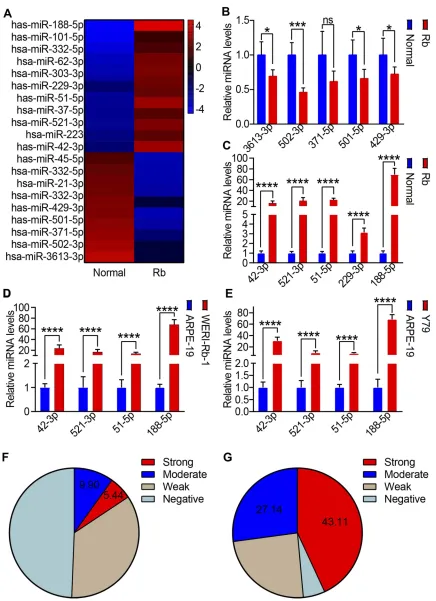
To examine the differences in miRNA expression, the total RNA of 35 Rb tissues and the corresponding adjacent normal tissues was collected and analyzed using a miRNA micro-array. Our results showed that 9 miRNAs were downregu-lated and 11 miRNAs were significantly upregulated in Rb tissues (Figure 1A, log|FC|>2, p<0.05). These results were confirmed using qRT-PCR; where, the expression of 5 miRNAs, whether up or downregulated, was analyzed. The qRT-PCR results showed that the expression of miR-3613-3p, miR-502-miR-3613-3p, miR-371-1–5p, miR-501–5p, and miR-429–3p was decreased (Figure 1B), whereas the expression of miR-42-3p, miR-521-3p, miR-51-5p, miR-229-3p, and miR-188-5p was significantly increased in Rb tissues (Figure 1C). Thus, potential miRNAs were further examined using the Rb cell lines WERI-Rb-1 and Y79 and the human retinal epithelial cells, ARPE-19. Consistent with human tissue samples, miR-42-3p, miR-521-3p, miR-51-5p, and miR-188-5p were increased in WERI-Rb-1 cells (Figure 1D) and Y79 (Figure 1E). The distribution of miR-188-5p in Rb tissues and the corresponding adjacent normal tissues were then identified as score 0 (negative), score 1 (weak), score 2 (moderate), and score 3 (strong). Score 3 (strong) and score 2 (moderate) for miR-188-5p expression were observed in 5.44% and 9.90% of the 35 retina tissues, respec-tively (Figure 1F). Score 3 (strong) and score 2 (moderate) for miR-188-5p expression were observed in 43.11% and 27.14% of the Rb tissues, respectively (Figure 1G). MiR-188-5p expression in the Rb tissues was significantly higher than that in the corresponding adjacent normal tissues.
ID4 Is the Target Gene of miR-188-5p
To investigate the role of miR-188-5p in Rb cells, further gain-of-function and loss-of-function studies were performed using the WERI-Rb-1 cell line. WERI-Rb-1 cells were trans-fected with miR-188-5p mimics (Figure 2A), inhibitors (Figure 2B), or a negative control. miR-188-5p expression was confirmed using qRT-PCR. To further identify the func-tional targets of miR-188-5p, we performed an mRNA profile analysis on miR-188-5p mimic- and inhibitor-treated WERI-Rb-1 cells. Here, 20 genes (log|FC|>2, p<0.05) were found to be dysregulated in the mimic-treated WERI-Rb-1 cells, com-pared to the control cells (Figure 2C), and the inhibitor-treated WERI-Rb-1 cells, compared to the control cells (Figure 2D). Computationally predicted target genes werethen overlapped with the genes obtained from the mRNA profile; accordingly, 17 overlapped targets were identified (Figure 2E); of these, ID4 was selected as a potential target gene. Furthermore, the possible miR-188-5p binding sites of ID4 were calculated (Figure 2F).To determine whether miR-188-5p repressed ID4 by targeting its potential binding site, PCR products containing either wild-type or mutant ID4 3′ -UTR sequences were cloned downstream of a luciferase open reading frame (Figure 2F). The overexpression of miR-188-5p suppressed the luciferase activities of the ID4 3′-UTR reporter constructs; this effect was abolished when mutations were introduced into its seed sequences (Figure 3G). Furthermore, qPCR analysis (Figure 3H) and Western blot analysis (Figure 3I) revealed that ectopic miR-188-5p expres-sion reduced the protein and mRNA levels of ID4, whereas miR-188-5p knockdown increased ID4 expression. Collectively, these results suggest that miR-188-5p reduced ID4 expression by directly targeting the ID4 3′-UTR.
ID4 Inhibits Retinoblastoma Metastasis
In the analysis of The Cancer Genome Atlas (TCGA) database of 24 cancers, it was found that ID4 expression was lower in several cancer tissues than in normal tissues (Figure 4A). In total, 35 Rb tissues and the corresponding adjacent normal tissues were also analyzed for ID4 expres-sion; the qRT-PCR results revealed a lower expression of ID4 in the Rb tissues compared to the normal tissues (Figure 4B). Ten Rb mice were sacrificed; the expression of ID4 in Rb tissues and the corresponding adjacent normal tissues was detected using qRT-PCR. Our results showed that ID4 was decreased in Rb tissues compared to the corresponding adjacent normal tissues (Figure 4C). Next, the ID4 protein level was analyzed. The Western blot results showed that the protein level of ID4 in the Rb tissues was lower than that in the corresponding adjacent normal tissues (Figure 4D); similar results were obtained with mice Rb tissues as well (Figure 4E). The function of ID4 in retino-blastoma was further analyzed via gain-of-function studies in WERI-Rb-1 cells. Cells were transfected with ID4 over-expression lentivirus; the ID4 over-expression was confirmed using qRT-PCR (Figure 4F) and Western blot (Figure 4G). The results showed that ID4 mRNA expression (Figure 4F) and protein levels (Figure 4G) were significantly increased by the ID4 overexpression lentivirus. The wound healing assay (Figure 4H) and cell invasion assay (Figure 4I) per-formed after transfection for 48 h showed that the over-expression of ID4 inhibited the migration ability and cell invasion ability of the WERI-Rb-1 cells.OncoTargets and Therapy downloaded from https://www.dovepress.com/ by 118.70.13.36 on 25-Aug-2020
Figure 1The level of miR-188-5p is significantly increased in retinoblastoma cells.(A) Heat map of differential miRNA expression between 32 retinoblastoma tissues and the corresponding adjacent normal tissues. Gene expression data were obtained using a human miRNA array. Red: increased expression, blue: decreased expression. (B, C) Top 5 downregulated miRNAs (B) and upregulated (C) miRNAs were tested using qRT-PCR. (D, E) qRT-PCR was applied to test target miRNA retinoblastoma cell lines WERI-Rb-1 (D) and Y79€compared to ARPE-19. (F, G) Score 0 (negative), score 1 (weak), score 2 (moderate), and score 3 (strong) for miR-188-5p expression were observed in 35 adjacent normal tissues and Rb cancer tissues, respectively. Means ± SEM of experiments performed in triplicates are shown. *P < 0.05; ***p < 0.005; ****p < 0.001; ns, not significant.
OncoTargets and Therapy downloaded from https://www.dovepress.com/ by 118.70.13.36 on 25-Aug-2020
Figure 2ID4 is the target gene of miR-188-5p. (A, B) WERI-Rb-1 cells were transfected with miR-188-5p mimics (A) or inhibitors (B) at afinal concentration of 100 and 200 nM. miR-188-5p expression was detected using qRT-PCR at 48 h post-transfection. U6 was used as the internal control in the qRT-PCR of miR-188-5p. (C, D) Whole genome expression profiles for WERI-Rb-1 cells treated with mimics (C) or inhibitors (D) (200μM) for 24 h. Heat map illustrating the global differences in gene expression between mimic- (C) or inhibitor- (D) treated WERI-Rb-1 cells and control (fold change >2.0; p<0.05). (E) Potential target genes of miR-188-5p were predicted using micro T, PITA, PicTar, and TargetScan. (F) A schematic representation of the ID4 3′-UTR. G. Mutations were generated at the predicted miR-188-5p-binding sites. The wild-type or mutant reporter plasmids were co-transfected with miR-188-5p or NC in WERI-Rb-1 cells; relative luciferase activity was calculated. (H, I) ID4 expression was determined by qPCR (H) and Western blot (I) analysis in WERI-Rb-1 cells after transfection with miR-188-5p mimics or miR-188-5p inhibitors 72 h post-transfection. GAPDH levels were used as the internal control in immunoblots. Means ± SEM of experiments performed in triplicates are shown. **P < 0.01; ****p < 0.001; ns, not significant.
OncoTargets and Therapy downloaded from https://www.dovepress.com/ by 118.70.13.36 on 25-Aug-2020
Figure 3miR-188-5p/ID4 promotes EMT. (A) The levels of E cadherin,αcatenin,fibronectin and vimentin were analyzed by Western blot analysis in WERI-Rb-1 cells after transfection with miR-188-5p mimics and/or ID4 overexpression lentivirus. (B) Western blot analysis showed the expression of EMT-related proteins in WERI-Rb-1 cells after transfection with miR-188-5p inhibitors and/or ID4 inhibition lentivirus. (C, D) Phase contrast images showing the migration of WERI-Rb-1 cells transfected with miR-188-5p mimics and/or ID4 overexpression lentivirus (C) and miR-188-5p inhibitors and/or ID4 inhibition lentivirus (D). (E–G) After miR-188-5p mimic and/or ID4 overexpression treatment, the wound healing assay (E), in vivo imaging (F), and survival statistics (G) were used to evaluate tumor progression. (H–J) After miR-188-5p inhibitor and/or ID4 inhibition treatment, the wound healing assay (H), in vivo imaging (I), and survival statistics (J) were used to evaluate tumor progression.
OncoTargets and Therapy downloaded from https://www.dovepress.com/ by 118.70.13.36 on 25-Aug-2020
MiR-188-5p/ID4 Regulated Metastasis by
Promoting EMT
As previously mentioned, EMT plays a crucial role in cancer metastasis. Additionally, E-cadherin andα-catenin are well known positive indicators of tumor metastasis whereasfi bro-nectin and vimentin are negative indicators. To verify the mechanism of Rb metastasis, we transfected WERI-Rb-1
cells with ID4 overexpressing lentivirus or miR-188-5p mimics and analyzed the markers of EMT through Western blotting. Our results showed that miR-188-5p promoted EMT in Rb; moreover, overexpression of ID4 could reverse miR-188-5p-induced EMT (Figure 3A). The experiment was repeated using a miR-188-5p inhibitor or an ID4 knockdown lentivirus to confirm these results. The results revealed that Figure 4ID4 promotes retinoblastoma proliferation. (A) The level of the ID4 in 24 tumor types in the TCGA database was analyzed (blue: Normal, red: Cancer). (B) ID4 expression was determined using qRT-PCR in 35 retinoblastoma tissues and the corresponding adjacent normal tissues. GAPDH RNA was used as the internal control in the qRT-PCR of ID4. After sacrificing 10 model mice, ID4 expression was determined using qRT-PCR in retinoblastoma and the corresponding adjacent normal tissues (C) GAPDH RNA was used as the internal control in the qRT-PCR of ID4. (D, E) ID4 protein levels were examined by Western blot analysis in human (D)/mice (E) retinoblastoma and the corresponding adjacent normal tissues. (F, G) The retinoblastoma cell line WERI-Rb-1 was transfected with miR-188-5p-5p mimics at afinal concentration of 200 nM. ID4 expression was detected using qRT-PCR (F) and Western blot at post-transfection. (H) Phase contrast microscope images of wound healing assay showing the cell migration pattern in ID4-deficient retinoblastoma cell lines. (I) ID4 overexpressing cells were unable to migrate, whereas the untransfected and control cells showed increased migration. Means ± SEM of experiments performed in triplicates are shown. ***P < 0.005; ****p < 0.001.
OncoTargets and Therapy downloaded from https://www.dovepress.com/ by 118.70.13.36 on 25-Aug-2020
inhibiting miR-188-5p also inhibited EMT in Rb; moreover, ID4 knockdown could reverse these results (Figure 3B). In vitro transwell migration assays showed that miR-188-5p mimics promoted invasion of Rb and overexpression of ID4 could reverse this observation (Figure 3C). In contrast, inhibiting miR-188-5p promoted invasion of Rb and knock-down of ID4 reversed this result (Figure 3D). Wound healing assays, mice imaging in vivo, and survival statistics were used to evaluate the influence of ID4 and miR-188-5p on metastasis. The results showed that overexpression of ID4 prohibited migration (Figure 3E), inhibited tumor progres-sion (Figure 3F), and prolonged survival time (Figure 3G), whereas simultaneous upregulation of miR-188-5p reversed these effects. Correspondingly, inhibiting ID4 promoted migration (Figure 3H), accelerated tumor progression (Figure 3I), and reduced the survival time (Figure 3J), whereas miR-188-5p knockdown reversed these results.
ID4 Regulates Metastasis of Retinoblastoma
Through the Wnt/
β
-Catenin Signaling
Pathway
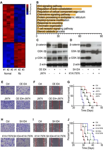
To investigate the mechanism underlying the miR-188-5p/ ID4-mediated regulation of metastasis, the differences in gene expression between three retinoblastoma tissues and the corresponding normal tissues were identified using GeneChip® technology. A heat map was used to reveal differential genes (Figure 5A); signal path analysis was used to classify differential genes. As shown, the Wnt/ β-catenin signaling pathway aggregates the most differential genes (Figure 5B). The results of chip analysis were
con-firmed using Western blot, and markers of Wnt/β-catenin signaling were analyzed. The results showed that overex-pressing ID4 decreasedβ-catenin and inhibited phosphory-lation of GSK-3β, whereas the Wnt signaling activator JW74 could reverse these results (Figure 5C). Further, inhibiting ID4 increased β-catenin and accelerated phos-phorylation of GSK-3β, whereas the Wnt signaling inhibi-tor KYA1797K could reverse these results (Figure 5D). Transwell migration assays, wound healing assays, and survival statistics were used to evaluate the influence of ID4 and Wnt/β-catenin signaling on metastasis. The results showed that overexpressing ID4 inhibited tumor invasion (Figure 5E), prohibited migration (Figure 5F), and pro-longed survival time (Figure 5G), whereas simultaneous JW74 treatment could reverse these observations. In con-trast, inhibiting ID4 promoted migration (Figure 5H), accel-erated tumor progression (Figure 5I), and decreased
survival time (Figure 5J), whereas simultaneous knockdown of miR-188-5p reversed these changes.
Discussion
Retinoblastoma (Rb), a rare form of cancer that rapidly develops from the immature cells, or cone precursor cells, of the developing retina, is almost exclusively found in young children, accounting for approximately 3% of all childhood malignancies.17,18Nevertheless, the mechanism underlying Rb progression remains unclear. In this study, we identified a pathway involving the upregulation of miR-188-5p and its downstream target ID4. This axis mediated the induction of EMT via Wnt/β-catenin signal-ing and promoted metastasis in Rb.
Previous studies have reported that miR-188-5p is a tumor suppressor in several cancers, including gliomas, non-small-cell lung cancer, prostate cancer, and hepatocel-lular carcinoma.19 MiR-188-5p has also been reported as an oncogenic miRNA that promotes proliferation, inva-sion, and aggressive progression of prostate cancer cells.20 In this study, we identified the miRNA profile of Rb in a miRNA microarray; here, upregulation of several miRNAs was observed. We determined that miR-188-5p was frequently upregulated in Rb tissues and cell lines. To examine the function of miR-188-5p in the progression of Rb, miR-188-5p was inhibited or overexpressed in the Rb cell line, WERI-Rb-1. The invasive and migratory abilities of WERI-Rb-1 cells were significantly enhanced by the 5p mimic but were suppressed by the miR-188-5p inhibitor. Substantiating our results, in vivo metastasis experiments confirmed that miR-188-5p significantly pro-moted the progression of Rb, leading a poor prognosis. These results indicate the crucial role of miR-188-5p as a tumor promoter in Rb cell invasion and metastasis. Furthermore, our data revealed that miR-188-5p promotes Rb cell metastasis in vitro and in vivo by inducing EMT via Wnt/β-catenin signaling by targeting ID4.
Recent studies have demonstrated that ID4 is a miRNA-regulated protein, which is repressed by miR-342, miR-335, and miR-485-5p.21Despite the fact that inhibitors of DNA binding (ID) act as cancer suppressor genes in several cancers, characterization of the ID isoforms are limited. ID proteins are dominant-negative transcriptional regulators of basic helix–loop–helix (bHLH) transcription factors, which are expressed by essentially all cell lineages.22ID expression is highest in undifferentiated, pro-liferating populations and is subsequently downregulated in adipocytes, prostate epithelial cells, neurons, and osteoblasts;
OncoTargets and Therapy downloaded from https://www.dovepress.com/ by 118.70.13.36 on 25-Aug-2020
Figure 5ID4 regulates the metastasis of retinoblastoma through the Wnt/β-catenin signaling pathway. (A) Heat map of differential genes between retinoblastoma tissues and the corresponding normal tissues. (B) Diagram of gene clustering of signaling pathways. (C, D) Western blot was used to test the markers of Wnt/β-catenin signaling after overexpressing ID4 and/or treating with JW74 (C) and inhibiting ID4 and/or treating with KYA1797K (D). (E–G) Trans-well assays (E), wound healing assay (F), and survival statistics (G) were used to evaluate tumor progression after overexpressing ID4 and/or treating with JW74. (H–J) Trans-well assays (H), wound healing assay (I), and survival statistics (J) were used to evaluate tumor progression after overexpressing ID4 and/or treating with JW74.
OncoTargets and Therapy downloaded from https://www.dovepress.com/ by 118.70.13.36 on 25-Aug-2020
this supports its role as a pro-differentiation factor. It is easy to understand why a high level of ID4 is present in the retina cells of children but not in Rb cells. The absence of ID4 has been previously reported in many cancers, implying that ID4 could suppress the occurrence of cancer at some point during a lifetime. ID4 appears to act primarily as a tumor suppressor in most cancers, in contrast to ID1, ID2, and ID3, which act as tumor promoters or supporting oncogenes. However, inac-tivation of ID4 leads to unfavorable prognosis and poor differentiation in colorectal carcinoma. In breast cancer, the absence of ID4 is associated with recurrence-free survival and increased tumor relapse. Acute myeloid leukemia (AML) patients with myelodysplastic syndrome (MDS) exhibited a significantly higher frequency of ID4 methylation with shorter survival.23
In summary, ID4 acts as a miRNA sponge in retino-blastoma progression and provides a potential therapeutic strategy for Rb treatment. Our data suggest that reducing miR-188-5p levels could help improve performance during Rb, introducing this novel axis as a potential indicator to detect retinoblastoma, and elicit beneficial therapeutic effects in this rare cancer (Figure 6).
Disclosure
The authors report no conflicts of interest in this work.
References
1. Li F, Kitajima S, Kohno S, et al. Retinoblastoma inactivation induces a protumoral microenvironment via enhanced CCL2 secretion. Cancer Res.2019;79(15):3903–3915. doi:10.1158/0008-5472.CAN-18-3604
2. Munier FL, Beck-Popovic M, Chantada GL. et al. Conservative management of retinoblastoma: challenging orthodoxy without com-promising the state of metastatic grace.“Alive, with good vision and no comorbidity”.Prog Retin Eye Res;2019. 100764. doi:10.1016/j. preteyeres.2019.05.005
3. Topacio BR, Zatulovskiy E, Cristea S, et al. Cyclin D-Cdk4,6 drives cell-cycle progression via the retinoblastoma protein’s C-terminal helix.Mol Cell.2019;74(4):758–770.e4. doi:10.1016/j.molcel.2019. 03.020
4. Rajasekaran S, Nagarajha Selvan LD, Dotts K, et al. Non-coding and coding transcriptional profiles are significantly altered in pediatric retinoblastoma tumors.Front Oncol.2019;9:221. doi:10.3389/fonc. 2019.00221
5. Castro-Magdonel BE, Orjuela M, Camacho J, et al. miRNome land-scape analysis reveals a 30 miRNA core in retinoblastoma.BMC Cancer.2017;17(1):458. doi:10.1186/s12885-017-3421-3
6. Liu R, Chen Y, Shou T, Hu J, Qing C. miRNA-99b-5p targets FZD8 to inhibit non-small cell lung cancer proliferation, migration and invasion.Onco Targets Ther.2019;12:2615–2621. doi:10.2147/OTT. S199196
7. Sun B, Zhang Y, Zhou L, et al. The proliferation of cervical cancer is promoted by miRNA-125b through the regulation of the HMGA1. Onco Targets Ther. 2019;12:2767–2776. doi:10.2147/ OTT.S197740
8. Liu SS, Wang YS, Sun YF, et al. Plasma microRNA-320, microRNA-let-7e and microRNA-21 as novel potential biomarkers for the detection of retinoblastoma.Biomed Rep.2014;2(3):424–428. doi:10.3892/br.2014.246
9. Zhao JJ, Yang J, Lin J, et al. Identification of miRNAs associated with tumorigenesis of retinoblastoma by miRNA microarray analysis. Childs Nerv Syst.2009;25(1):13–20. doi:10.1007/s00381-008-0701-x 10. Sreenivasan S, Thirumalai K, Danda R, Krishnakumar S. Effect of curcumin on miRNA expression in human Y79 retinoblastoma cells. Curr Eye Res. 2012;37(5):421–428. doi:10.3109/02713683.2011.64 7224
11. Perk J, Iavarone A, Benezra R. Id family of helix-loop-helix proteins in cancer.Nat Rev Cancer.2005;5(8):603–614. doi:10.1038/nrc1673 12. Fontemaggi G, Dell’Orso S, Trisciuoglio D, et al. The execution of the transcriptional axis mutant p53, E2F1 and ID4 promotes tumor neo-angiogenesis. Nat Struct Mol Biol. 2009;16(10):1086–1093. doi:10.1038/nsmb.1669
13. Wen YH, Ho A, Patil S, et al. Id4 protein is highly expressed in triple-negative breast carcinomas: possible implications for BRCA1 downregulation. Breast Cancer Res Treat. 2012;135(1):93–102. doi:10.1007/s10549-012-2070-0
14. Junankar S, Baker LA, Roden DL, et al. ID4 controls mammary stem cells and marks breast cancers with a stem cell-like phenotype.Nat Commun.2015;6:6548. doi:10.1038/ncomms7548
15. Chien MH, Lin YW, Wen YC, et al. Targeting the SPOCK1-snail/ slug axis-mediated epithelial-to-mesenchymal transition by apigenin contributes to repression of prostate cancer metastasis.J Exp Clin Cancer Res.2019;38(1):246.
16. Esposito M, Mondal N, Greco TM, et al. Bone vascular niche E-selectin induces mesenchymal-epithelial transition and Wnt activa-tion in cancer cells to promote bone metastasis. Nat Cell Biol.
2019;21(5):627–639. doi:10.1038/s41556-019-0309-2
17. Ray A, Gombos DS, Vats TS. Retinoblastoma: an overview.Indian J Pediatr.2012;79(7):916–921. doi:10.1007/s12098-012-0726-8 18. García-Chequer AJ, Méndez-Tenorio A, Olguín-Ruiz G, et al.
Overview of recurrent chromosomal losses in retinoblastoma detected by low coverage next generation sequencing. Cancer Genet.2016;209(3):57–69. doi:10.1016/j.cancergen.2015.12.001 19. Xue M, Cheng Y, Han F, et al. Triptolide attenuates renal tubular
epithelial-mesenchymal transition via the MiR-188-5p-mediated PI3K/AKT pathway in diabetic kidney disease. Int J Biol Sci.
2018;14(11):1545–1557. doi:10.7150/ijbs.24032
Figure 6Schematic of the topic. MiRNA-188-5p can promote EMT by targeting ID4 through the Wnt/β-catenin signaling pathway.
OncoTargets and Therapy downloaded from https://www.dovepress.com/ by 118.70.13.36 on 25-Aug-2020
20. Zhang H, Qi S, Zhang T, et al. miR-188-5p inhibits tumour growth and metastasis in prostate cancer by repressing LAPTM4B expression. Oncotarget.2015;6(8):6092–6104. doi:10.18632/oncotarget.3341 21. Zhou JD, Li XX, Zhang TJ, et al. MicroRNA-335/ID4 dysregulation
predicts clinical outcome and facilitates leukemogenesis by activating PI3K/Akt signaling pathway in acute myeloid leukemia. Aging (Albany NY).2019;11(10):3376–3391. doi:10.18632/aging.101991
22. Nasif D, Campoy E, Laurito S, et al. Epigenetic regulation of ID4 in breast cancer: tumor suppressor or oncogene. Clin Epigenetics.
2018;10(1):111. doi:10.1186/s13148-018-0542-8
23. Zhou JD, Zhang TJ, Li XX, et al. Epigenetic dysregulation of ID4 predicts disease progression and treatment outcome in myeloid malignancies.J Cell Mol Med.2017;21(8):1468–1481. doi:10.1111/ jcmm.13073
OncoTargets and Therapy
Dove
press
Publish your work in this journal
OncoTargets and Therapy is an international, peer-reviewed, open access journal focusing on the pathological basis of all cancers, potential targets for therapy and treatment protocols employed to improve the management of cancer patients. The journal also focuses on the impact of management programs and new therapeutic
agents and protocols on patient perspectives such as quality of life, adherence and satisfaction. The manuscript management system is completely online and includes a very quick and fair peer-review system, which is all easy to use. Visit http://www.dovepress.com/ testimonials.php to read real quotes from published authors.
Submit your manuscript here:https://www.dovepress.com/oncotargets-and-therapy-journal
OncoTargets and Therapy downloaded from https://www.dovepress.com/ by 118.70.13.36 on 25-Aug-2020