Effect of changing lumbar stiffness by single facet
joint dysfunction on the responsiveness of lumbar
muscle spindles to vertebral movement
William R. Reed,
DC, PhD*1Joel G. Pickar,
DC, PhD1Cynthia R. Long,
PhD11 Palmer Center for Chiropractic Research, Davenport, IA
* Corresponding Author: William Reed DC, PhD
Palmer Center for Chiropractic Research 741 Brady Street
Davenport, IA 52803 563-884-5145
This project was made possible by grant number K01AT005935 from the National Center for Complementary and Alternative Medicine to WRR and was conducted in a facility constructed with support from Research Facilities Improvement Grant number C06 RR15433 from the National Center for Research Resources, National Institutes of Health. Its contents are solely the responsibility of the authors and do not necessarily represent the official views of the National Center for Complementary and Alternative Medicine or the National Institutes of Health. No conflict of interest was reported by any of the authors.
©JCCA 2014
Objective: Individuals experiencing low back pain often
present clinically with intervertebral joint dysfunction. The purpose of this study was to determine whether relative changes in stiffness at a single spinal joint alters neural responsiveness of lumbar muscle spindles to either vertebral movement or position.
Methods: Muscle spindle discharge was recorded in
response to 1mm L6 ramp and hold movements (0.5mm/s) in the same animal for lumbar laminectomy-only (n=23), laminectomy & L5/6 facet screw (n=19), laminectomy & L5/6 facetectomy (n=5) conditions. Mean instantaneous frequency (MIF) was calculated for the ramp-up, hold, ramp-down and post-ramp phases during each joint condition.
Objectif : La lombalgie se manifeste souvent
cliniquement sous forme de dysfonction articulaire intervertébrale. Cette étude a pour objet de déterminer si des changements relatifs dans la rigidité d’une seule articulation vertébrale modifieraient la réactivité des fuseaux musculaires lombaires envers le mouvement ou la position des vertèbres.
Méthodologie : Les décharges des fuseaux
Introduction
Aberrant neuromuscular control of the trunk along with the inability of individuals with LBP to adopt optimal postural control strategies is thought to be involved in the etiology of low back pain (LBP).1-10 Individuals with
LBP demonstrate reduced lumbar muscle activation or earlier onsets of muscle activation following predictable and unpredictable trunk loading.6 It has been reported that
individuals experiencing an active episode of LBP dem-onstrate inadequate trunk muscle activation or inappro-priate trunk muscle co-activation in response to rapid and/ or unexpected perturbation.11-13 In addition, LBP patients
exhibit altered movement patterns between recurrent epi-sodes but it is unclear whether the patterns develop pri-or to pri-or following the first LBP episode.10 These altered
neuromuscular responses that accompany LBP have been attributed to a number of mechanisms including segment-al neursegment-al circuitry and cognitive responses due to stress, pain avoidance and/or anticipation of pain.14,15
Muscle spindles are proprioceptors which provide a continuous sensory input to the central nervous system related to muscle length and rate of change in muscle length, and thereby potentially supply information re-garding joint position and movement. This sensory input may help to optimize neuromuscular control of the trunk and intervertebral motion during intended movement tra-jectories. Compared to muscle spindles in appendicular muscles, much less is known about the functional
char-acteristics of these proprioceptors in trunk musculature. However differences in structural complexity, organ-ization and response to changes in muscle length have been described in muscle spindles of the trunk relative to appendicular muscles.16-21 For example, we have
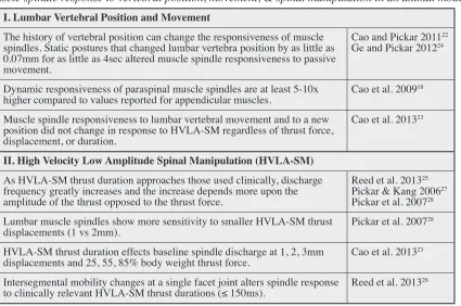
re-cently shown that measures of dynamic responsiveness in trunk muscles are 5-10x higher than values reported for appendicular muscles.18 Table 1 provides an
abbrevi-ated summary of recent findings regarding the responses of paraspinal muscle spindles to changes in both vertebral position and movement as well as to high velocity low amplitude spinal manipulation using variations of the ex-perimental model employed in the present study.
Impaired spinal biomechanics are thought to have ad-verse physiological consequences by producing less than optimal neuromuscular control of the trunk. Individuals experiencing acute or chronic LBP episodes often present clinically with intervertebral joint dysfunction.29-31 The
relationship between intervertebral joint mobility and alterations in trunk mechanoreception has received little direct investigation but is of clinical interest due to the frequent assessment of intervertebral joint mobility by manual therapy practitioners during their clinical decision making process when treating patients experiencing LBP. There is evidence suggesting that clinical identification of spinal joint hypo- and hypermobility subgroups along with correspondingly tailored manual therapy treatment approaches can lead to more successful therapeutic Results: Mean MIFs were not significantly different
between the laminectomy-only and the other two types of joint dysfunction for the ramp-up, hold, ramp-down, or post-ramp phases.
Conclusion: Stiffness changes caused by single facet
joint dysfunction failed to alter spindle responses during slow 1mm ramp and hold movements of the L6 vertebra.
(JCCA 2014;58(2):160-169)
k e y w o r d s: stiffness, joint, muscle spindle,
chiropractic
Résultats : Les FIM n’étaient pas significativement différentes entre le groupe laminectomie seule et les deux autres types de dysfonction articulaires pour les phases d’intensification, de maintien, d’atténuation et post-rampe.
Conclusion : Les changements de rigidité causés par une dysfonction articulaire à facette unique n’ont pas réussi à modifier la réponse des fuseaux au cours de mouvements lents de rampe et de maintien de 1 mm de la vertèbre L6.
(JCCA 2014;58(2):160-169)
m o t s c l é s : rigidité, articulation, fuseau musculaire,
comes.32-34 In a randomized clinical trial categorizing 131
LBP patients with respect to the clinical determination of spinal joint hypo- and hypermobility, it was reported that individuals with spinal joint hypomobility had greater improvement with spinal manipulation than individuals with spinal joint hypermobility.33 This clinical study
highlights the need not only to understand the under-lying biological mechanisms of manual therapy interven-tion but suggests that the physiological response to the same therapeutic intervention differs based on the clinic-ally identified types of spinal joint dysfunction (hypo- or hypermobility).
Motivated by the lack of knowledge regarding how different types of spinal joint dysfunction affect trunk mechanoreceptor activity and possibly clinical outcomes to the same manual therapeutic intervention, we under-took a series of basic science experiments investigating the effect of spinal joint dysfunction on sensory input related to vertebral movement and spinal manipulation.
We previously reported the effects that single facet joint dysfunction has on sensory input during spinal manipula-tion.26 The purpose of this paper is to report the effects
that single facet joint dysfunction have on the mean in-stantaneous frequency of muscle spindles located in trunk musculature during 1mm ramp and holdmovements of the L6 lumbar vertebra derived from secondary analyses
of the previous study involving facet joint dysfunction and spinal manipulation.26
Methods
Electrophysiological recordings were made from lumbar paraspinal muscle spindles in 23 Nembutal-anesthesized male cats weighing an average of 4.46kg (SD 0.31). All experiments were reviewed and approved by the Institu-tional Animal Care and Use Committee and comply with the Canadian Council on Animal Care. One neuron was investigated per animal because of the irreversible nature of the L5/6 facetectomy surgical procedure. The
experi-Table 1
Muscle spindle response to vertebral position, movement, & spinal manipulation in an animal model
I. Lumbar Vertebral Position and Movement
The history of vertebral position can change the responsiveness of muscle spindles. Static postures that changed lumbar vertebra position by as little as 0.07mm for as little as 4sec altered muscle spindle responsiveness to passive movement.
Cao and Pickar 201122
Ge and Pickar 201224
Dynamic responsiveness of paraspinal muscle spindles are at least 5-10x
higher compared to values reported for appendicular muscles. Cao et al. 200918 Muscle spindle responsiveness to lumbar vertebral movement and to a new
position did not change in response to HVLA-SM regardless of thrust force, displacement, or duration.
Cao et al. 201323
II. High Velocity Low Amplitude Spinal Manipulation (HVLA-SM)
As HVLA-SM thrust duration approaches those used clinically, discharge frequency greatly increases and the increase depends more upon the amplitude of the thrust opposed to the thrust force.
Reed et al. 201325
Pickar & Kang 200627
Pickar et al. 200728
Lumbar muscle spindles show more sensitivity to smaller HVLA-SM thrust
displacements (1 vs 2mm). Pickar et al. 200728
HVLA-SM thrust duration effects baseline spindle discharge at 1, 2, 3mm
displacements and 25, 55, 85% body weight thrust force. Cao et al. 201323 Intersegmental mobility changes at a single facet joint alters spindle response
mental approach has been described previously in de-tail25,26,35 and is presented only briefly.
A mixture of O2 and isoflurane was delivered through
a facemask (2L/min and 2%) in order to place catheters in a common carotid artery and an external jugular vein to monitor blood pressure and introduce fluids respect-ively. Following catheterization, deep anesthesia was maintained throughout the experiment with Nembutal (35 mg/kg, iv). Deep anesthesia was identified by absence of withdrawal reflex to noxious pinching of the toe pad, mean arterial pressures less than 120mmHg and the ab-sence of a pressor response to surgical manipulation. The proximal portion of the L6 dorsal roots (cats have
7 lumbar vertebrae) was exposed after a bilateral laminec-tomy at the L5 vertebra. The musculature on the right side
of the spinal column (multifidus, longissimus and iliocos-talis muscles) remained intact except for any attachments to the posterior portions of the L4-5 vertebrae and for small slit incisions (3mm) on either side of the L6 spinous
pro-cess for forceps attachment by which the vertebra was moved. Most of the multifidus muscle remained attached to the L6 vertebra using this method because it’s apo-neurotic tendon inserts onto the process’s caudal edge.36
In addition, the L6 dorsal root enters the spinal cord 1 to 1½ vertebral segments cranial to the L6 paraspinal soft
tis-sues. The L6 dorsal root was cut close to its entrance into the spinal cord and placed on a small platform. Thin fila-ments from the cut proximal dorsal rootlets were teased apart until muscle spindle activity from a single neuron with the most sensitive part of its receptive field being in the low back could be identified. At the end of the experi-mental protocols several approaches were used to confirm receptor location and its identity as a muscle spindle
in-cluding: (1) vonFrey filaments (Stoelting, USA) to con-firm the most sensitive area for mechanically activating the neuron was in the multifidus or longissimus muscles (the intervening lumbococcygeus muscle innervated by sacral nerves was removed; (2) a sustained increase in discharge response to succinylcholine injection (100 ug/ kg, ia); (3) a sustained increase in response to a fast vibra-tory stimulus and (4) decreased discharge to paraspinal muscle electrically induced muscle twitch.
Ramp and hold movement of the L6 vertebra was controlled using an electronic feedback control system (Lever System Model 310; Aurora Scientific) under dis-placement control. Attached to the control system’s lever arm was a pair of adjustable tissue forceps which were clamped tightly onto the lateral surfaces of the L6
spin-ous process. Ramp and hold movements of 1mm peak amplitude were applied at a rate of 0.5mm/s. Due to the facetectomy, testing order for the three joint conditions in the same animal was fixed. Therefore, determination of muscle spindle responses to the 1mm ramp and hold dis-placements was conducted in the following order: lamin-ectomy-only, laminectomy & facet screw, laminectomy & facetectomy (Table 2). It should be noted that ramp test-ing for each spinal joint condition was performed prior to conducting a series of 5 randomized spinal manipu-lative thrust protocols (time control-0 ms, 75, 100, 150, 250ms) each separated by 5 minute intervals as previ-ously described in detail.26 In addition, insertion of the
facet screw and facetectomy procedure typically required 30-35 minutes to accomplish equating to approximately 1 hour elapsing between ramp and hold testing per spinal joint condition once a paraspinal muscle spindle was iso-lated.
Table 2
Laminectomy
Only & Facet ScrewLaminectomy & FacetectomyLaminectomy
Experimental
Order 1st 2nd 3rd
Vertebra
Movement L6 L6 L6
Number of L6
Creating Spinal Joint Conditions and Determining of Lumbar Stiffness
Changes in spinal stiffness were created by unilateral (left) L5/6 facet-fixation (to increase intervertebral
stiff-ness) or L5/6 facetectomy (to decrease intervertebral stiff-ness). The left L5/6 facet joint was fixated by inserting a
single 10mm titanium endosteally-anchored mini-screw (tomas®-pin; Dentaurum, Germany) through the articular pillars of the L5/6 facet joint. For the facetectomy, the left L5 inferior facet and left L6 superior facet were removed
using bone rongeurs.
Lumbar stiffness testing was first performed in the laminectomy-only condition, as opposed to the intact spine, as this was the spinal condition in which neural recordings were first obtained. To determine lumbar stiffness in each joint condition, a 1mm ramp and hold movement of the L6 vertebra was applied in the dorsal ventral direction at a rate of 0.5mm/s using the feedback-controlled motor. During the 1mm ramp and hold, forces and displacements were being measured so that force-displacement curves of the ramp portion could be con-structed. The slope of the most linear portion of the force-displacement curve (between 2.16 – 8.83N) of the 1mm ramp was calculated and represented the spinal joint stiff-ness for each condition. Ramp pre-conditioning was not performed in order to minimize the total number of facet screw/bone engagements.
Twenty-three animals were used in this study. As de-scribed previously,26 animals in which the laminectomy
& facet screw (n=4) failed to increase ramp stiffness by at least 2 % when compared to the laminectomy-only condition were excluded from further analysis. Similarly, animals in which the laminectomy & facetectomy (n=8) failed to decrease ramp stiffness by at least 2% when compared to laminectomy-only were also excluded. In addition, during the laminectomy & facetectomy condi-tion the neural recording was lost in 10 animals due to facetectomy-associated bleeding. Therefore, of the 23 animals used in this study, 20 neurons were included in the analysis: 4 had data for all 3 conditions (laminec-tomy-only, laminectomy & facet screw, laminectomy & facetectomy), 15 had data for the laminectomy-only and laminectomy & facet screw conditions, and 1 had data for the laminectomy-only and laminectomy & facetectomy conditions (Table 2).
Data Analysis
Muscle spindle activity was converted to instantaneous frequency (IF) by taking the reciprocal of the time inter-val between successive action potentials. IFs during the constant velocity ramp movement and hold position were used to obtain the following 5 measures of afferent re-sponse: (a) baseline during the 2 seconds that immedi-ately preceded each ramp-up; (b) ramp-up; (c) during the last 2 seconds of the hold phase; (d) ramp-down and (e) post-ramp during 2 seconds that immediately follow the ramp-down (Fig. 1). Mean IF (MIF) was calculated over the durations of baseline, ramp-up, hold, ramp-down and the post-ramp phase.
Changes from baseline MIF due to the laminectomy-only, laminectomy & screw and the laminectomy & facetectomy condition during the 1mm ramp and hold constituted the response measures. All neural activity was reported in impulses per second (imp/s). Data were ana-lyzed in SAS System for Windows (Release 9.2) (SAS Institute Inc., Cary, NC). Statistical significance was set at 0.05. Each response variable was analyzed with a linear mixed effects longitudinal regression model including individual random effects to account for correlation be-tween repeated measurements for an individual neuron based on a compound symmetry covariance structure. Adjusted means and 95% confidence intervals based on this model are reported.
Results
Muscle spindle recordings were analyzed from neurons with receptive fields located in the multifidus muscle (n=3) and longissimus muscle (n=17). In the cat, these lumbar paraspinal muscles are the two most medial to the spinous process.36 In response to succinylcholine
injec-tion, all neurons exhibited long lasting high frequency discharges relative to baseline. In addition, all neurons exhibited a sustained response to vibratory stimulus and were silenced by muscle twitch during bipolar muscle stimulation (amplitude 0.1-0.3mA: 50 µs).
Figure 1.
An example of a 1mm ramp and hold movement of a L6 vertebra in a laminectomy-only preparation. Force, displacement, primary afferent activity and instantaneous frequency recordings are shown. Baseline, ramp-up, hold,
ramp-down, and post-ramp regions used to calculate mean instantaneous frequencies are demarcated. Note the increase in afferent activity during the ramp-up and hold phase and the cessation of discharge due to unloading of the
muscle spindle during the ramp-down phase.
Figure 2.
The mean change in mean instanteous frequency relative to baseline discharge during (A) up, (B) hold, (C) ramp-down, and (D) post-ramp for laminectomy-only, laminectomy-facet fixation, and lamectomy-facetectomy conditions.
(range: −0.69 to −2.26N/mm; −3.25% to −21.09%) as re-ported previously.26
Figure 1 shows an original recording from a muscle spindle with a receptive field in the longissimus muscle during a 1mm ramp and hold experimental protocol in the laminectomy-only condition. There was an increase in neural activity during the ramp-up and hold phases which was typically followed by a cessation of muscle spindle discharge due to spindle unloading and a resumption of resting discharge.
Figure 2 shows the adjusted mean MIF and 95% con-fidence intervals for each response measure during the ramp and hold movements for each facet joint condition. Mean MIFramp-up was not significantly different among the
3 conditions (Fig. 2A; F2,22=1.71, p=.20). The adjusted mean difference in MIFramp-up between the
laminectomy-only and the laminectomy & facet screw condition was 1.82imp/s (-1.61, 11.04) and 4.99imp/s (-1.07, 11.04) be-tween the laminectomy-only condition and the laminec-tomy & faceteclaminec-tomy condition. Mean MIFhold (Fig. 2B;
F2,22=0.27, p=.76), MIFramp-down (Fig. 2C; F2,22=0.56, p=.58) and MIFpost-ramp (Fig. 2D; F2,22=0.33, p=.72) were also not
significantly different among conditions.
Discussion
The potential for interactive effects between interverte-bral joint mobility and sustained changes in sensory sig-naling from peripheral paraspinal tissues is of fundamen-tal importance to all researchers and clinical practition-ers interested in optimizing neuromuscular control of the trunk. Spinal manipulation and/or spinal mobilization are typically delivered to patients at anatomical locations ex-hibiting signs and symptoms of biomechanical dysfunc-tion.37-39 The present study is a first step toward
investigat-ing the relationship between muscle spindle signalinvestigat-ing and acute spinal joint dysfunction during passive movements applied to the lumbar spine.
This study indicated that acute biomechanical dys-function (laminectomy & facet screw, laminectomy & facetectomy) at a single facet joint failed to alter mech-anoreceptive afferent response during slow (0.5mm/s) 1mm dorsal-ventral ramp and hold movements of a lum-bar vertebra. These findings mirror results from the re-cent study investigating the effects spinal manipulation thrust durations under the same spinal joint conditions in which the longest thrust duration (250ms) also failed to
demonstrate changes between conditions.26 Acute spinal
joint dysfunction at multiple joints, chronic spinal joint dysfunction, increased vertebral displacement, rotary dis-placement, and/or a faster ramp rate may be required to affect neuromuscular sensory input from trunk muscle proprioceptors during slow ramp and hold movements and/or longer spinal manipulative thrust durations.
It is interesting to note that in two previous feline stud-ies using the laminectomy-only condition, muscle spindle responses to ramp and hold movements (1, 2, and 3mm; 0.5mm/s), both similar to and greater than the hold ampli-tude used in the current study (1mm, 0.5mm/s) were not affected by an interposed high velocity low amplitude spinal manipulative thrust;23 yet the afferents were almost
twice as sensitive during the manipulative thrust itself when the peak amplitude was 1mm compared to 2mm.28
These previous studies along with the present study sug-gest that mechanoreceptive trunk responses to slow ver-tebral movements (0.5mm/s) are neither affected by acute single facet joint dysfunction nor by high velocity low amplitude spinal manipulation regardless of ramp ampli-tude.
Limitations
musculo-skeletal inflammatory milieu that frequently accompanies spinal joint dysfunction clinically. One or more of these factors may be required to physiologically affect chan-ges in sensory input from trunk muscle spindles during slower and/or small intervertebral joint movements. Failure to create a minimum 2% change in lumbar stiffness in a dozen preparations is likely due to a number of factors including but not limited to the greater inherent flexibility of the feline spinal column, inadequate place-ment of the facet screw, partial splintering of the facet joint, incomplete facetectomy, and/or lack of a rotary or lateral displacement component of the spine during bio-mechanical testing. Dorsal-ventral ramp testing was the only direction used in current study due to the increased risk of tearing the afferent fiber off the recording elec-trode that accompanies rotary or lateral movements in this type of experimental preparation.
Neurophysiological and biomechanical studies using anesthetized animals where measurements from the spinal tissues can be obtained directly are of growing importance in the quest to understanding the underlying mechanisms of spinal manipulation despite certain inherent limitations of this work. Since the chiropractic profession’s first basic science white paper was published in 1997,40 much basic
work has been accomplished (see 41-44 for review), and
yet there remains a great need for more and better animal models if the goal is to identify the biological mechan-isms involved in spinal manipulation intervention. Once mechanisms are identified, this knowledge can then be translated into providing better clinical care for individ-uals seeking chiropractic services. As shown in Table 1, much information relevant to the practice of chiropractic has been learned over a relatively short period using slight variations of the animal model used in the current study.
Conclusion
Coordination of paraspinal muscles is required to pro-vide optimal neuromuscular control of dynamic inter-vertebral mobility during intended bodily movements. It is possible that distorted proprioceptive input related to acute or chronic spinal joint dysfunction could result in suboptimal neuromuscular trunk control; however, the results of this study indicate that changes in lumbar stiffness due to dysfunction at a single facet joint fails to alter paraspinal muscle spindle responses during slow (0.5mm/s) 1mm ramp and hold movements. Spinal joint
dysfunction at multiple joints, chronic joint dysfunction, and/or more rotary/combinatorial motions in a facet dys-functional model may be necessary to alter responses of trunk spindle afferents during small slow movements of the lumbar spine.
References
1 Cholewicki J, Greene HS, Polzhofer GK, Galloway MT, Shah RA, Radebold A. Neuromuscular function in athletes following recovery from a recent acute low back pain injury. J Orthop Sports Phys Ther. 2002; 32: 568-75. 2 Brumagne S, Cordo P, Lysens R, Verschueren S,
Swinnen S. The role of paraspinal muscle spindles in lumbosacral position sense in individuals with and without low back pain. Spine. 2000; 25: 989-94.
3 Claeys K, Brumagne S, Dankaerts W, Kiers H, Janssens L. Decreased variability in postural control strategies in young people with non-specific low back pain is associated with altered proprioceptive reweighting. Eur J Appl Physiol. 2011; 111: 115-23.
4 Hodges P. Altered trunk muscle recruitment in people with low back pain with upper limb movement at different speeds. Arch Phys Med Rehabil. 1999; 80: 1005-12. 5 Johanson E, Brumagne S, Janssens L, Pijnenburg M,
Claeys K, Paasuke M. The effect of acute back muscle fatigue on postural control strategy in people with and without recurrent low back pain. Eur Spine J. 2011; 20: 2152-9.
6 MacDonald D, Moseley GL, Hodges PW. People with recurrent low back pain respond differently to trunk loading despite remission from symptoms. Spine. 2010; 35: 818-24.
7 Reeves NP, Cholewicki J, Lee AS, Mysliwiec LW. The effects of stochastic resonance stimulation on spine proprioception and postural control in chronic low back pain patients. Spine. 2009; 34: 316-21.
8 Reeves NP, Cholewicki J, Narendra KS. Effects of reflex delays on postural control during unstable seated balance. J Biomech. 2009; 42: 164-70.
9 Radebold A, Cholewicki J, Polzhofer GK, Greene HS. Impaired postural control of the lumbar spine is associated with delayed muscle response times in patients with chronic idiopathic low back pain. Spine. 2001; 26: 724-30. 10 Jones SL, Hitt JR, DeSarno MJ, Henry SM. Individuals
with non-specific low back pain in an active episode demonstrate temporally altered torque responses and direction-specific enhanced muscle activity following unexpected balance perturbations. Exp Brain Res. 2012; 221: 413-26.
12 Stokes IA, Fox JR, Henry SM. Trunk muscular activation patterns and responses to transient force perturbation in persons with self-reported low back pain. Eur Spine J. 2006; 15: 658-67.
13 D’hooge R, Hodges P, Tsao H, Hall L, MacDonald D, Danneels L. Altered trunk muscle coordination during rapid trunk flexion in people in remission of recurrent low back pain. J Electromyogr Kinesiol. 2013; 23: 173-81. 14 Moseley GL, Nicholas MK, Hodges PW. Pain differs from
non-painful attention demanding or stressful tasks in its effect on postural control patterns of trunk muscles. Exp Brain Res. 2003; 156: 64-71.
15 Moseley GL, Nicholas MK, Hodges PW. Does anticipation of back pain predispose to back trouble? Brain. 2004; 127: 2339-47.
16 Bakker DA, Richmond FJR. Muscle spindle complexes in muscles around upper cervical vertebrae in the cat. J Neurophysiol. 1982; 48: 62-74.
17 Cao DY, Pickar JG, Ge W, Ianuzzi A, Khalsa PS. Position sensitivity of feline paraspinal muscle spindles to vertebral movement in the lumbar spine. J Neurophysiol. 2009; 101: 1722-9.
18 Cao DY, Khalsa PS, Pickar JG. Dynamic responsiveness of lumbar paraspinal muscle spindles during vertebral movement in the cat. Exp Brain Res. 2009; 197: 369-77. 19 Durbaba R, Taylor A, Ellaway PH, Rawlinson S.
Classification of longissimus lumborum muscle spindle afferents in the anaesthetized cat. J Physiol. 2006; 571: 489-98.
20 Reed WR, Cao DY, Ge W, Pickar JG. Using vertebral movement and intact paraspinal muscles to determine the distribution of intrafusal fiber innervation of muscle spindle afferents in the anethesized cat. Exp Brain Res. 2013; 225: 205-15.
21 Richmond FJ, Abrahams VC. Physiological properties of muscle spindles in dorsal neck muscles of the cat. J Neurophysiol. 1979 March; 42: 604-17.
22 Cao DY, Pickar JG. Lengthening but not shortening history of paraspinal muscle spindles in the low back alters their dynamic sensitivity. J Neurophysiol. 2011; 105: 434-41.
23 Cao DY, Reed WR, Long CR, Kawchuk GN, Pickar JG. Effects of thrust amplitude and duration of high-velocity, low-amplitude spinal manipulation on lumbar muscle spindle responses to vertebral position and movement. J Manipulative Physiol Ther. 2013; 36: 68-77.
24 Ge W, Pickar JG. The decreased responsiveness of lumbar muscle spindles to a prior history of spinal muscle lengthening is graded with the magnitude of change in vertebral position. J Electromyogr Kinesiol. 2012; 22: 814-20.
25 Reed WR, Cao DY, Long CR, Kawchuk GN, Pickar JG. Relationship between biomechanical characteristics of spinal manipulation and neural responses in an animal
model: effect of linear control of thrust displacement versus force, thrust amplitude, thrust duration, and thrust rate. Evid Based Complement Altern Med. 2013; http:// dx.doi.org/10.1155/2013/492039.
26 Reed WR, Long CR, Pickar JG. Effects of unilateral facet fixation and facetectomy on muscle spindle responsiveness during simulated spinal manipulation in an animal model. JMPT. 2013; 36:585-94.
27 Pickar JG, Kang YM. Paraspinal muscle spindle responses to the duration of a spinal manipulation under force control. J Manipulative Physiol Ther. 2006; 29: 22-31. 28 Pickar JG, Sung PS, Kang YM, Ge W. Response of
lumbar paraspinal muscles spindles is greater to spinal manipulative loading compared with slower loading under length control. Spine J. 2007; 7: 583-95.
29 Lundberg G, Gerdle B. The relationships between spinal sagittal configuration, joint mobility, general low back mobility and segmental mobility in female homecare personnel. Scand J Rehab Med. 1999; 31: 197-206. 30 Lundberg G, Gerdle B. Correlations between joint and
spinal mobility, spinal sagittal configuration, segmental mobility, segmental pain, symptoms and disabilities in female homecare personnel. Scand J Rehab Med. 2000; 32: 124-33.
31 Thompson RE, Pearcy MJ, Downing KJ, Manthey BA, Parkinson IH, Fazzalari NL. Disc lesions and the mechanics of the intervertebral joint complex. Spine. 2000; 25: 3026-35.
32 Childs JD, Fritz JM, Flynn TW et al. A clinical prediction rule to identify patients with low back pain most likely to benefit from spinal manipulation: a validation study. Annals Internal Medicine. 2004; 141: 920-8.
33 Fritz JM, Whitman JM, Childs JD. Lumbar spine
segmental mobility assessment: an examination of validity for determining intervention strategies in patients with low back pain. Arch Phys Med Rehabil. 2005; 86: 1745-52. 34 Hicks GE, Fritz JM, Delitto A, McGill SM. Preliminary
development of a clinical prediction rule for determining which patients with low back pain will respond to a stabilization exercise program. Arch Phys Med Rehabil. 2005; 86: 1753-62.
35 Pickar JG. An in vivo preparation for investigating neural responses to controlled loading of a lumbar vertebra in the anesthetized cat. J Neurosci Methods. 1999; 89: 87-96. 36 Bogduk N. The dorsal lumbar muscles of the cat. Anatomy
Anz. 1980; 148: 55-67.
37 Greenman PE. Principles of manual medicine. Baltimore: Williams & Wilkins, 1989.
38 Haldeman S. Principles and Practice of Chiropractic. 3rd. ed. New York: McGraw-Hill, 2005.
39 Leach RA. The chiropractic theories. 4th. ed. Philadelphia: Lippincott Williams & Wilkins, 2004.
recommendations for a research agenda. J Manipulative Physiol Ther. 1997 March; 20: 150-68.
41 Cramer G, Budgell B, Henderson C, Khalsa P, Pickar J. Basic science research related to chiropractic spinal adjusting: the state of the art and recommendations revisited. J Manipulative Physiol Ther. 2006 November; 29: 726-61.
42 Henderson CN. The basis for spinal manipulation:
chiropractic perspective of indications and theory. J Electromyogr Kinesiol. 2012; 22: 632-42.
43 Khalsa PS, Eberhart A, Cotler A, Nahin R. The 2005 conference on the biology of manual therapies. J Manipulative Physiol Ther. 2006 June; 29: 341-6. 44 Pickar JG. Neurophysiological effects of spinal