after a motor vehicle collision:
a case report
Maja Stupar,
BSc, DCPeter SY Kim,
BSc, DC, FCCS(C)*
Introduction: Headaches are common after a motor vehicle accident (MVA). Post-traumatic headaches share many clinical symptoms including the clinical course of primary headaches. Secondary headaches (including those resulting from a subdural hematoma) are not as common, but should be considered in cases of post-traumatic events particularly if clinical symptoms progress.
Clinical Features: A case of a patient with a post-traumatic subdural hematoma demonstrates the
importance of carefully examining, properly diagnosing and managing patients that experience headaches after MVAs. This patient presented with uncomplicated low back pain, neck pain and headache which progressed at one month to include focal neurological deficits. Since clinical examination alone may not be sufficient to diagnose secondary headaches, immediate referral to the emergency department may be required.
Conclusion: Primary contact practitioners should be aware of the various causes of headaches that result after a MVA. Headaches, which do not respond or progress, should be followed aggressively to determine their source.
(JCCA 2007; 51(2):83–90)
k e y wo r d s : headache, subdural hematoma, post-traumatic
Introduction : Les céphalées sont communes après un accident d’automobile. Les céphalées post-traumatiques partagent de nombreux symptômes cliniques, dont l’évolution cliniques des céphalées primaires. Les céphalées secondaires (dont celles résultant d’un hématome sous-dural) ne sont pas aussi communes, mais elles devraient être considérées selon le cas
d’événements post-traumatiques, particulièrement si les symptômes sont en progression.
Caractéristiques cliniques : Le cas d’un client ayant un hématome sous-dural post-traumatique a prouvé
l’importance d’un examen consciencieux, d’un bon diagnostique et de directives justes pour les patients qui souffrent d’une céphalée causée par un accident d’automobile. Ce patient présentait des douleurs non compliquées du bas du dos, du cou et des migraines qui ont progressé en un mois et qui ont occasionné des déficits neurologiques en foyer. Puisque l’examen clinique à lui seul peut ne pas être suffisant pour diagnostiquer une céphalée secondaire, une orientation immédiate du patient au service des urgences peut être nécessaire.
Conclusion : Les praticiens de première ligne devraient être informés des nombreuses causes de céphalées qui résultent des suites d’un accident d’automobile. Les migraines qui ne répondent pas aux traitements, devraient faire l’objet d’un suivi dynamique pour déterminer leur source.
(JACC 2007; 51(2):83–90)
m o t s c l é s : migraine, sous-dural, hématome, post-traumatique.
* Canadian Memorial Chiropractic College, Division of Clinical Education, 6100 Leslie Street, Toronto, ON M2H 3J1. Corresponding Author: Maja Stupar BSc, DC, 115–140 Leeward Glenway, Toronto, ON M3C 2Y9.
Introduction
Many individuals commonly experience head pain of var-ying character, onset, duration and location. The lifetime population prevalence of all types of headache is approx-imately 21.2%,1 and in any one-year period, 13.4% of the
population may be affected by this condition. Headaches can cause a significant health burden with a direct impact on the quality of life of individuals, affecting their rela-tionships, work attendance, productivity and their ability to plan for the future.1
The International Headache Society (IHS), with the re-cently updated International Classification of Headache Disorders,2 categorizes headaches into primary and
sec-ondary. Primary headaches can be diagnosed and treated conservatively, while secondary headaches require more immediate intervention. Such headaches often have a more immediate threat of mortality or higher morbidity. Since many patients use alternative therapies, including chiropractic for the relief of their headaches3, a proper
and prompt diagnosis of secondary headaches by all pri-mary contact practitioners is crucial in ensuring proper management. Post-traumatic headaches resulting from motor vehicle accidents (MVA) are secondary headaches that can be caused by an acceleration/deceleration injury and that often occur with other whiplash-related symp-tomatology.4 In assessing post-traumatic headache
pa-tients, rare but more serious differential diagnoses such as subdural hematoma must be considered, particularly if the individual’s symptoms worsen during the clinical course.
The incidence of subdural hematoma in adults after mild head injury presenting with a normal neurological status is approximately 0.5–1%.5 Headache resulting
from subdural hematoma can be nonspecific, ranging from mild headache to a more severe paroxysmal or con-stant pain. Acceleration/deceleration injuries without head trauma resulting in subdural hematoma have been reported previously in four case reports of roller coaster headache6–9 and one case after whiplash traffic injury.10
One case of subdural hematoma presenting with a head-ache and cognitive impairment after minor head trauma has also been published.11
A case of a post-traumatic headache, following a MVA is presented. This patient required immediate medical at-tention, after her headache worsened over several weeks. A discussion is provided with an emphasis on differential
diagnosis and the critical thought process in managing such cases.
Case
A 65-year-old retired female presented to a chiropractic clinic, six days after a motor-vehicle collision (MVA), complaining of neck pain, lower back pain and head-ache. She stated that she was a right, rear and belted pas-senger in a vehicle, which was slowing down, when it was rear-ended. Her seat was not equipped with a head-rest. She reported no loss of consciousness and she did not hit any body parts in the interior of the vehicle. She did not seek immediate medical attention. She took one Advil that evening, which provided her with some pain relief. At the time, she had more pain along the left side of her body. Neck Disability and Headache indexes were not completed due to language barriers but she stated that she was able to perform most of her daily activities, although she was experiencing dull, lower back pain along with occipital headaches. In fact, she stated that she was able to and remembered having a number of so-cial functions prior to presenting for therapy, which was suggestive of a good level of function, particularly with regard to her memory and cognitive abilities. She de-scribed her headaches as a pressure-like sensation, with-out any referral pain pattern.
Past history indicated that she was involved in a prior MVA six years ago. She received a few chiropractic treat-ments and her symptoms resolved completely. She re-ported no previous medical problems or significant neck/ lower back pain.
limits. Plantar responses were down going. She was able to ambulate on her toes and heels, but complained of pain in the lower back with heel walking.
She was diagnosed with a post-MVA strain/sprain inju-ry of her neck and low back, along with cervicogenic headache. The plan of management included soft tissue therapy to the neck (she wanted to try a course of spinal mobilization, prior to consenting to manipulative thera-py) as well as soft tissue therapy and spinal manipulation to the lower back region. She was also instructed on a home-stretching program for her neck and lower back. On her sixth visit, three weeks after the initial treatment, she reported feeling approximately 50% better subjec-tively and demonstrated some objective improvement in cervical and lumbar ranges of motion, but her headaches were getting more intense. Her blood pressure was 120/ 74. Her pulse was regular. She received three more treat-ments and by this time, she reported feeling 80% im-provement.
Approximately one month after the initial treatment, she reported feeling numbness in her right arm. She felt her right leg was weak and also noticed that her right leg was dragging. She noticed that she started to lose her bal-ance and stated that on several occasions, her daughter caught her, as she was “falling.” On this visit, her blood pressure was 112/70. Her pulse was still regular. Howev-er, neurological examination demonstrated 3+ reflex of the right upper limb, while the left side was still at 2+. She had three beats of clonus on the right foot on Achil-les reflex testing that was not sustained. Due to the al-tered physical findings, she was advised to go to hospital immediately. She was somewhat reluctant, as she was planning to leave for her vacation the following day. However, the urgency of her condition was relayed to her and she proceeded to the emergency department. No chi-ropractic treatment was provided on this date.
A computerized tomography (CT) scan was performed at the emergency department and a tentative diagnosis of subdural hematoma was made. She was subsequently transported to another hospital for surgical decompres-sion. She returned to the chiropractic clinic approximate-ly 2 weeks later. Examination at this time failed to demonstrate any abnormal neurological findings. She did not complain of any significant physical impairment after decompression surgery and as such, she was discharged from chiropractic treatment.
Discussion
Post-traumatic Headache
Headache is one of the most prominent symptoms result-ing from MVAs, even in cases of minor head trauma and whiplash injuries.4,12 Theories on pathogenesis of
post-traumatic headaches are still controversial because there are no current objective measures to determine the physi-cal neck or head injury, but current opinion is that the in-jury is organic and not psychological as previously proposed.4
There is still some debate as to whether mild traumatic brain injury (MTBI) results only from direct head trauma or can arise from whiplash injuries. Some authors include acceleration/deceleration injuries as part of mild traumatic brain injury while others keep these categories separate. Mild traumatic brain injury after traffic collisions, not in-cluding whiplash injury, has been studied by the WHO Collaborating Centre Task Force on Mild Traumatic Brain Injury.13 The population-based incidence of these mild
traumatic brain injuries due to direct head trauma and pos-sible-to-definite loss of consciousness was approximately 600/100,000. These represented 70–90% of all treated brain injuries. They are commonly caused by falls and mo-tor-vehicle collisions and they are more common in teen-agers, younger adults and the male gender. The prognosis is favourable in adults with resolution of cognitive deficits and symptoms within 3–12 months. However, recovery may be prolonged by litigation and compensation claims.14 With the lack of consensus in the literature on
whether a whiplash mechanism of injury can cause MT-BIs and the fact that this patient did not experience loss of or dazed consciousness or other indicative signs and symptoms, the index of suspicion was low for MTBI.
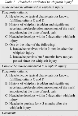
The new IHS classification published in 20042, unlike
the original 1988 publication, has also developed a cate-gory for headaches attributed to whiplash injuries [Table 1] separate from the post-traumatic headaches.
The current diagnostic features of post-traumatic head-ache due to minor head trauma2 require the following:
• Lack of moderate-to-severe head trauma.
• Headache is new or that if there was a pre-existing headache, it has worsened after the trauma.
• Glasgow Coma Scale of 13 or above.
• Signs or symptoms diagnostic of a concussion.
The new classification has a stricter definition on the onset of symptoms from the date of the trauma as well as the definition on the chronicity of the symptoms. Specifi-cally, the symptoms must appear within seven days post-trauma in the new classification, not within 14 days as previously defined, and the timeframe for chronicity is
set at three months, as opposed to eight weeks in the pre-vious classification.
The type of post-traumatic headache that results from traffic collisions varies among individuals. A recent clini-cal study reported that 37% of the post-traumatic head-ache patients following cranio-cervical acceleration/ deceleration trauma had tension-type headaches, 27% had migraine, 18% cervicogenic headaches and 18% did not fulfill criteria for any particular headache category.12
One study found the incidence of unilateral cervicogenic headaches after whiplash injury to be at approximately 8% at 6 weeks, 3% at one year and that both the headache and associated disability follow an expected course of re-covery.15 Physical impairments such as decreased neck
mobility have been shown to be more frequent in cervico-genic headaches especially with a traumatic onset.16,17
Post-traumatic cervicogenic patients experience limited active cervical range of motion and decreased strength of neck flexors and extensors, significantly more frequently than other headache types.17
The patient in this case presented with a decreased ac-tive range of motion in all directions and posiac-tive palpato-ry findings following an acceleration/deceleration injupalpato-ry without head trauma or loss of consciousness as a rear-seat passenger. The vehicle she was in did not have a headrest. Studies have shown that most drivers do not maintain the proper headrest height or the distance between the anterior headrest and the back of their head. Improper use of this assistive device can increase their risk of whiplash injury by 1.6 to 6 times.18 The most favourable headrest position
is with the top of the headrest above the ear and with the post-cranial gap of less than four inches.18
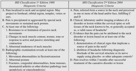
Cervicogenic headaches have also been redefined by the new IHS criteria [Table 2]. Patients with a similar presentation as reported in this case, with myofascial ten-der points, and without any positive clinical signs that are valid and reliable, should be classified under the tension-type headache with associated pericranial tenderness, ac-cording to the new classification. In this case, the patient presented with a post-MVA headache of a cervicogenic type that progressed over the course of conservative treat-ment requiring further investigation and immediate medi-cal referral for management of a subdural hematoma.
Subdural Hematoma
Subdural hematoma is a rare condition with an incidence Table 1 Headache attributed to whiplash injury2
Acute headache attributed to whiplash injury
Diagnostic criteria:
A. Headache, no typical characteristics known, fulfilling criteria C and D
B. History of whiplash (sudden and significant acceleration/deceleration movement of the neck) associated at the time of neck pain
C. Headache develops within 7 days after whiplash injury
D. One or the other of the following:
1. headache resolves within 3 months after the whiplash injury
2. headache persists but 3 months have not yet passed since the whiplash injury
Chronic headache attributed to whiplash injury
Diagnostic criteria:
A. Headache, no typical characteristics known, fulfilling criteria C and D
B. History of whiplash (sudden and significant acceleration/deceleration movement of the neck) associated at the time of neck pain
C. Headache develops within 7 days after whiplash injury
D. Headache persists for > 3 months after the whiplash injury
Comment:
rate of 1.72 per 100,000 population per year.19 The
inci-dence does increase with age to approximately 7.35 per 100,000 population in the 70 to 79 year old individuals, but it remains a rare condition with possibly higher risk of mortality and higher morbidity.19,20 The bridging cerebral
veins play a central role in the pathophysiology of
subdur-al hematoma especisubdur-ally in the older individusubdur-als due to cer-ebral atrophy that predisposes these veins to tearing stresses.19,20,21 Other risk factors for developing subdural
hematoma include history of trauma, male gender, coagu-lopathy, thrombocytopenia and alcoholism.19,20,22 History
of trauma can be elicited in a large number of patients, but Table 2 Cervicogenic headache definition changes2
IHS Classification 1st Edition 1988 Diagnostic Criteria
IHS Classification 2nd Edition 2004 Diagnostic Criteria
A. Pain localized to neck and occipital region. May project to forehead, orbital region, temples, vertex or ears.
B. Pain is precipitated or aggravated by special neck movements or sustained neck posture.
C. At least one of the following:
1. Resistance to or limitation of passive neck movements
2. Changes in neck muscle contour, texture, tone or response to active and passive stretching and contraction
3. Abnormal tenderness of neck muscles
D. Radiographic examination reveals at least one of the following:
1. Movement abnormalities in flexion/extension 2. Abnormal posture
3. Fractures, congenital abnormalities, bone tumours, rheumatoid arthritis or other distinct pathology (not spondylosis or osteochondrosis)
A. Pain, referred form a source in the neck and perceived in one or more of the head and/or face, fulfilling C and D
B. Clinical, laboratory and/or imaging evidence of a disorder or lesion within the cervical spine or soft tissues of the neck known to be, or generally accepted as, a valid cause of headache1
C. Evidence that the pain can be attributed to the neck disorder or lesion based on at least one of the following:
1. demonstration of clinical signs that implicate a source of pain in the neck2
2. abolition of headache following diagnostic blockade of the cervical structure or its nerve supply using placebo- or other adequate controls3
D. Pain resolves within 3 months after successful treatment of the causative disorder or lesion
Notes for 2004:
1. Tumours, fractures, infections and rheumatoid arthritis of the upper cervical spine have not been validated formally as causes of headaches, but are nevertheless accepted as valid causes when demonstrated to be so in individual cases. Cervical spondylosis and osteochondritis are not accepted as valid causes fulfilling criterion B. When myofascial tender spots are the cause, the headache should be coded under 2. Tension-type headache.
2. Clinical signs acceptable for criterion C1 must have demonstrated reliability and validity. The future task is the identification of such reliable and valid operational tests. Clinical features such as neck pain, focal neck tenderness, history of neck trauma, mechanical exacerbation of pain, unilaterality, coexisting shoulder pain, reduced range of motion in the neck, nuchal onset, nausea, vomiting, photophobia etc are not unique to cervicogenic headache. These may be features of cervicogenic headache, but they do not define the relationship between the disorder and the source of the headache.
it can also be trivial trauma that older individuals may not even recall that can contribute to the development of sub-dural hematoma.19–23 In one report, 24% of patients did
not report any obvious trauma, suggesting either sponta-neous causes or inability to recall trivial traumatic events.22 In any event, males may be more predisposed
due to their higher risk of trauma.19,20 Alcoholic
individu-als have a higher risk of falling and may have liver disease with secondary coagulopathy and thrombocytopenia. An-ticoagulant and antiplatelet therapies including aspirin, coumadin and warfarin medications increase the risk of developing subdural hematoma.19,20 Some subdural
he-matomas can resolve spontaneously while others progress and enlarge.21,23,24 They are classified into acute, subacute
and chronic.
The acute form usually occurs within 48 hours of the injury and it is composed of a blood clot.21,23 The
suba-cute form develops between 2 and 14 days post-injury and the chronic can occur weeks or months after the inju-ry. The composition of the subacute and chronic forms is a mixture of clotted and fluid blood and completely fluid blood, respectively.21,23
The symptoms associated with subdural hematoma [Table 3] can be subtle and, therefore, may be overlooked by practitioners. An overview study of 2300 chronic cases over a 30 year period has demonstrated a male preponder-ance in a ratio of 5:1.22 In this retrospective report, 14.7%
of patients presented fully conscious with a headache due to raised intracranial pressure. Behavioural disturbances were present in 17.6%, focal and generalized seizures in 12%, visual disturbances in 11.7%, stroke in 29.1% and 14.9% of patients were comatose. The headaches had pe-riods of remission and exacerbation. Exacerbations oc-curred on standing up from a prone position and vomiting increased with greater severity of headache. The patients also had papilloedema on examination but no neurological deficits. Behavioural changes included emotional out-bursts, garrulous periods, sleep disturbance, lack of concentration, depressive and mania-like states and di-sheveled appearance. Reported visual disturbances in-cluded blurring due to severe papilloedema and double vision due to isolated sixth and third cranial nerve deficits. Weakness in one side of the body as a symptom was present in the largest single group in this report. The au-thors suggested that in patients with a history of trauma that present with intermittent hemiparesis even in the
ab-sence of signs of raised intracranial pressure, chronic subdural hematoma should be strongly suspected.22
Al-though the natural history is not known, acute subdural hematoma may resolve spontaneously prior to diagnosis or treatment.24 In some cases, they may progress to
chronic subdural hematomas but they are usually a differ-ent histopathologic differ-entity from chronic subdural hemato-mas.23 Chronic subdural hematomas are more common in
older age groups and in alcoholics.20 In patients over 60
years of age, the most common presenting symptoms are headache, altered mental status, hemiparesis, a distur-bance in gait and aphasia.20,22
A definite diagnosis of subdural hematoma is estab-lished with the use of neuroimaging. Computerized tom-Table 3 Headache attributed to subdural haematoma2
Headache attributed to subdural haematoma
Diagnostic criteria:
A. Acute or progressive headache, no other typical characteristics known, fulfilling criteria C and D B. Neuroimaging evidence of subdural haematoma C. Headache develops within 24–72 hours after the
development of the haematoma D. One or other of the following
1. headache resolves within 3 months after evacuation of the haematoma
2. headache persists but 3 months have not yet passed evacuation of the haematoma
Comments:
ography (CT) scans or magnetic resonance imaging (MRI) may be the imaging of choice but CT scanning is usually more accessible. Although guidelines for imaging have not been specifically developed for headache, cer-tain symptoms such as reversible causes of dementia, due to subdural hematomas may be an indication for neu-roimaging studies and a referral to a medical doctor or a hospital.25 Indications for CT scanning should be
consid-ered in the progression of an existing headache or the on-set of a new headache, rapid unexplained deterioration of mental status, appearance of focal neurological signs, gait disturbance and if there is history of recent head trau-ma.19,20 In the presence of these symptoms and a positive
CT scan identifying a subdural hematoma, surgery to re-lieve the intracranial pressure would be the indicated treatment.20
Although the patient in this case was experiencing short-term relief with soft tissue therapy for approximate-ly one month, a referral was indicated once the patient developed focal neurological deficits with her headaches. Prognosis with surgical decompression for subdural he-matoma should result in a resolution of symptoms with low recurrence rates.20,22
Differential Diagnosis
While a patient presenting with headache can have more than 300 differential diagnoses, individuals suffering from subdural hematoma will have fewer similar clinical presentations to consider.5 Differential diagnoses for
sub-dural hematoma, requiring neuroimaging studies include epidural hematoma/intracranial hematomas, cerebrovas-cular insults, neoplasms, normal-pressure hydrocephalus and Alzeimer’s dementia.5,20,21
Intracranial hematomas due to traumatic events can be localized in different locations of the cranium. The extra-dural or epiextra-dural hematomas usually develop in the epi-dural space from arterial sources and therefore, develop rapidly.21 They strip the dura from the scalp and develop
into a circumscribed ovoid mass that progressively in-dents the brain but with little damage to the brain tissue. Within four to six weeks they may decrease in size and resolve. The epidural hematomas are usually caused by more severe traumatic events with associated cranial frac-tures and are more likely to be fatal. The symptomatolo-gy may be similar to acute subdural hematomas with an acute onset and progressive nature of symptoms unlike
the chronic subdural hematomas.19 In contrast, chronic
subdural hematomas are usually associated with minor injuries with a slower onset of focal neurological deficits and changes in mental status. Other intradural hemato-mas such as subarachnoid, intracerebral or intracerebellar hematomas should also be differentiated from subdural hematoma. Usually they are associated with more severe traumatic events and fractures of the cranial bones. The computerized tomography scanning would be indicated to differentiate between the named conditions.21
Patients that experience cerebrovascular insults also experience focal neurological deficits and cognitive im-pairments with an acute onset of a few hours to days. Acute subdural hematomas would, therefore, have to be differentiated using neuroimaging studies, but chronic subdural hematomas would develop more progressively over a period of weeks.20,21 Cerebrovascular insults can
occur at the time of a traumatic event or after the event and practitioners should be cognizant of any change in mental status or appearance of focal neurological symp-toms and signs.21
Intracranial neoplasms can also present with similar symptomatology and may be difficult to differentiate from chronic subdural hematomas clinically.20 These
pa-tients can present with all the signs of increased intracra-nial pressure including headache, focal neurological signs and cognitive impairments.20,21 Neuroimaging
stud-ies can differentiate the conditions with MRI being the diagnostic study of choice but often CT scanning is ob-tained due to accessibility.21
Normal-pressure hydrocephalus has similar symp-tomatology to chronic subdural hematoma. The related symptoms include dementia, gait ataxia and urinary in-continence.20,21 Urinary incontinence does not occur in
chronic subdural hematoma and can therefore be used as a clinical differentiating factor but neuroimaging is the definitive diagnostic test in differentiating these two con-ditions.21
Patients with Alzeimer’s dementia will develop cogni-tive impairments over a period of months to years.20 In
acute subdural hematoma, the onset of symptoms is acute and in chronic subdural hematoma, the onset occurs with-in a period of weeks.19,20,21 Focal neurological signs and
The case presented represents a subdural hematoma, which was successfully treated with decompression. Al-though such conditions can occur in the elderly without any significant trauma, this case presents the aforemen-tioned condition which occurred a month following a motor vehicle collision. Although definitive causation cannot be established, it is nevertheless interesting that her initial headaches symptoms, which were minimal, progressed with development of neurological deficits. In-itial cranial nerve examination was not conducted, due to her presenting complaints, but retrospectively, such as-sessments should be entertained in elderly patients pre-senting with headaches, particularly after trauma.
Conclusion
Post-traumatic headaches are a common condition pre-senting to a chiropractor’s office. Rare differential diag-noses such as subdural hematoma must be considered in post-traumatic headaches, which progress and demon-strate new signs/symptoms such as focal neurological def-icits, cognitive impairment or progression of headaches. Neuroimaging studies are required to identify the source, with immediate medical referral and management.
References
1 Kryst S, Scherl E. A population-based survey of the social and personal impact of headache. Headache 1994; 34(6):344–50.
2 Headache classification subcommittee of the International Headache Society. The International Classification of Headache Disorders 2nd ed. Cephalalgia 2004; 24 Suppl 1:1–159.
3 von Peter S, Ting W, Scrivani S, Korkin E, Okvat H, Gross M, Oz C, Balmaceda C. Survey on the use of
complementary and alternative medicine among patients with headache symptoms. Cephalalgia 2002; 22:395–400. 4 Goadsby PJ, Silberstein SD. Blue Books of Practical
Neurology: Headache. United States: Butterworth-Heinemann, 1997; 253–277.
5 Evans RW. Diagnostic testing for the evaluation of headaches. Neurol Clin 1996; 14(1):1–26.
6 Fukutake T, Mine S, Yamakami I, Yamaura A, Hattori T. Roller coaster headache and subdural hematoma. Neurol 2000; 54(1):264.
7 Snyder RW, Sridhaaran ST, Pagnanelli DM. Subdural hematoma following roller coaster ride while anticoagulated. Am J Med 1997; 102:488–489.
8 Bo-Abbas Y, Bolton CF. Roller-coaster headache. N Engl J Med 1995; 332:1585.
9 Fernandes CMB, Daya MR. A roller coaster headache: case report. J Trauma 1994; 37(6):1007–1010.
10 Deci DM. Chronic subdural hematoma presenting as headache and cognitive impairment after minor head trauma. W V Med J 2004; 100(3):106–107.
11 Ommaya AK, Yarnel P. Subdural hematoma after whiplash injury. Lancet 1969; 2:237–239.
12 Radanov BP, Di Stefano G, Augustiny KF. Symptomatic approach to posttraumatic headache and its possible implications for treatment. Eur Spine J 2001; 10:403–407. 13 Cassidy JD, Carroll LJ, Peloso PM, Borg J, von Holst H,
Holm L, Kraus J, Coronado VG. Incidence, risk factors and prevention of mild traumatic brain injury: results of the WHO collaborating centre task force on mild traumatic brain injury. J Rehab Med 2004; Suppl 43:28–60. 14 Carroll LJ, Cassidy JD, Peloso PM, Borg J, von Holst H,
Holm L, Paniak C, Pepin M. Prognosis for mild traumatic brain injury: results of the WHO collaborating centre task force on mild traumatic brain injury. J Rehab Med 2004; Suppl 43:84–105.
15 Drottning M, Staff PH, Sjaastad O. Cervicogenic headache (CEH) after whiplash injury. Cephalalgia 2002; 22:165– 171.
16 Zwart JA. Neck mobility in different headache disorders. Headache 1997; 37:6–11.
17 Dumas JP, Arsenault AB, Boudreau G, Magnoux E, Lepage Y, Bellavance A, Loisel P. Physical impairments in cervicogenic headache: traumatic vs. nontraumatic onset. Cephalalgia 2001; 21:884–893.
18 Viano DC, Gargan MF. Headrest position during normal driving: implications to neck injury risk in rear crashes. Accid Anal Prev 1996; 26(6):665–674.
19 Chen JCT, Levy ML. Causes, epidemiology and risk factors of subdural hematoma. Neurosurg Clin N Am 2000; 11(3):399–406.
20 Karnath B. Subdural hematoma: presentation and
management in older adults. Geriatrics 2004; 59(7):18–23. 21 Rosenthal M, Griffith ER, Kreutzer JS, Pentland B.
Rehabilitation of the adult and child with traumatic brain injury. 3rd ed. Philadelphia: F.A. Davis Company, 1999; 22–27.
22 Sambasivan M. An overview of chronic subdural
hematoma: experience with 2300 cases. Surg Neurol 1997; 47:418–422.
23 Lee KS, Bae WK, Doh JW, Bae HG, Yun IG. Origin of chronic subdural hematoma and relation to traumatic subdural lesions. Brain Injury 1998; 12(11):901–910. 24 Parlato C, Guarracino A, Moraci A. Spontaneous
resolution of chronic subdural hematoma. Surg Neurol 2000; 53:312–317.