Windup of Nociceptive Flexion Reflex Depends on Synaptic and Intrinsic Properties of Dorsal Horn Neurons in Adult Rat
Franck Aby 1,2, Rabia Bouali-Benazzouz1,2, Marc Landry1,2 and Pascal Fossat1,2,*
1Université de Bordeaux.
2Institut interdisciplinaire des neurosciences, IINS CNRS UMR 5297
*Corresponding Author:
Pr. Fossat Pascal,
University of Bordeaux
Interdisciplinary institute for neurosciences, CNRS UMR 5297
pascal.fossat@u-bordeaux.fr
+33630168681
Abstract :
Windup, a progressive increase in spinal response to repetitive stimulations of nociceptive peripheral fibres, is a useful model to study central sensitization to pain. Windup is expressed by neurons in of both dorsal and ventral horn of the spinal cord. In juvenile rats, it has been demonstrated both in vivo and in vitro that windup depends on calcium-dependent intrinsic properties and their modulation by synaptic components. However, the involvement of these two components in the adult remain controversial. In the present study, by means of electromyographic and extracellular recordings, we show that windup in adult, in vivo, depends on a synaptic balance between excitatory NMDA receptors and inhibitory glycinergic receptors. We also demonstrate the involvement of L-type calcium channels in both the dorsal and ventral horn of the spinal cord. These results indicate that windup in adults is similar to juveniles rats and that windup properties are the same regardless spinal network, i.e. sensory or motor.
Keywords: l-type calcium channels; nociception; spinal cord; central sensitization; windup
Introduction
The dorsal horn of the spinal cord is the first relay of nociceptive inputs raising from
the periphery. Dorsal horn neuronal networks integrate nociceptive information and
output neurons project both to supraspinal areas and motoneurones. Within this
network, deep dorsal horn neurons (DHNs) receive convergent inputs from tactile
and nociceptive fibers and express a form of short term activity-dependent plasticity,
so-called windup. Windup is a form of early onset spinal sensitization [1, 2] and is
considered as a simple tool to study pain processing plasticity in the spinal cord (for
reviews see [3, 4]. In clinics, windup protocol serves to evaluate temporal summation
of pain, and is considered as a good predictor for clinical pain intensity in idiopathic
pain syndromes such as fibromyalgia [5, 6]. Windup is also expressed in motor
network and repetitive nociceptive stimulations of the hindpaw elicit windup of
segmental reflexes [7]. Moreover, repetitive stimulations of the sciatic nerve elicit
windup of ventral roots [4]. Finally, motor neurons express windup in different animal
models such as rats or turtle [8, 9].
At the level of the dorsal horn, windup is expressed by wide dynamic range neurons
and has been extensively studied. In adult rats, it has been shown that windup
depends on excitatory synaptic properties as NMDA glutamate receptors and
neurokinin receptors. [10]. Windup also depends on DHNs intrinsic properties since
windup is blocked by L-type calcium channels (LTCs) antagonists and increase by
agonist of these channels [11]. Other studies performed in spinal slices from juvenile
rats confirmed the role of LTCs in this phenomenon and also show the involvement of
another cationic non specific conductance (CAN) [12]. DHNs project to the ventral
nociceptive flexion reflex [7]. Repetitive nociceptive stimulations of the ipsilateral paw
elicit a windup of nociceptive flexion. It has been shown that windup of the flexion
reflex in juvenile rats depends on a synaptic balance between excitation and
inhibition that allow expression of intrinsic properties [11, 13, 14]. However, the
flexion reflex is the output of a neural circuit that comprise both DHNs and motor
neurons and the latter ones also exhibit a windup of their discharge that present the
same molecular sensitivity as windup of DHNs [8, 9, 15]. Because of the different
model used (juvenile versus adult, turtles), the site of recordings (DHNs, motor
neurons, flexion reflex), our understanding of windup remains partial. For instance, it
is still unknown if the balance between excitation and inhibition also control windup at
the level of the dorsal horn of the spinal cord. The involvement of intrinsic properties
in the windup of the flexion reflex in adult rats is still controversial [4]. Finally,
previous studies on reflex have been performed in juvenile rats and we cannot
exclude developmental modifications altering windup molecular sensitivity.
We propose in this study to analyze in anesthetized rats, the windup for DHNs by
means of extracellular recordings and the windup of flexion reflex with
electromyographic recordings in the biceps femoris muscle of the ipsilateral hindpaw
and compared its molecular sensitivity between adult and juvenile rats.
Methods
Experimental procedures
We used both juvenile (14 to 21 days old (55g)) and adult wistar rats in this study
(250-350g). All experimental procedures were approved by the local ethic
(agreement number : APA3650) committee according to the International Association
for the Study of Pain (IASP) ethical guidelines for experimental research were
followed. At the end of each experiment, rats were killed with an overdose of
anaesthetics.
Drug administration
Rats were anaesthetized with intraperitoneal urethane (1.1 mg/kg). A catheter (PE-10;
Phymep, France) was inserted into the sub-arachnoid space, and the tip was pushed
forward to the L4-L5 region. During recordings, drugs were injected using the
catheter connected to a Hamilton syringe and flushed with 10 μl saline (dead volume
of the catheter). Nicardipine and verapamil (Sigma Aldrich, France) were freshly
dissolved in saline. AP-5 and strychnine (Sigma Aldrich, France) came from 10 mM
stock solutions stored at -20°C. Flufenamic acid (FFA) were freshly dissolved in
dimethylsulfoxide (DMSO, Sigma Aldrich), then diluted with saline. For dose
response curve, the lower dose was first intrathecally injected and responses
measured. Each doses was separated by 30min.
In vivo electromyographic recordings
Recordings were made in the ipsilateral biceps femoris muscle with two teflon-coated
stainless steel electrodes (Phymep, France; diameter 200 μm). Electrical stimuli (500
μs, single chocks, master 8 stimulator, AMPI, Israel) were delivered with electrodes
obtained for a stimulus intensity of 60 to 80 V, as determined with single shocks
delivered at 30 s intervals to avoid central sensitization. At this stimulus intensity, a
slight muscle contraction was elicited without movement of the limb. Data were
acquired by a CED 1401 interface, and analyzed with Spike 2 software (CED, UK).
In vivo extracellular recordings
After urethane anesthesia, rats were placed in a stereotaxic frame to ensure stability
during electrophysiological recordings. A laminectomy was performed on lumbar
vertebrae L1-L3, for exposition of the L4-L5 segments of the spinal cord. Extracellular
recordings of wide dynamic range dorsal horn neurons (DHNs) were made with
borosilicate glass capillaries (2 MΩ, filled with 4% NaCl) (Harvard apparatus, USA).
Electrodes were positioned with a microdrive (unimecanique, France). The depth of
the neurons from the dorsal surface of the spinal cord was monitored (recordings
were performed between 500 to 1000μm from the surface). The response to various
natural stimuli (brush, pressure, pinch) in the most responsive part of the receptive
field of the neurons was characterized to ensure the wide dynamic range properties
of the recorded neurons. We recorded the neurons’ responses following
transcutaneous electrical stimulation (see above) of the centre of the receptive field.
The criterion for the selection of a neuron was the presence of an Aβ-fiber evoked
response followed by a C fiber-evoked response. For analysis, we performed post
stimulus histogram (PSTH) and separate in 4 zones: an Aβ zone between 0 and
20ms an Aδ zone between 20 and 90 ms, a C zone between 90 and 300 ms and a
post discharge zone (PD) between 300 ms and 1s.
Windup protocol
Sequences of 15 stimuli were delivered at 1 Hz, except in experiment of Fig.1, in
which the stimulation frequency was varied from 0.1 to 1 Hz as indicated on the
graph. For Flexion reflex, each stimulus was a single shock (500 μs) at ~80% of the
intensity eliciting a maximal response (see above). For extracellular recordings,
single shock 3 fold the threshold for C-fibres.
Data Analysis and statistics :
The electromyographic signals were integrated by measuring the area under the
curve after rectification of the signal. Windup plots (e.g. Fig 1A) were normalized to
the first response of the series. For extracellular recordings, spikes in the C+PD part
of the response were counted and Data were plotted with the Prism software (v 5.0,
GraphPad Inc.). All values are mean ± SEM, and N indicates the number of tested
animals. A windup coefficient was calculated by dividing the sum of the 15 responses
by 15 times the first recorded response. Comparison of windup plots were performed
using a two-way ANOVA. Windup coefficient comparison between two populations
was performed using non parametric Mann Whitney test. Windup coefficient
comparison between before and after drug applications were compared using paired
t test or a one way ANOVA when 2 drugs were applied. A value of p<0.05 was
considered significant.
Results
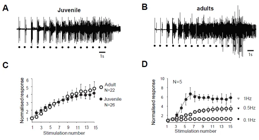
Windup of WDR neurons and of the flexion reflex share similar characteristics. In juvenile rats, electrical stimulation of high threshold fibers in the paw triggers a
muscular contraction of the flexor ipsilateral muscle. This response called RIII flexion
reflex or nociceptive flexion reflex, appears in the electromyogram with a latency of
80-300 ms with high stimulations (Figure 1A). The same type of reflex is also
observed in adult rats (figure 1B). Repetitive stimulation at low frequency (here 1Hz)
stimulation, with a progressive development of an after-discharge (Fig. 1A and B).
This increased response is so-called windup. The amplitude of the windup is similar
in juvenile and adult rats since the last response of the series represents 467+/-97%
in the adult and 453 +/- 56% (N=22 and 26 respectively, p>0.05, Mann-Whitney) in
juvenile rats. Windup coefficient defined as the sum of the 15 responses substracted
by 15 times the first response is also very similar (Figure 1C, windup coefficient is
30.38 ± 5.77 in adult et de 32.04 ± 6.99 in juvenile; p>0.05, Mann-Whitney). Windup
of the flexion reflex is frequency dependent in adult rats (Figure 1D). Together these
results show that windup phenomenon is not modified during development and
exhibits the same characteristics at the level of the muscle or the dorsal horn of the
spinal cord.
Windup of the nociceptive flexion reflex in adult rats is controlled by synaptic and intrinsic components.
We next study the molecular mechanisms controlling the windup of the flexion reflex
in adult rats. First we focused on synaptic components and we observed that
application of 100µg AP5 strongly decreased the flexion reflex windup (Figure 2A, B
and C). This effect is dose dependent with an EC50 of 10,26 µg (not shown). The
baseline response was also modified with a significant decrease at 57% of the
control. This result is comparable to previous study in juvenile rats but the
sensitivity to NMDA blockers is strongly decreased in adult rats since the dose
necessary to obtain a complete suppression of the windup is 10 fold higher than in
juvenile rats.
In juvenile rats, windup of the nociceptive flexion reflex is always present if we
suppress both excitatory and inhibitory synaptic component (Fossat et al, 2007). We
tested for this possibility in adult rats. We first applied an inhibitory dose of 100μg AP5
to suppress the windup (Figure 2D, E and G), we further applied both AP5 and
strychnine and the windup was recovered (Figure 2D, F and G). Thus, we
demonstrated in adult rats that windup of the nociceptive flexion reflex depends on a
synaptic balance between excitatory and inhibitory component.
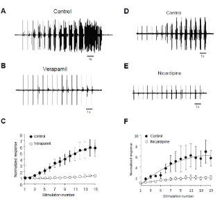
Windup of the flexion reflex is sensitive to IL blockers in adult rats:
We assessed the role of ILs in windup of the flexion reflex using two different families
of blockers (Lapirot et al, 2018). First, we applied 100μg verapamil a blocker of the
phenylalkylamine family. Verapamil blocked the windup of the flexion reflex (Figure
3A-C). The effect was dose dependant with an EC50 of 26μg. Because of lack of
specificity of phenylalkylamine, we used dihydropyridine to confirm the involvement of
LTCs. Intrathecal application of 50μg nicardipine also suppressed the windup of the
flexion reflex (Figure 3D-F). Again, the effect was dose dependant with an EC50 of
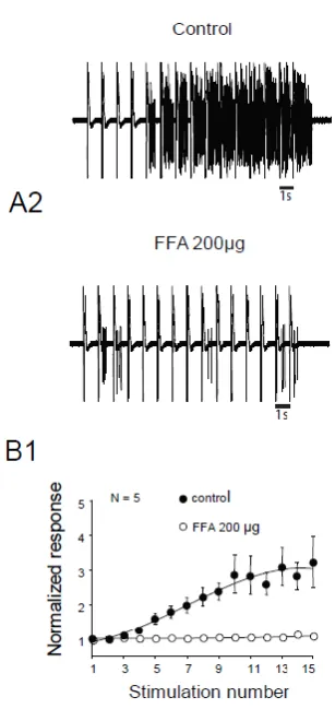
16.4μg. Finally, we assessed for the involvement of CAN in windup of the flexion
reflex. Intrathecal Application of 200μg flufenamic acid (FFA) completely suppress
windup of the flexion reflex (Figure 4 A and B). Effect of FFA was dose dependant
with an EC50 of 13.6 μg. These results demonstrate in adult rats that LTCs and CAN
are two important elements in the expression of windup of the nociceptive flexion
reflex.
Synaptic component of windup in DHNs of adult rats.
The nociceptive flexion reflex is the output of a reflex network that comprises
two neuronal levels i.e. dorsal and ventral horn. We then studied the properties of the
windup of the discharge of DHNs and its sensitivity to a synaptic balance between
excitation and inhibition. We first evaluated the characteristics of the windup in DHNs
neurons in adult rats. We observed a progressive increase in neuronal discharge in
53% (68/128) of the recorded neurons (Figure 5A). When present, windup was also
frequency dependent with no windup at 0.1 Hz and an increased amplitude until 1Hz
stimulations (Figure 5B).
Next, we wanted to determine if a balance between excitation and inhibition
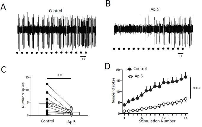
modulate windup of DHNs. We first confirmed that windup is influenced by NMDA
receptor blockers (Figure 6). We showed that intrathecal application of 100μg AP-5
significantly decreases DHNs excitability (Figure 6C) and Windup amplitude (Figure 6
A,B and D). As already shown previously for the flexion reflex, inhibition control the
amplitude of windup. To study this phenomenon in DHNs, we used strychnine to
block glycinergic receptors (Figure 7). Intrathecal application of 170μg strychnine
increased the excitability of DHN (Figure 7C). Windup was also increased (Figure 7A,
B and D). Therefore, windup of DHNs of adult rats is potentiated by synaptic
excitations and decreased by synaptic inhibitions. In the next step, we wondered if
we could restore a windup after blockade of NMDAr by suppressing inhibitory
influence (Figure 8). To that issue, we suppressed windup of DHNs by applying 100
μg AP-5 and then we applied both 100μg AP-5 and 170μg Strychnine. 100μg AP-5
almost completely suppressed the windup (Figure 8B D and E) and subsequent
application of AP-5 and Strychnine restore a windup of DHNs (Figure 8C, D and E).
Together, these results demonstrate that expression of windup by DHNs in adult rats
depends on a synaptic balance between excitations and inhibitions.
Discussion :
This study shows that windup of a nociceptive flexion reflex depends on both
synaptic and intrinsic components in adult rats in vivo. We confirmed the role of LTCs
and CAN in this form of short term sensitization to pain. We also show similar
characteristics between the windup of nociceptive flexion reflex and the windup of
DHNs and we observed a global decrease in sensitivity to blockers of the molecular
components of windup in adult rats.
Adult and juvenile windup
These results demonstrates that the windup of the flexion reflex is not different
between juvenile and adult rats. Indeed, as in juvenile rats, windup in adult also
depends on stimulation frequency and is sensitive to synaptic modulators and
plateau potential blockers. Windup in adult rats is also sensitive to synaptic NMDA
blockers and its onset depends on a balance between synaptic inhibitory and
excitatory inputs. However, we show here a general decrease in drug sensitivity in
adult rats as compared to juvenile. These modifications in drug sensitivity have
already been found for NMDA receptors blockers [16]. One may consider that
differences between juveniles and adults could reflect a modification of neuronal
phenotypes with altered expression in channels or receptors subunits. However,
such a general effect for all tested substances is more probably due to an increased
difficulty for any drugs to reach their targets as the areas concerned are deep and
adult tissues have a complex extracellular matrix and are enriched in myelin.
Importance of LTCs in windup.
Windup is an activity dependent short term plasticity involved in pain processing. It
represents a form of input/ouput amplification mechanism, which strengthened
contrast between background activity and relevant stimuli. Windup is known to
depend on synaptic plasticity exerted through NMDAr and neurokinin[10, 17, 18]. In
vitro recordings in juvenile rats also revealed the role of intrinsic properties of deep
expression of plateau potentials are also necessary to trigger windup of the
discharge. These two channels are LTCs and CAN. LTCs is necessary in the early
phase of plateau whereas CAN promote afterdischarge [19]. However, the role of
LTCs in short term central sensitization to pain in adult rats is not fully demonstrated.
For instance, in the formalin model of short term sensitization, LTCs blockers have no
effect [20] while LTCs blockers suppress DHNs windup [11]. Here, we show that, in
adult rats, windup of a flexion reflex depends on LTCs since windup is blocked by
blockers of LTCs belonging to two different families in a dose dependent manner.
Moreover, a recent study suggest that one specific type of LTCs is involved in windup
in juvenile rats. Indeed, Two LTCs forming channels are expressed in dorsal horn of
the spinal cord, Cav1.2 and Cav1.3. Blocking Cav1.3 expression with an antisens
strategy suppress the windup of the flexion reflex [13, 14]. The participation of LTCs
in pain depends on the type of pain. For instance, they are not involved in acute
nociception but they are clearly important for windup, central sensitization resulting
from joint inflammation [21] and in long term changes that accompany nerve injury
[22]but not in sensitization induced by formalin injection [20].
We also show here that CAN are also involved in the windup of the flexion reflex in
adult rats. CAN promote another important conductance of plateau potentials. Indeed,
CAN is triggered following LTCs activation and elicit a larger depolarisation
responsible for prolonged after discharge [12, 19]. The role of CAN in windup has
been demonstrated in DHNs neurons in slices from juvenile rats [19] or in
anesthetized juvenile rats [11] and we show here their involvement in the windup of
flexion reflex in adult rats.
Synaptic component of windup
The other major component of windup in vivo is synaptic. NMDAr and neurokinin
receptors are activated during trains of stimuli and this lead to the onset of windup.
Here, we show that both windup of flexion reflex and windup of DHNs are completely
suppressed by NMDAr blockers in vivo in adult rats, thus confirming classical results
of the literature [10]. This blockade is also accompanied by a decrease in the
baseline response and DHNs excitability indicated an effect of NMDAr not only in
sensitization but also in acute nociception [11]. We also show here that windup
depends on inhibitory synaptic influences. Blocking glycine receptors increase the
amplitude of windup of DHNs. This effect was accompanied by an increase in DHNs
excitability. This effect is comparable to that previously observed in nociceptive
flexion reflex [7, 11]. This result confirm that inhibitory interneurons control the onset
of central sensitization by exerting a tonic inhibitory tone. Finally, we show that
windup depends in adult rat on a dynamic balance between excitation and inhibition.
Indeed, when blocking NMDAr, we suppressed windup that can be restored by the
subsequent blockade of glycine receptor. This suggest that intrinsic properties of
spinal neurons are a the key element that elicits windup as suggested in juvenile rats
[11].
Neural substrate of windup
Two neuronal levels can elicit windup of their discharge within the flexion reflex
circuit: motoneurone and DHNs neurons [8, 9, 12, 15, 23]. We previously showed
that in control conditions, the windup of the flexion reflex was strictly correlated with
DHNs windup [11]. In this condition, motor neurons seems to follow amplification
properties expressed by DHNs neurons. Here, we show that the windup of the
suggesting that the dorsal level is crucial to elicit reflex windup. Indeed, we show that
both windup of flexion reflex and windup of DHNs are modulated by NMDAr, Glycine
and controlled by LTCs and CAN. We cannot exclude a role of windup of motor
neurons in other motor tasks but our results strongly suggest that the flexion reflex is
first controlled at the dorsal horn level.
Windup and central sensitization to pain.
Windup has long been studied in various models of pain. It has been defined as a
model of short term sensitization to pain and it is considered as a good model for the
study of mechanisms leading to central sensitization to pain. Moreover, windup is still
used to assess level of pain in many models. For instance, several evidences show
the usefulness of windup as a cue of central sensitization in patients suffering of
idiopathic pain [5, 6]. However, windup is a short term sensitization with a time scale
of few seconds and cannot be responsible for long term central modifications leading
to neuropathic pain [24]. On the one hand, time scale of windup is too short to trigger
neuronal modifications appearing in long term changes leading to an increase in
spontaneous activity and an increased response to both innocuous and noxious
stimuli. On the other hand, it seems that windup is not affected by nerve
lesions-induced neuropathy or slightly decreased [13, 25].
Nevertheless, recent studies have shown that in SNL rats, the number of neurons
expressing plateau potentials increases dramatically as compared to naïve animals
[26]. In conclusion, even if windup is not responsible for the deep changes appearing
in central sensitization to pain, an increased capacity to generate windup in such
pathology cannot be excluded on chronic pain syndromes and controlling windup
onset in the dorsal horn of the spinal cord is a potential way to limit exaggerate pain
in chronic pain syndrome.
References
1. Li J, Simone DA, Larson AA. Windup leads to characteristics of central sensitization. Pain. 1999 Jan;79(1):75-82.
2. Ji RR, Kohno T, Moore KA, Woolf CJ. Central sensitization and LTP: do pain and memory share similar mechanisms? Trends Neurosci. 2003 Dec;26(12):696-705. 3. Baranauskas G, Nistri A. Sensitization of pain pathways in the spinal cord: cellular
mechanisms. Prog Neurobiol. 1998 Feb;54(3):349-65.
4. Herrero JF, Laird JM, Lopez-Garcia JA. Wind-up of spinal cord neurones and pain sensation: much ado about something? Prog Neurobiol. 2000 Jun;61(2):169-203. 5. Price DD, Staud R, Robinson ME, Mauderli AP, Cannon R, Vierck CJ. Enhanced
temporal summation of second pain and its central modulation in fibromyalgia patients. Pain. 2002 Sep;99(1-2):49-59.
6. Staud R, Vierck CJ, Robinson ME, Price DD. Spatial summation of heat pain within and across dermatomes in fibromyalgia patients and pain-free subjects. Pain. 2004 Oct;111(3):342-50.
7. Gozariu M, Bragard D, Willer JC, Le Bars D. Temporal summation of C-fiber afferent inputs: competition between facilitatory and inhibitory effects on C-fiber reflex in the rat. J Neurophysiol. 1997 Dec;78(6):3165-79.
8. Russo RE, Hounsgaard J. Short-term plasticity in turtle dorsal horn neurons mediated by L-type Ca2+ channels. Neuroscience. 1994 Jul;61(2):191-7.
9. Baranauskas G, Nistri A. NMDA receptor-independent mechanisms responsible for the rate of rise of cumulative depolarization evoked by trains of dorsal root stimuli on rat spinal motoneurones. Brain Res. 1996 Nov 4;738(2):329-32.
10. Woolf CJ, Thompson SW. The induction and maintenance of central sensitization is dependent on N-methyl-D-aspartic acid receptor activation; implications for the treatment of post-injury pain hypersensitivity states. Pain. 1991 Mar;44(3):293-9. 11. Fossat P, Sibon I, Le Masson G, Landry M, Nagy F. L-type calcium channels and
NMDA receptors: a determinant duo for short-term nociceptive plasticity. Eur J Neurosci. 2007 Jan;25(1):127-35.
12. Morisset V, Nagy F. Plateau potential-dependent windup of the response to primary afferent stimuli in rat dorsal horn neurons. Eur J Neurosci. 2000 Sep;12(9):3087-95. 13. Radwani H, Lopez-Gonzalez MJ, Cattaert D, Roca-Lapirot O, Dobremez E,
Bouali-Benazzouz R, Eiriksdottir E, Langel U, Favereaux A, Errami M, Landry M, Fossat P. Cav1.2 and Cav1.3 L-type calcium channels independently control short- and long-term sensitization to pain. J Physiol. 2016 Nov 15;594(22):6607-26.
14. Roca-Lapirot O, Radwani H, Aby F, Nagy F, Landry M, Fossat P. Calcium signalling through L-type calcium channels: role in pathophysiology of spinal nociceptive transmission. Br J Pharmacol. 2018 Jun;175(12):2362-74.
15. Johnson KP, Tran SM, Siegrist EA, Paidimarri KB, Elson MS, Berkowitz A. Turtle Flexion Reflex Motor Patterns Show Windup, Mediated Partly by L-type Calcium Channels. Front Neural Circuits. 2017;11:83.
17. Ma QP, Woolf CJ. Tachykinin NK1 receptor antagonist RP67580 attenuates progressive hypersensitivity of flexor reflex during experimental inflammation in rats. Eur J Pharmacol. 1997 Mar 19;322(2-3):165-71.
18. Nagy I, Miller BA, Woolf CJ. NK1 and NK2 receptors contribute to C-fibre evoked slow potentials in the spinal cord. Neuroreport. 1994 Oct 27;5(16):2105-8.
19. Morisset V, Nagy F. Ionic basis for plateau potentials in deep dorsal horn neurons of the rat spinal cord. J Neurosci. 1999 Sep 1;19(17):7309-16.
20. Diaz A, Dickenson AH. Blockade of spinal N- and P-type, but not L-type, calcium channels inhibits the excitability of rat dorsal horn neurones produced by subcutaneous formalin inflammation. Pain. 1997 Jan;69(1-2):93-100.
21. Vanegas H, Schaible H. Effects of antagonists to high-threshold calcium channels upon spinal mechanisms of pain, hyperalgesia and allodynia. Pain. 2000 Mar;85(1-2):9-18.
22. Fossat P, Dobremez E, Bouali-Benazzouz R, Favereaux A, Bertrand SS, Kilk K, Leger C, Cazalets JR, Langel U, Landry M, Nagy F. Knockdown of L calcium channel subtypes: differential effects in neuropathic pain. J Neurosci. 2010 Jan 20;30(3):1073-85.
23. Thompson SW, King AE, Woolf CJ. Activity-Dependent Changes in Rat Ventral Horn Neurons in vitro; Summation of Prolonged Afferent Evoked Postsynaptic Depolarizations Produce a d-2-Amino-5-Phosphonovaleric Acid Sensitive Windup. Eur J Neurosci. 1990;2(7):638-49.
24. Woolf CJ. Windup and central sensitization are not equivalent. Pain. 1996 Aug;66(2-3):105-8.
25. Chapman V, Suzuki R, Dickenson AH. Electrophysiological characterization of spinal neuronal response properties in anaesthetized rats after ligation of spinal nerves L5-L6. J Physiol. 1998 Mar 15;507 ( Pt 3):881-94.
26. Reali C, Fossat P, Landry M, Russo RE, Nagy F. Intrinsic membrane properties of spinal dorsal horn neurones modulate nociceptive information processing in vivo. J Physiol. 2011 Jun 1;589(Pt 11):2733-43.
Figure legends : Figure 1 :
Flexion reflex in adult and juvenile rats (A et B) Electromyographic recordings (EMG)
of windup triggerred by a series of electrical shocks in the sural nerve peripheral
receptive field in juvenile (A) and in adult rats (B). Each dot indicates an electric
stimulation (500 μs, 80% of the maximal response). (C) Normalised windup curves in
juvenile (open dots) and adult rats (filled dots). Note that the two curves are strictly
superimposed showing a comparable sensitization. (D) In adult rats, windup is
frequency dependant with no windup for frequance <0.1Hz and a progressive
increase until at least 1Hz.
Figure 2 :
Windup of the nociceptive flexion reflex in adult rats is controlled by NMDAr and
glycine receptors. A) EMG in control or B) after application of 100µg AP5. C) Windup
is significantly decreased by AP5 (windup coefficient : 20.4±4 in control vs 1.2±0.5 in
AP5, n=10, p<0.01, Paired t test). D) EMG in control, E) after 100µg AP5 and F) after
100µg AP5 + 170 µg strychnine. G) Application of AP5 suppressed the windup that
was restored after subsequent application of AP5 and Strychnine (windup coefficient;
41±13.8 in control vs -1.39±1 in AP5 and 24,2±7,2 in AP5+Strychnine, n=6,
Figure 3 :
Flexion reflex windup depends on LTCs in adult rats. A and B) EMG in control and
after application of 100µg Verapamil. C) Normalised response showing a significant
decrease of flexion reflex windup (windup coefficient; 31±9.8 in control vs 5.4±4 in
verapamil, n=5, p<0.05, paired t test). D and E) EMG in control and after application
of 100µg Nicardipine. F) Normalised response showing a significant decrease of
flexion reflex windup (windup coefficient; 62.5±12 in control vs 11±3.3 in nicarpidine,
n=5, p<0.05, paired t test).
Figure 4
Flexion reflex in adult rats is sensitive to CAN currents. A and B) EMG in control or
after application of 200µg of FFA C). B1 Normalised response showing a significant
decrease of flexion reflex windup with 200µg FFA (Windup Coefficient ; 16.9±4.7 in
Figure 5
(A) Repetitive stimulations of the paw at three times the threshold for C-fibre induce a
progressive increase in DHNs discharge showing up a windup. (B) windup of DHNs
is frequency dependant. Windup Coefficient was 130±15.7 at 1Hz, 86.7±24 at 0.5 Hz
and 29±7 at 0.1Hz, n=7, p1Hz vs 0.1Hz <0.05, Dunns multiple comparison test).
Figure 6 :
DHNs windup depends on NMDAr. A and B) Extracellular recordings of DHN
response to a series of electric shocks before and after 100μg AP5. C) AP5
decreases DHNs excitability, since the response to the first nociceptive stimulation is
significantly decrease (Response to the first stimulation 4.7±1.2 spikes in control vs
1.3±0.25 spikes in AP5, N=10, p<0.01, Paired t test). D) Amplitude of windup is
significantly decrease (windup coefficient; 103±15.7 in control vs 31±9.2 in AP5,
N=10, p<0.001, paired t test).
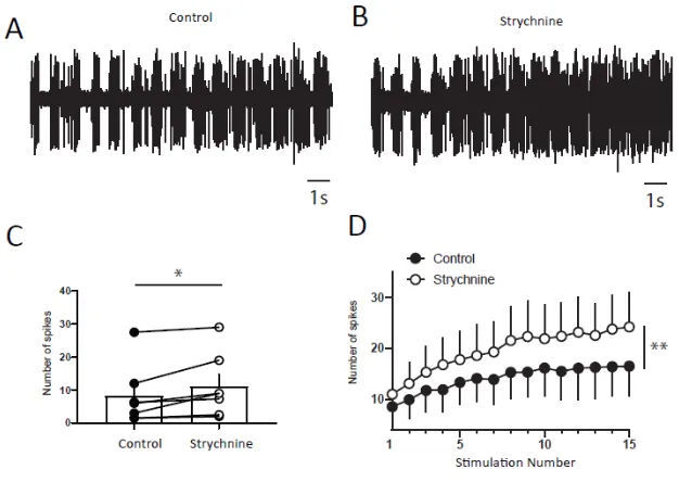
Figure 7 :
DHNs windup depends on glycinergic receptors. A and B) Extracellular recordings of
DHN response to a series of electric shocks before and after 170μg strychnine. C)
strychnine increases DHNs excitability, since the response to the first nociceptive
stimulation is significantly increase (8.5±3.5 spikes in control vs 11±3.6 spikes in
Strychnine, N=7, p<0.01, Paired t test). D) Amplitude of windup is significantly
increased (windup coefficient; 83±28.6 in control vs 130±43.3 in Strychnine, N=7,
Flexion reflex windup in adult rats
Figure 8 :
Dynamic synaptic balance control DHNs windup. A) Extracellular recordings of DHN
in control showing a windup. B) After 100μg application of AP5, windup is decreased.
C) Subsequent co-application of AP5 and strychnine restored a windup. D) Windup
curve showing difference between control and AP5 and recovery with AP5 and
Strychnine. E) Windup coefficient is significantly decreased after AP5 and partially
restored after AP5 and strychnine (windup coefficient 139,0±47,07 in control;
0,2500±5,155 in AP5; 84.33±42.77 in AP5+Strychnine, N=8, pcontrol vs AP5<0.001,
pcontrol vs AP5+Strychnine>0.05, Dunn's post hoc test.)