Article Type: Case Report
DOI: 10.1102/1470-5206.2009.0003
ß2009 e-MED Ltd
Large bowel obstruction secondary to
gallstone impaction at a sigmoid diverticular
stricture: the radiological features
Saravanan Munusamy, Kumar Subramanian and Chris Loughran
Department of Neuroradiology, Barts and the London NHS Trust, London, UK
Corresponding address: Saravanan Munusamy, 10 Mere Bank Close, Worsley, Manchester, M28 0AS, UK.
E-mail: msaravanan75@hotmail.com
Date accepted for publication 2 February 2009
Abstract
Intestinal obstruction secondary to displacement of a stone from the gall bladder into the intestinal tract is relatively uncommon. The commonest site of calculus impaction is at the ilio-caecal valve. Occasionally, however, the gall stone may either pass through the valve into the colon or perforate directly into the transverse colon and impact in the distal colon. The extruded calculus is often only faintly calcified and may be difficult to identify on plain radiographs. We describe a case where multislice computed tomography of the abdomen enabled an accurate diagnosis to be made. Prompt surgical treatment was subsequently undertaken. We report the imaging findings with particular emphasis on the importance of computed tomography in establishing the diagnosis.
Keywords
Gallstone; diverticular stricture; large bowel obstruction.
Introduction
Intestinal obstruction secondary to displacement of a stone from the gall bladder into the intestinal tract is relatively uncommon. The commonest site of calculus impaction is at the ilio-caecal valve. Occasionally, however, the gall stone may either pass through the valve into the colon or perforate directly into the transverse colon and impact in the distal colon. The extruded calculus is often only faintly calcified and may be difficult to identify on plain radiographs.
We describe a case where multislice computed tomography (MSCT) of the abdomen enabled an accurate diagnosis to be made. Prompt surgical treatment was subsequently undertaken. We report the imaging findings with particular emphasis on the importance of computed tomography (CT) in establishing the diagnosis.
Case report
A seventy-five year old female presented with a 4 week history of colicky lower abdominal pain which had become localised to the left iliac fossa. She felt feverish and had vomited once since
the onset of the pain. She denied any blood or mucus per rectum. Her general practitioner had commenced her on oral ciprofloxacin 1 week earlier. She had undergone an appendicectomy 50 years ago. On examination she was tender in the left iliac fossa with minimal guarding but no signs of peritonism. Bowel sounds were present. A rectal examination was unremarkable.
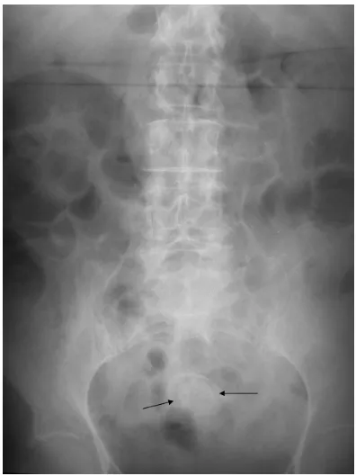
Although a plain abdominal radiograph demonstrated gas in the biliary tree, acute diverticulitis was thought a more likely clinical diagnosis (Fig. 1). A dynamic multislice enhanced CT scan of the abdomen and pelvis was performed. (Toshiba Asteion, enhancement with 100 ml of ioversol 300 at 3 ml/s). The scan confirmed gas within the biliary system and a laminated calculus impacted in a markedly thickened loop of the sigmoid colon (Fig. 2). A putative diagnosis of gall stone impaction in a segment of diverticular change was made. A water-soluble contrast enema confirmed complete obstruction at the site of calculus impaction (Fig. 3). The patient subsequently proceeded to laparotomy and underwent a Hartmann’s procedure. The resected
Fig. 1. Abdominal radiograph. Gas is shown in the biliary tract. There are dilated bowel loops and a radio-opaque stone is visible in the pelvis (arrows).
segment of sigmoid colon revealed an impacted gallstone proximal to a diverticular stricture. Recovery was uneventful.
Discussion
Gall stone ileus is a condition in which gall bladder calculi erode into the intestinal tract. The fistulous communication so created permits passage of intestinal gas into the biliary system, a characteristic feature sometimes seen on plain radiographs. Usually the calculi erode into the adjacent duodenum and migrate distally. The stones then impact – usually at the ileo-caecal valve – and cause a distal small bowel obstruction. The stone itself is sometimes demonstrated in one of the terminal ileal loops on plain abdominal films. A combination of these findings is diagnostic of a gallstone ileus[1,2].
Other sites for impaction include the duodeno-jejunal flexure or at any small bowel stricture[3].
If large enough, they may impact proximally and cause duodenal obstruction. (Bouveret’s syndrome). However, it is well recognised that calculi may rarely pass directly into the transverse colon. The larger calibre of the colon permits passage of the stone into the distal large bowel. The calculus is liable to impact if there is any distal colonic stricture. In this case the impacted endoluminal gallstone occurred at a diverticular stricture and incited an acute diverticulitis.
The primary CT findings in uncomplicated diverticular change include thickening of the wall of the involved segment of colon with visualisation of colonic diverticula[4,5]. In cases where there is a complicating acute inflammatory change, there may be more marked bowel wall thickening, stranding of the mesenteric fat and, in the most severe cases, peri-colic abscess formation. Other visualised complications include free perforation, colo-vesical fistula and ureteric obstruction[4,5]. Pericolonic inflammation and segment involvement greater than 10 cm are very suggestive of acute diverticulitis[6].
pericolonic soft tissue stranding include inflammatory bowel disease, pseudo membranous colitis and other infectious colitides. Other rarer causes include eosinophilic enteritis, lymphocytic colitis and lupus colitis[9].
Teaching points
MSCT enabled precise identification of the location of the calculus in the intestinal tract. Although a standard open laparotomy was pursued in this instance, the knowledge that the obstructing stone was in the distal colon could have led to consideration of calculus fragmentation via an endoscope and a lithotripter device – thus saving an open laparotomy.
A recent review of the use of CT scanning in the acute abdomen suggested that if used more liberally there may be benefits, including a shorter hospital stay and reduced patient morbidity and mortality[10]. This case reflects that opinion and highlights the potential for MSCT to establish
an accurate diagnosis even in unusual conditions such as that described.
References
1. Oikarinen H, Paivansalo M, Tikkakoski T, et al. Radiological findings in biliary fistula and gallstone ileus. Acta Radiol 1996; 37–917–22.
Fig. 3.Water-soluble contrast enema. The flow of contrast medium is obstructed at the site of impaction of the laminated stone (arrows). Associated sigmoid diverticular change is shown.
2. Seal EC, Creagh MF, Finch PJ. Gallstone ileus: a new role for abdominal computed tomography. Postgrad Med J 1995; 71: 313–15.
3. Reisner RM, Cohen JR. Gallstone ileus: a review of 1001 reported cases. Am Surg 1994; 60: 44–46.
4. Birnbaum BA, Balthazar EJ. CT of appendicitis and diverticulitis. Radiol Clin North Am 1994; 32: 885–98.
5. Doringer E. Computerized tomography of colonic diverticulitis. Crit Rev Diagn Imaging 1992; 33: 421–35.
6. Chintapalli KN, Chopra S, Ghiatas AA, Esola CC, Fields SF, Dodd GD. Diverticulitis versus colon cancer: differentiation with helical CT findings. Radiology 1999; 210: 429–35.
7. Cho KC, Morehouse HT, Alterman DD, Thornhill BA. Sigmoid diverticulitis: diagnostic role of CT – comparison with barium enema studies. Radiology 1990; 176: 111–15.
8. Smith TR, Cho KC, Morehouse HT, Kratka PS. Comparison of computed tomography and contrast enema evaluation of diverticulitis. Dis Colon Rectum 1990; 33: 1–6.
9. Turner DR, Markose G, Arends MJ, Ng C, Freeman AH. Unusual causes of colonic wall thickening on computed tomography. Clin Radiol 2003; 58: 191–200.