Poly (ε-caprolactone) Fiber: An Overview
Bahareh Azimi1, Parviz Nourpanah1, Mohammad Rabiee2, Shahram Arbab3
1
Amirkabir University of Technology, Textile Engineering Department, Tehran, IRAN
2
Amirkabir University of Technology, Biomedical Engineering Department, Tehran, IRAN
3
ATMT Research Institute, Faculty of Textile Engineering, Amirkabir University of Technology, Tehran, IRAN
Correspondence to:
Parviz Nourpanah email: [email protected]
ABSTRACT
Poly (ε-caprolactone), (PCL) or simply
polycaprolactone as it is usually referred to, is a synthetic biodegradable aliphatic polyester which has attracted considerable attention in recent years, notably in the biomedical areas of controlled-release drug delivery systems, absorbable surgical sutures, nerve guides, and three-dimensional (3-D) scaffolds, for use in tissue engineering. Various polymeric devices like microspheres, microcapsules, nanoparticles, pellets, implants, and films have been fabricated using this polymer. It can be transformed by spinning into filaments for subsequent fabrication of desirable textile structures. Spinning may be accomplished by various approaches. The fibers may be fabricated into various forms and can be used for implants and other surgical applications such as sutures. Although numerous studies have investigated different properties and applications of PCL, there is no comprehensive study investigating different fabrication methods of PCL fibers and their biomedical applications. The present article presents a review on the production of PCL fiber via various methods, along with correlations between structure and properties of the fibers. The applications of these fibers in biomedical domains are also discussed.
Keywords: PCL, Fiber, Spinning, Medical
application.
INTRODUCTION
In recent years, biodegradable polymers have attracted considerable attention as biomaterials in pharmaceutical, medical, and biomedical engineering applications, including drug delivery systems, artificial implants, and functional materials in tissue engineering. Aliphatic polyesters, due to their favorable features of biodegradability and biocompatibility, comprise one of the most important classes of synthetic biodegradable polymers. The advantage of these polyesters is their biocompatibility and higher hydrolysability in the human body [1].
Poly (ε-caprolactone) (PCL) is a family member of biodegradable aliphatic polyesters which have found important use as biomaterials in prosthetics, sutures, and drug delivery systems. As a commercial material, the main attractions of PCL are (1) its approval by the Food and Drug Administration (FDA) for use in humans, (2) its biodegradability, (3) its compatibility with a wide range of other polymers, (4) its good processibility which enables fabrication of a variety of structures and forms, (5) its ease of melt processing due to its high thermal stability and (6) its relatively low cost [2-3]. It can also be transformed by spinning into filaments for subsequent fabrication of desirable textile structures. Due to excellent characteristics, such as biodegradability, biocompatibility, mild undesirable host reactions, and three-dimensional and directional porous structures, PCL fiber, whose diameter range from nanometer to millimeter, is broadly studied. In fiber form, PCL and its copolymers have been investigated for usage in drug delivery systems [4], ‘long-lasting’ absorbable sutures [5-8] and, 3-D scaffolds for tissue engineering applications [9]. For example, to use in absorbable nerve guides, ε-caprolactone has been copolymerized with DL-lactide [10] and trimethylene carbonate [11]. PCL has received relatively comprehensive attention in the literature [3, 12-13], however, there are few studies investigating different fabrication methods and biomedical application of PCL fibers. The present article presents a review on the chemistry and different properties of PCL, production of PCL fiber by various methods and correlations between structure and properties of the fibers. The applications of these fibers in biological and medical domains are also discussed.
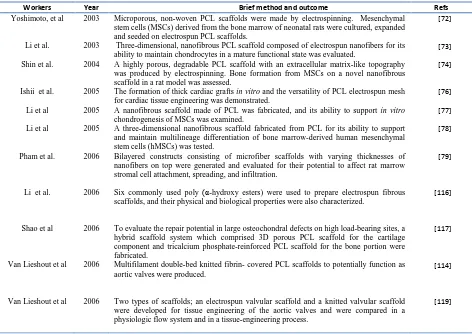
wide range of catalysts for the ring opening polymerization of caprolactone has been reviewed [14].
FIGURE 1. Ring opening polymerization of ε-caprolactone to polycaprolactone.
Catalysts such as stannous octoate are used to catalyze the polymerization and low molecular weight alcohols can be used to control the molecular weight of the polymer [15]. There are various mechanisms which affect the polymerization of PCL and these are anionic, cationic, co-ordination and radical. Each method affects the resulting molecular weight, molecular weight distribution, end group composition and chemical structure of the copolymers [16]. The number average molecular weight of PCL samples generally vary from 3,000 to 80,000 g/mol and can be graded according to the molecular weight [17].
It is a semi-crystalline polymer with a melting point of 59–64 οC and a glass-transition temperature of 60οC [18]. PCL is soluble in chloroform, dichloromethane, carbon tetrachloride, benzene, toluene, cyclohexanone and 2-nitropropane at room temperature. It has a low solubility in acetone, 2-butanone, ethyl acetate, dimethylformamide and acetonitrile and is insoluble in alcohol, petroleum ether and diethyl ether [12]. The versatility of PCL is due to the fact that, it allows modification of its physical, chemical and mechanical properties by co-polymerization or blending with many other polymers efficiently. It has been observed that co-polymerization alters the chemical property that indirectly affects all other properties such as crystallinity, solubility, and degradation pattern resulting in a modified polymer with intended properties for drug delivery [13]. Whereas, blending that leads to altered physical property and biodegradation along with highly influenced mechanical properties is preferred for formulations of tissue engineering such as scaffolds, fibers and films. A number of polymers have been studied for their compatibility to modify the thermal, rheological as well as biophysical properties of PCL, based on its application. PCL is reported to be compatible with natural polymers like starch, hydroxy apatite (HA), chitosan and synthetic polymers namely
poly ethylene glycol (PEG), poly urethane (PU), oxazolines, poly ethylene oxide (PEO), poly vinyl alcohol (PVA), polylactic acid and polylactic co-glycolic acid (PLGA) [19-26]. These PCL modifications satisfy the required biophysical properties for most of the formulation currently used in drug delivery [13].
Biodegradation
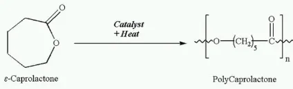
PCL is degraded by hydrolysis of its ester linkages in physiological conditions (such as in the human body) and has therefore received a great deal of attention in order to be used as an implantable biomaterial. In particular it is especially interesting for the preparation of long term implantable devices, due to its degradation which is even slower than that of polylactide. From degradation studies presented in the literature it can be concluded that PCL undergoes a two-stage degradation process: firstly the non-enzymatic hydrolytic cleavage of ester groups and secondly, when the polymer is more highly crystalline and has a low molecular weight (less than 3000) the polymer is shown to undergo intracellular degradation as this was observed during experiments of PCL fragments uptake in phagosomes of macrophages and giant cells and within fibroblasts [27]. This supports the theory that PCL may be completely resorbed and degraded via an intracellular mechanism once the molecular weight was reduced to 3000 or less. It was also noted that in the first stage the degradation rate of PCL is essentially identical to the in vitro hydrolysis at 40°C and obeyed first-order kinetics. It was concluded that the mechanism of PCL degradation could be attributed to random hydrolytic chain scission of the ester linkages, which caused a decrease in molecular weight. The homopolymer PCL has a total degradation of two to four years (depending of the starting molecular weight of the device or implant) [28-29]. The rate of hydrolysis can be altered by copolymerization with other lactones or glycolides/lactides. Surprisingly, more than 1000 papers have been published during the last decade in the biomaterials and tissue engineering literatures which use PCL-based-scaffolds. Among these studies only a small number of groups have included a study of the degradation and resorption kinetics of the PCL scaffolds [12]. Table I shows comprehensive data on PCL fiber degradation.
Spinning of PCL Fibers
3-D scaffolds for tissue engineering applications. Extrusion of the PCL into monofilament and multifilament may be achieved by fiber formation
mechanisms such as melt spinning, solution spinning, and electrospinning. There are distinct features of each of these processes that are subsequently reflected in fiber properties.
TABLE I. Comprehensive data on PCL fiber degradation.
Workers Year .... Brief method and outcome Refs
N. BÖLGEN et al. 2005 In vitro& in vivo In vitro and in vivo degradation studies of non-woven materials made of PCL nanofibers showed that electrospun PCL materials were degraded much faster in vivo as compared with in vitro due to the enzymatic degradation of PCL in addition to the hydrolytic degradation.
[30]
Lam. C.X.F. et al. 2007 In vivo Over 6 months, composite PCL/ b-Tri-calcium phosphate (TCP)
scaffolds degrade faster than PCL homopolymer scaffolds in vivo.
[31]
Pektok, E, et al. 2008 In vivo In vivo healing and degradation characteristics of small-diameter vascular grafts made of PCL nanofibers compared with expanded polytetrafluoroethylene (ePTFE) grafts were evaluated.
[32]
Lam et al. 2008 In vitro PCL and PCL/ TCP scaffolds degraded via a surface degradation
pathway in the alkaline accelerated setting; however, this appeared to switch to a bulk degradation pathway under the long term simulated condition.
[33]
Wan et al. 2008 In vitro Degradation of the PCL component with chitosan could be
accelerated at various rates depending on the compositions of the scaffolds and the media, and the chitosan component could effectively buffer the acidic degradation products of the PCL component.
[34]
Mobarakeh et al. 2008 In vitro By increasing gelatin content the biodegradability of PCL/ gelatin nanofibrous scaffolds increased in PBS over 2-week period.
[35]
Tillman, B.W. et al.
2009 In vivo PCL/collagen electrospun scaffolds maintain a high degree of
patency and structural integrity in vivo without eliciting abnormal inflammatory response over the course of 1 month.
[36]
Johnson et al. 2009 In vitro The net effects of biological and non-biological environments on PCL electrospun structures following 7 and 28 days of in vitro
exposure are established. Material degradation, as well as biological deposition, was responsible for the changes in mechanical properties.
[37]
Vieira et al. 2011 In vitro Hyper elastic constitutive models were used to predict the
mechanical behavior and biodegradation of a blend composed of PLA and PCL fiber.
[38]
Melt Spinning
Since PCL is thermoplastic in nature, it is possible to melt the polymer under reasonable conditions. In the melt spinning process polymer is melted, filtered, and extruded through the spinneret. The melt is drawn from the spinneret hole at a melt temperature. In the draw zone the extruded filaments are cooled to the solidification temperature and further to below the glass transition temperature. Finally, the filaments come to the take-up bobbins, and the temperature of the filaments are less than the Tg. Various research groups have studied the melt spinning of PCL fibers under various processing conditions [39-45].
Charuchinda et al. [2] studied some of the main factors affecting the small-scale melt spinning of PCL, monofilament fibers. These factors included spinning temperature, extrusion rate, take-up rate and draw ratio. The underlying influence of the polymer’s own characteristic properties, were also interpreted within the context of the melt spinning process. A
under the various processing conditions is given in
associated with changes in morphological parameters. In another study, lidocaine release from PCL suture threads produced by micro-extrusion was investigated to assess the reliability of the manufacturing process. The full and rapid release of the loaded drug demonstrates that the extrusion process does not alter the drug and that the loaded amount is embedded in an open structure porosity that allows it to be available for the release [45]. An et al. [46] have reported a novel technique, named microfiber melt drawing, to fabricate a bundle of three dimensionally aligned PCL microfibers of about 10 μm fiber diameters without using any organic solvent. Orifice diameter, temperature and take-up speed have shown significant effects on the linear density of fabricated microfibers. Each microfiber bundle has a stiffness of about 1382.5 N/tex and a maximum load of 30.7 N, which can be used as a building block for larger microfiber bundles. Mechanical properties of these microfiber bundles can thus be adjusted by the number of fibers or the number of bundles. In order to prepare monofilaments to be applied as threads for surgical sutures and possessing antimicrobial properties, Scaffaro et al. [47] have used an “online” method to combine the physicochemical and biological property of PCL and chlorhexidine (CHX), respectively. A piston pressed the molten polymer from a cylindrically shaped reservoir to the capillary. At the exit of the capillary, the filaments were drawn at constant speed and free cooling at room temperature. Under these conditions, the threads had a diameter of 300±10 μm.
Wet Spinning
The solution spinning methods, dry spinning and wet spinning, are usually utilized for polymers that do not melt. In both methods polymer is dissolved into a suitable solvent and the polymer solution is filtrated, dearated, and pumped through the spinneret [48]. In dry spinning, solvents are removed by thermal evaporation while in wet spinning the coagulation of the polymer is carried out in another fluid that is compatible with the spinning solvent. However, is not itself a solvent for the polymer [49]. In practice, as the polymer solution enters into the coagulation bath, phase separation begins due to solvent out-flow and nonsolvent in-flow and the polymer precipitates as fibrils [50-51]. Table III shows comprehensive data on wet spun PCL fiber [52-64].
Electrospinning
Electrospinning is another interesting technique for spinning PCL (and other polymers). In the electrospinning process, a polymer solution or melt is
subjected to strong electric fields, and then the liquid-phase polymer is ejected from a nozzle. The diameter of the ejected fibers is significantly reduced as they travel toward a collector. Figure 2 shows schematically an electrospinning system. The electrospinning process of the PCL has been widely studied. Table IV reviews some fabrication process parameters and fiber diameter of electrospun PCL fibers.
FIGURE 2. Schematic of electrospinning system [67].
Medical Application of PCL Fiber
Biodegradability, biocompatibility, pliability, good solubility, low melting point and exceptional blend-compatibility of PCL have stimulated extensive research into its potential application in the biomedical field [16, 68-69]. Following are the major applications of the PCL fibers in biomedical domain.
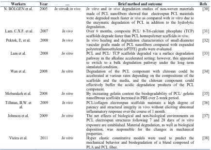
Suture
infection through the interstices of the braid structure. This block copolymer offers reduced stiffness compared to pure polyglycolide, which is being sold
as a monofilament suture by Ethicon, Inc. (Somerville, NJ), under the trade name Monacryl (scheme1)[105].
TABLE II. Summary of the effects of processing variables on the as-spun monofilament fiber diameter [2]. .
Spinning temperature
(οC)
Ram speed (mm min-1)
Extrusion rate (m min-1)
Take-up rate (m min-1)
On-line draw ratio
Fiber diameter (mm)
85 2 0.12 0.6 5 0.91
90 2 0.12 0.6 5 0.81
100 2 0.12 0.6 5 0.80
110 2 0.12 0.6 5 0.80
120 2 0.12 0.6 5 0.79
85 0.5 0.03 0.6 20 0.50
85 1 0.06 0.6 10 0.66
85 2 0.12 0.6 5 0.91
85 2 0.12 0.6 5 0.91
85 2 0.12 1 8.3 0.67
85 2 0.12 2 16.7 0.49
SCHEME 1. Synthesis of copolymer of e-caprolactone and glycolide [18].
Bezwada et al. showed that Monocryl sutures displayed excellent handling properties, minimal resistance during passage through tissue, and excellent tensile properties. Absorption data on these sutures indicate that absorption is complete between the 91st and 119th days of implantation, with slight or minimal tissue reaction [106].
Pharmaceutical
The drug delivery system was developed for the purpose of bringing, uptaking, retaining, releasing, activating, localizing and targeting the drugs at the right timing, period, dose and place. The biodegradable polymer can contribute largely to this technology by adding its own characters to the drugs. The history of biodegradable polymers in drug delivery systems dates back to 1970 when PLGA was used to control the release of narcotics. PCL is suitable for controlled drug delivery due to a high permeability to many drugs, excellent biocompatibility and its ability to be fully excreted from the body once bioresorbed. Biodegradation of PCL is slow in comparison to other polymers, so it is more suitable for long-term delivery, which extends over a period of more than one year. PCL also has the ability to form compatible blends with other polymers which can affect the degradation kinetics which can be in turn tailored to fulfill desired release profiles [107-108].
Several drug delivery vehicles composed of PCL, such as microspheres, microcapsules, nanospheres and micro and nanofibers have been developed for the controlled release of drugs or protein. The biodegradability of PCL fibers has inspired several studies on controlled drug delivery systems. Williamsona et al. [55] have incorporated a hydrophilic macromolecule (ovalbumin (OVA)) and a lipophilic drug (progesterone) in PCL fibers by gravity spinning using particulate dispersions and co-solutions of PCL and steroid, respectively. PCL
fibers loaded with 1% (w/w) OVA powder displayed a pronounced burst release phase (60% of the protein load) over 2 days in PBS at 37οC. The release profile then tended to plateau. In contrast, OVA nanoparticle-loaded fibers exhibited delayed protein release initially and then a major increase at day 14. The amount of progesterone release from PCL fibers in PBS increased with drug loading but the cumulative release profiles (% w/w) were little affected by the initial drug loading of the fibers or the concentration of the PCL spinning solution. Gravity spinning shows potential for producing PCL fibers-based platforms for programmed delivery of bioactive molecules of utility for tissue engineering and drug delivery. Chang et al. [56] have reported on the incorporation of gentamicin sulphate (GS) in gravity-spun PCL fibers to illustrate the potential for controlled, local delivery of antibiotics from wound closure materials, and tissue substitutes such as textile vascular grafts. The production rate of GS-loaded PCL fibers was confined to the range 1–1.5 m/min and the fiber diameter to 170–220 µm. The kinetics of drug release could be adjusted by varying the GS loading of the fibers and the suspension preparation conditions. Puppi et al. [61] optimized the wet-spinning conditions to obtain 3D *PCL scaffolds loaded with Enrofloxacin (EF) and Levofloxacin (LF) antibiotics. Most of the antimicrobial agent added to the polymer solution was found in the coagulation bath and the loading efficiency was in the range of 18%–27% depending on the type of antibiotic and its concentration. Both the EF-loaded and LF-loaded meshes, after a fast release at the early stages, provided sustained release for up to five weeks (Figure 3).
TABLE IV. Fabrication process parameters: polymer solution properties, electrospinning process parameters.
Luong-Van et al. [80] incorporated heparin into electrospun PCL fiber mats for assessment as a controlled delivery device. The fiber diameter was found to be dependent on the concentration of heparin added, while increasing heparin concentration lead to a decreased fiber diameter, which can be attributed to an increased ionic charge of the spinning solution (Figure 4). A sustained release of heparin could be achieved from the fibers over 14 days with the release diffusion controlled during this time. Merrell et al. [94] have investigated the feasibility and potential of PCL nanofibers as a delivery vehicle for curcumin for wound healing applications. The fibers showed sustained release of curcumin for 72 h and could be made to deliver a dose much lower than the reported cytotoxic concentration while remaining bioactive. The in vivo
wound healing capability of the curcumin loaded PCL nanofibers was demonstrated by an increased rate of wound closure in a streptozotocin-induced diabeticmicemodel. In another study, nanofiber PCL scaffolds were loaded with two concentrations of rifampicin (RIF), and the RIF release kinetics and bactericidal efficacies of the scaffolds were evaluated compared to RIF-free control scaffolds.
There were significant differences between the RIF release profiles, though both scaffolds showed an initial burst release, and RIF release was completed after 8 hours. Approximately, 50% of the loaded RIF remained entrapped within the scaffolds [104]. Kanawung et al. [109] used electrospinning to fabricate ultrafine fiber mats from PCL and PCL solution that contained diclofenac sodium (DS) as the model drug. The effects of solution and process parameters (i.e., solution concentration, applied electrical potential, and collection distance) on morphological appearance and size of the as-spun PCL were investigated. Incorporation of the model drugs caused the resulting as-spun fibers to be larger in their diameters. As shown in Figure 5, the cumulative release of the model drug from drug-loaded as-spun PCL fiber mats increased monotonically with increasing immersion it became practically constant at long immersion times.
FIGURE 5. Cumulative amount of DS released from DS-loaded PCL fiber mats that were electrospun from PCL solutions loaded with 10, 30 and 50 mg of DS over an immersion period of 465 min [109].
In another attempt, Tammaro et al. [110] have prepared new fibrous composites by using electrospinning technique, obtained by fixing an anti-inflammatory drug, diclofenac sodium, into a lamellar inorganic compound, and incorporating the obtained nanohybrid into a biodegradable PCL. The structure, morphology, and thermal behavior of the electrospun fibers were analyzed. The release of the active diclofenac molecules was found much slower in comparison to the release of the fibers in which the drug was directly incorporated into the polymer. Liu et al. [111] produced PCL electrospun fibers containing ampicillin sodium salt and twisted them into nanofiber yarns. The fiber diameters and crystallinity, the in vitro antimicrobial properties of the yarns, and the in vitro release of ampicillin from yarns containing various ampicillin concentrations were studied. Figure 6 shows the smooth surface of a fiber.
FIGURE 6. Representative SEM image showing the smooth surface of a fiber (The scale bar represents 500 nm.) [111].
Tissue Engineering
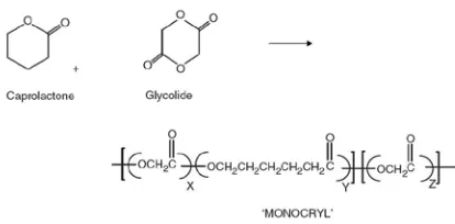
different classes of biodegradable polymers, PCL is suitable for scaffold fabrication. PCL is an incredibly versatile bioresorbable polymer and by way of its superior rheological properties it can be used by almost any polymer processing technology to produce an enormous array of scaffolds. A number of fabrication technologies have been applied to process PCL into 3D polymeric scaffolds of high porosity and surface area. Cell-invasive fiber-based scaffolds can be produced using methodologies developed for the textile industry, but with structures specifically designed for tissue engineering applications. Tissue engineering textiles have a relatively high surface area and their ‘value added’ application, from an industry with established techniques, is an advantage. Additionally, textiles are typically formed into thin meshes and therefore the permeability is high, allowing the necessary nutrients to reach the seeded cells. Traditional fabrication of non-woven textiles is based on the production of continuous micron diameter; fibers by extruding a polymer melt or polymer solution through a spinneret which is then mechanically drawn onto a winder, or a series of winders, and collected onto a spool. The fiber of the
diameter is determined by the extrusion rate and the speed(s) of the winder(s) with a constant drawing rate paramount for attaining uniform diameter, continuous fibers [12]. Van Lieshout et al produced multifilament double-bed knitted, fibrin- covered PCL scaffolds to potentially function as aortic valves. On testing, it demonstrated good durability, proper opening and it showed coaptation upon closing, but had higher associated leakage compared to those of tested porcine valves [114]. Electrospinning is of great interest as a scaffold fabrication technique, since the resulting fiber diameters are in the size range (submicron to nanometer) of the extracellular matrix (ECM) microstructures, particularly the higher-ordered collagen microfibrils [115]. The flexibility of the electrospun fibers, due to the very high aspect ratio (length/diameter), is also beneficial, allowing seeded cells to remodel their surroundings. A plethora of research papers have focused on specific applications of PCL scaffolds in various tissue engineering applications. Table V reviews some studies, investigated the fabrication of PCL scaffold in various tissue engineering applications.
TABLE V. Fabrication of PCL scaffold in various tissue engineering applications.
Workers Year Brief method and outcome Refs
Yoshimoto, et al 2003 Microporous, non-woven PCL scaffolds were made by electrospinning. Mesenchymal stem cells (MSCs) derived from the bone marrow of neonatal rats were cultured, expanded and seeded on electrospun PCL scaffolds.
[72]
Li et al. 2003 Three-dimensional, nanofibrous PCL scaffold composed of electrospun nanofibers for its ability to maintain chondrocytes in a mature functional state was evaluated.
[73]
Shin et al. 2004 A highly porous, degradable PCL scaffold with an extracellular matrix-like topography was produced by electrospinning. Bone formation from MSCs on a novel nanofibrous scaffold in a rat model was assessed.
[74]
Ishii et al. 2005 The formation of thick cardiac grafts in vitro and the versatility of PCL electrospun mesh for cardiac tissue engineering was demonstrated.
[76]
Li et al 2005 A nanofibrous scaffold made of PCL was fabricated, and its ability to support in vitro
chondrogenesis of MSCs was examined.
[77]
Li et al 2005 A three-dimensional nanofibrous scaffold fabricated from PCL for its ability to support and maintain multilineage differentiation of bone marrow-derived human mesenchymal stem cells (hMSCs) was tested.
[78]
Pham et al. 2006 Bilayered constructs consisting of microfiber scaffolds with varying thicknesses of nanofibers on top were generated and evaluated for their potential to affect rat marrow stromal cell attachment, spreading, and infiltration.
[79]
Li et al. 2006 Six commonly used poly (α-hydroxy esters) were used to prepare electrospun fibrous scaffolds, and their physical and biological properties were also characterized.
[116]
Shao et al 2006 To evaluate the repair potential in large osteochondral defects on high load-bearing sites, a hybrid scaffold system which comprised 3D porous PCL scaffold for the cartilage component and tricalcium phosphate-reinforced PCL scaffold for the bone portion were fabricated.
[117]
Van Lieshout et al 2006 Multifilament double-bed knitted fibrin- covered PCL scaffolds to potentially function as aortic valves were produced.
[114]
Van Lieshout et al 2006 Two types of scaffolds; an electrospun valvular scaffold and a knitted valvular scaffold were developed for tissue engineering of the aortic valves and were compared in a physiologic flow system and in a tissue-engineering process.
Li, et al 2007 The fabrication of biodegradable nanofibrous scaffolds composed of aligned fibers via electrospinning onto a rotating target was reported and their mechanical anisotropy as a function of the production parameters was characterized.
[81]
Oh et al. 2007 PCL cylindrical scaffolds with gradually increasing pore size along the longitudinal direction were fabricated by a novel centrifugation method to investigate pore size effect on cell and tissue interactions.
[115]
Pektok et al. 2008 The degradation and healing characteristics of small diameter PCL vascular grafts, produced via electrospinning, in the rat systemic arterial circulation was investigated.
[32]
Choi et al. 2008 PCL/collagen nanofibers of different orientations, to engineer functional muscle tissue for restoring large skeletal muscle tissue defects were produced.
[121]
Guarino et al. 2008 Scaffolds comprising PLLA fibers embedded in a porous PCL matrix were obtained by synergistic use of phase inversion/particulate leaching technique and filament winding technology.
[122]
Bregy et al. 2008 The use of fused diode laser soldering of vascular tissue using PCL scaffolds doped with bovine serum albumin (BSA) and Indocyanine green (ICG) was investigated.
[123]
Chen et al. 2009 A vacuum seeding technique on PCL electrospun scaffolds was used to prevent congregate and proliferate of cells on the surface of scaffold.
[91]
Nisbet, et al. 2009 The extent of microglial and astrocytic response was measured following implantation of electrospun PCL scaffolds into the caudate putamen of the adult rat brain.
[92]
Wise et al 2009 Electrospun and oriented PCL scaffolds were created, and hMSCs were cultured on these scaffolds. Cell viability, morphology, and orientation on the fibrous scaffolds were quantitatively determined as a function of time.
[93]
Li et al 2009 Cell-seeded nanofibrous PCL scaffolds for cartilage repair using 7 mm full-thickness cartilage defects in a swine model was evaluated. Biodegradable nanofibrous scaffolds seeded with MSCs could effectively repair cartilage defects in vivo, and that this approach is promising for cartilage repair.
[89]
Balguid et al. 2009 Electrospun sheets comprising PCL with different fiber diameters (3-12 μm) were investigated for penetration depth using human venous myofibroblastsas as a means to optimize cell delivery during cardiovascular tissue engineering applications.
[125]
Ruckh et al. 2010 Nanofiber PCL scaffolds were fabricated by electrospinning, and their ability to enhance the osteoblastic behavior of MSCs in osteogenic media was investigated.
[96]
Wu et al. 2010 A novel electrospinning technique was demonstrated to fabricate small diameter 3-D nanofibrous tubular scaffold with controllable nanofiber orientations so as to regulate the macroscopic mechanical property of the scaffolds and benefit cell responses along different directions for vascular grafts applications.
[97]
Cao et al. 2010 The biocompatibility with regard to scaffold architecture and topographical effect of PCL nanofibrous scaffolds on the in vivo and in vitro foreign body reaction was investigated.
[99]
Puppi et al. 2010 Three-dimensional electrospun microfibrous meshes of (*PCL) as potential scaffolds for tissue engineering applications was developed. Cell culture experiments employing MC3T3-E1 osteoblast like cells showed good cell viability adhesion and collagen production on the *PCL scaffolds.
[100]
Jha, et al 2011 The structural and functional properties of three-dimensional (3D) nerve guides fabricated from PCL using the air gap electrospinning process was describe.
[101]
Puppi et al. 2011 The cytocompatibility of the wet-spun *PCL and the influence of the scaffold architecture on cell behavior were performed with pre-osteoblast cells.
[61]
Puppi et al. 2012 Three-dimensional polymeric scaffolds, based on wet-spinning of PCL and PCL/ hydroxyapatite (HA) solutions, was developed. The developed scaffolds showed good reproducibility of the internal architecture characterized by highly porous, aligned fibers with an average diameter in the range 200–250 μm.
[64]
Ruckh et al. 2012 PCL nanofiber scaffolds were fabricated to include both 10 or 20% (w/w) rifampicin (RIF), and the RIF release kinetics and bactericidal efficacies of the scaffolds were evaluated compared to RIF-free control scaffolds.
[104]
Diban et al. 2013 Hallow fibers were successfully manufactured from blends of PCL/PLGA by phase separation. These have adequate elongation characteristics to be applied in small-caliber blood vessel regeneration. The PCL/PLGA85/15 ratio yielded a miscible blend after processing, whereas higher PLGA contents in the blend led to separation of the polymer phases.
CONCLUSION
Featured with excellent characteristics, such as biodegradability, biocompatibility, mild undesirable host reactions, three-dimensional and directional porous structures, PCL fiber is broadly studied and used in different biomaterials. Therefore investigating of production of PCL fiber by various methods can be of much importance. This article presented a review on the production of PCL fiber by various methods including melt spinning, solution spinning and electrospinning. Correlations between structure and properties of the fibers and applications of them in biomedical domains such as sutures, drug loaded fibers and scaffold for use in tissue engineering were also discussed.
REFRENCES
[1] Bikiaris D N, Papageorgiou G Z, Achilias D S, Pavlidou E and Stergiou A. "Miscibility and enzymatic degradation studies of poly(e-caprolactone)/poly(propylene
succinate) blends". European Polymer Journal, Vol 43, 2007, pp. 2491–2503. [2] Charuchinda A, Molly R, Siripitayananon J,
Molloy N and Sriyai M. "Factors influencing the small-scale melt spinning of poly(ε-caprolactone) monofilament fibres".
Polymer International, Vol 52, pp. 1175– 1181.
[3] Cipitria A, Skelton A, Dargaville T R, Dalton P D and Hutmacher D W. "Design, fabrication and characterization of PCL electrospun scaffolds—a review". Journal of Materials Chemistry, Vol 21, 2011, pp. 9419–9453.
[4] Goodson J M, Holborow D, Dunn R L, Hogan P and Dunham L. "Monolithic tetracycline containing fibers for controlled delivery to periodontal pockets". J Periodontol, Vol 54, 1983, pp. 575-579.
[5] Barber F A, Click J N. "The Effect of Inflammatory Synivial Fluid on the Breaking Strength of New Long Lasting Sutures". J. Arthrosc. Relat. Surg, Vol 8,
1992, pp. 437–441.
[6] Nakamura T, Shimizu Y, Matsui T, Okumura N, Hyon S H and Nishiya K. "A novel bioabsorbable monofilament surgical suture made from (caprolacton , L Lactide) copolymer", in Degradation Phenomena on Polymeric Biomaterials, Springer: Berlin, 1992, pp. 153-162.
[7] Tomihata K, Suzuki M, Oka T, Ikada Y, "A new resorbable monofilament suture".
Polymer Degrad. Stability, Vol 59, 1998. pp. 13-18.
[8] Bezwada R S, Jamiolkowski D D, Lee I Y, Agarwal V, Persivale J, Erneta M, Trenka-Benthin S, Suryadevara J, Yang A and Liu S. "Monocryl suture, a new ultra-pliable absorbable monofilament suture".
Biomaterials, Vol 16, 1995, pp. 1141-1148. [9] Hutmacher D W, Schantz T, Zein I, Ng K W, Teoh S W and Tan K C, "Mechanical properties and cell cultural response of polycaprolactone scaffolds designed and fabricated via fused deposition modeling".
J Biomed Mater Res, Vol 55, 2001, pp. 203-216.
[10] Meek M F, d.D.W., Bartels H L, Pennings A J, Robinson and P.a.S. JM, "Peripheral nerve regeneration and functional nerve recovery after reconstruction with a thin-walled biodegradable poly(DL-lactide-e -caprolactone) nerve guide". Cells Mater, Val 7, 2007, pp. 53-62.
[11] Pego A P, Poot A A., Grijpma D W and Feijen J. "Copolymers of trimethylene carbonate and epsilon-caprolactone for porous nerve guides: Synthesis and properties". J Biomater Sci Polym Ed, Vol 12, 2001, pp. 35-53.
[12] Woodruff M A, Hutmacher DW. "The return of a forgotten polymer – Polycaprolactone in the 21st century". Prog. Polym. Sci, Vol 35, 2010, pp. 1217–1256.
[13] Dash T K, KonkimallaK V B. "Poly-є-caprolactone based formulations for drug delivery and tissue engineering: A review."
Journal of Controlled Release, Vol 158, 2012, pp. 15-33.
[14] Labet M and Thielemans W "Synthesis of polycaprolactone: a review". Chemical Society Reviews, Vol 38, 2009, pp. 3484– 3504.
[15] Storey R F, Taylor A. "Effect of stannous octoate concentration on the ethylene glycolinitiated polymerization of epsilon-caprolactone". Abstr Pap Am Chem Soc, Vol 211, 1996, pp. 114.
[16] Okada M. "Chemical syntheses of
biodegradable polymers". Progress in Polymer Science, Vol 27, 2002, pp. 87-133. [17] Hayash T. "Biodegradable polymers for
biomedical uses. Progress in Polymer Science, Vol 19, 1994, pp. 663-702. [18] Pillai, Chennakkattu K S and Sharma C P.
[19] Liu C B, Gong C Y, Huang M J, Wang J W, Pan Y F, Zhang Y D, Li G Z, Gou M L, Tu M J, Wang K, Wei Y Q and Qian Z Y. "Thermoreversible gel–sol behavior of biodegradable pcl-peg-pcl triblock copolymer in aqueous solutions". J. Biomed. Mater. Res. B Appl. Biomater. Vol 84, 2008, pp. 165-175.
[20] Kalambur S, Rizvi S S H. "Rheological behavior of starch–polycaprolactone (pcl) nanocomposite melts synthesized by reactive extrusion". Polym. Eng. Sci, Vol 46, 2006, pp. 650–658.
[21] Wan Y, Lu X, Dalai S and Zhang J, "Thermophysical properties of
polycaprolactone/ chitosan blend
membranes", Thermochim. Acta, Vol 487, 2009, pp. 33-38.
[22] Ma Z, Haddadi A, Molavi O, Lavasanifar A, Lai R and Samuel J. "Micelles of poly(- ethylene oxide)-b-poly(epsilon– caprolactone) as vehicles for the solubilization, stabilization, and controlled delivery of curcumin". J. Biomed. Mater. Res. A, Vol 86, 2008, pp. 300-310.
[23] Sheikh F A, Barakat N, Kanjwal M A, Aryal S, Khil M S, Kim H Y. "Novel selfassembled amphiphilic poly(epsilon– caprolactone)-grafted-poly(vinyl alcohol) nanoparticles: hydrophobic and hydrophilic drugs carrier nanoparticles". J. Mater. Sci. Mater. Med, Vol 20, 2009, pp. 821–831. [24] Chen C, Cai G, Zhang H, Jiang H and Wang
L. "Chitosan-poly(epsilon–caprolactone)- poly(ethylene glycol) graft copolymers: synthesis, self-assembly, and drug release Behavior". J. Biomed. Mater. Res. A, Vol 96, 2010, pp. 116–124.
[25] K. Gorna, Gogolewski, S. "In vitro degradation of novel medical biodegradable aliphatic polyurethanes based on epsilon–caprolactone and pluronics with various hydrophilicities".
Polym. Degrad. Stab, Vol 75, 2002, pp. 113-122.
[26] Pulkkinena M, Malin M, Böhmd J, Tarvainen T, Wirth T, Seppälä J, Järvinen K. "In vivo implantation of 2,2'-bis(oxazoline)-linked poly-epsilon–caprolactone: proof for enzyme sensitive surface erosion and biocompatibility". Eur. J. Pharm. Sci. Vol 36, 2009, pp. 310-319.
[27] Woodward C S, "The intracellular degradation of poly (epsilon-caprolactone". Journal of biomedical materials research, Vol 19, 1985, pp. 437-44.
[28] Gunatillake P A, Adhikari A. "Biodegradable synthetic polymers for tissue engineering".
European Cells and Materials, Vol 5, 2003, pp. 1-16.
[29] Middleton J C and Tipton A J. "Synthetic biodegradable polymers as orthopedic devices". Biomaterials, Vol 21, 2000, pp. 2335-46.
[30] N. BÖLGEN1, Y.Z.M.L., K. ACATAY2, I˙. VARGEL 3 and and E. PI ¸SKIN 1, In vitro and in vivo degradation of non-woven materials made of poly(ε-caprolactone) nanofibers prepared by electrospinning under different conditions. J. Biomater. Sci. Polymer Edn,, 2005. 16: p. 1537–1555. [31] Lam C X F, Hutmacher D W, Schantz J,
Woodruff M A and Teoh S H. "Evaluation of polycaprolactone scaffold degradation for 6 months in vitro and in vivo". Journal of Biomedical Materials Research Part A, 2008, pp. 906-919.
[32] Pektok E, Nottelet B, Jea, Tille J C, Gurny R, Kalangos A, Walpoth B H. "Degradation and Healing Characteristics of Small-Diameter Poly(-Caprolactone) Vascular Grafts in the Rat Systemic Arterial Circulation". Circulation. Vol 118, 2008, pp. 2563-2570.
[33] Lam C X F, Savalani M M, Teoh S H and Hutmacher D W. "Dynamics of in vitro polymer degradation of polycaprolactone-based scaffolds: accelerated versus simulated physiological conditions".
Biomed. Mater, Vol 3, 2008, pp. 1-15. [34] Wan, Y., Ying W, Hua C, Xiaoying D and
Siqin. "Compressive mechanical properties and biodegradability of porous poly(caprolactone)/chitosan scaffolds".
Polymer Degradation and Stability, Vol 93, 2008, pp. 1736-1741.
[35] Ghasemi-Mobarakeh L, Prabhakaran M P, Morshed M, Nasr-Esfahani M H, Ramakrishna, Se. "Electrospun poly (ε-caprolactone)/gelatin nanofibrous scaffolds for nerve tissue engineering". Biomaterials, Vol 29, 2008, pp. 4532-4539.
[36] Tillman B W, Yazdani S K, Lee S J, Geary R L, Yoo J J and Atala A. "The in vivo stability of electrospun
polycaprolactone-collagen scaffolds in vascular
[37] Johnson J, Niehaus A, Nichols S, Lee D, Koepsel J, Anderson D and Lannutti J, "Electrospun PCL in vitro: a microstructural basis for mechanical property changes". J. Biomater. Sci. Polymer Edn, Vol 20, 2009, pp. 467–481. [38] Vieiraa A C, Marquesb A T, Guedesb R M
and Titac V. "Material model proposal for biodegradable materials". Procedia Engineering, Vol 10, 2011, pp. 1597–1602. [39] Krishnanand K, Deopura B L and Gupta B.
"Determination of intrinsic birefringence values of polycaprolactone filaments".
Polym Int, Vol 62, 2013, pp. 49–53. [40] Meng Q, Hu J. "Study on poly
(ε-caprolactone)-based shape memory copolymer fiber prepared by bulk polymerization and melt spinning". Polymers for Advanced Technologies, Vol 19, 2008, 131-136.
[41] Mochizuki M, Havashi T, Nakatama K and MASUDA T. "Studies on biodegradable poly (hexane-6-lactone) fibers. Part 2: environmental degradation". Pure Appl. Chem, Vol 71, 1999, pp. 2177- 2188. [42] Mochizuki M, Nakajima K, Qian R, Jiang B Z,
Hirami M, Hayashi T, Masuda T and Nakajima A. "Studies on biodegradable poly(hexano-6-lactone) fibers. Part 1. Structure and properties of drawn PCL fibers". Pure Appl. Chem, Vol 69, 1997, pp. 2567-2575.
[43] Charuchinda A, Molloy R, Siripitayananon J, Molloy N and Sriyai M "Poly (-caprolactone) melt-spun monofilament fibers". Polymer International, Vol 52,
2003, pp. 1175-1181.
[44] Douglasa p, Andrewsb G, Jonesb D and Walkera G. "Analysis of in vitro drug dissolution from PCL melt extrusion".
Chemical Engineering Journal, Vol 164, 2010, pp. 359–370.
[45] Perale G, Casalini T, Barri V, Mu¨ ller M, Maccagnan S and Masi M. "Lidocaine Release from Polycaprolactone Threads". Journal of Applied Polymer Science, Vol 117, 2010, pp. 3610-3614.
[46] An J, Chua C K, Leong K F, Chen C H and Chen J P. "Solvent-free fabrication of three dimensionally aligned polycaprolactone microfibers for engineering of anisotropic tissues". Biomed Microdevices, Vol 14, 2012, pp. 863–872.
[47] Puglia, R.S.L.B.M.S.G.G.G.P.A.M.,
"Combining in the melt physical and biological properties of poly(caprolactone) and chlorhexidine to obtain antimicrobial surgical monofilaments". Appl Microbiol Biotechnol, Vol 97, 2013, pp. 99-109. [48] Fourne, F. "Synthetic Fibers – Machines and
Equipment, Manufacture, Properties". ohio: hanser/gardner. 1999, 930.
[49] Arbab S, Noorpanah P, Mohammadi N,
Soleimani M. "Designing index of void structure and tensile properties in wet-spun polyacrylonitrile (PAN) fiber. I. Effect of dope polymer or nonsolvent concentration". Journal of Applied Polymer Science, Vol 109, 2008, pp. 3461-3469. [50] Arbab S, Mohammadi N, Noorpanah P. "The
synergistic effect of dope concentration and jet-drawing on structure development of wet-spun poly (acrylonitrile)". epolymer, 2008, 80, pp. 1-11.
[51] Arbab S, Noorpanah P, Mohammadi N, Zeinolebadi A. "Simultaneous effects of polymer concentration, jet-stretching, and hot-drawing on microstructural development of wet-spun poly(acrylonitrile) fibers". Polymer Bulletin, Vol 66, 2011, pp. 1267-1280. [52] Polacco, G., et al., Biodegradable hollow
fibres containing drug-loaded nanoparticles as controlled release systems. Polymer International, 2002. 51(12): p. 1464-1472. [53] Williamson M R, Adams E F, Coombes A G
A. "Cell attachment and proliferation on novel polycaprolactone fibers having application in soft tissue engineering".
European Cells and Materials, Vol 4, 2002, pp. 62-63.
[54] Williamson M R, Coombes A G A. "Gravity spinning of polycaprolactone fibres for applications in tissue engineering".
Biomaterials, Vol 25, 2004, pp. 459–465. [55] Williamson M R, Changb H I, Coombes A G
A. "Gravity spun polycaprolactone fibres: controlling release of a hydrophilic macromolecule (ovalbumin) and a lipophilic drug (progesterone)".
Biomaterials, Vol 25, 2004, pp. 5053– 5060.
[57] Chiono V, Ciardelli G, Vozzi G, Sotgiu M G, Vinci B, Domenici C, Giusti P. "Poly(3-hydroxybutyrate-co-3-hydroxyvalerate)/ poly(e-caprolactone) blends for tissue engineering applications in the form of hollow fibers". Journal of Biomedical Materials Research Part A, Vol 85, 2008, PP. 938-953.
[58] Tuzlakoglu K, Pashkuleva I, Rodrigues M T, Gomes M E, Van Lenthe G H, Mu¨ ller R, Reis R L. "A new route to produce starch-based fiber mesh scaffolds by wet spinning and subsequent surface modification as a way to improve cell attachment and proliferation". Journal of Biomedical Materials Research Part A, 2008: p. 369-377.
[59] Puppi P, Piras A M, Chiellini F, Chiellini E, Martins A, Leonor I B, Neves N and Reis R. "Optimized electro- and wet-spinning techniques for the production of polymeric
fibrous scaffolds loaded with
bisphosphonate and hydroxyapatite". Journal of tissue engineering and regenerative medicine, Vol 5, 2011, pp. 253–263.
[60] Yilgor p, Sousa R A, Reis A L, Hasirci N and Hasirci V. "Effect of scaffold architecture and BMP-2/BMP-7 delivery on in vitro bone regeneration". J Mater Sci: Mater Med, Vol 21, 2010, pp. 2999-3008. [61] Puppi D, Dinucci D, Bartoli C, Mota C,
Migone C, Dini F, Barsotti G, Carlucci F and Chiellini F. "Development of 3D wet-spun polymeric scaffolds loaded with antimicrobial agents for bone engineering" . Journal of Bioactive and Compatible Polymers, Vol 26, 2011, pp. 478–492. [62] [62]. Wang Z, Wu H, Liao C, Zhou N, Cheng
W, Wan Y. "Sustained release of ketoprofen from fibrous chitosan-poly(ε-caprolactone) membranes". Carbohydrate Polymers, Vol 84, 2011, pp. 624-630. [63] Neves S C, Teixeira L S M, Moroni L, Reis R
L, Blitterswijk C A V, Alves N M, Karperien M, Mano J F. "Chitosan/Poly(3-caprolactone) blend scaffolds for cartilage repair". Biomaterials, Vol 32, 2011, pp. 1068-1079.
[64] Puppi D, Mota C, Gazzarri M, Dinucci D, Gloria A, Myrzabekova M, Ambrosio L and Chiellini F. "Additive manufacturing of wet-spun polymeric scaffolds for bone tissue engineering". Biomed Microdevices, Vol 14, 2012, pp. 1115–1127.
[65] Williamson M R, Adams E F, Coombes A G A. "Gravity spun polycaprolactone fibres for soft tissue engineering: Interaction with fibroblasts and myoblasts in cell culture".
Biomaterials, Vol 27, 2006, pp. 1019-1026. [66] Shen X X, Zheng Y, Ma Z H, Zhu L M,
Branford-White C. "Wet-Spinning Medicated PAN/PCL Fibers for Drug Sustained Release" Bioinformatics and Biomedical, Vol 13, 2008, 1375 - 1378 [67] Gupta B, Revagade N and Hilborn J. "Poly
(lactic acid) fiber: An overview". Progress in Polymer Science, Vol 32, 2007, pp. 455-482.
[68] Chandra R, Rustgi R. "Biodegradable polymers. Progress in Polymer Science", Vol 23, 1998, 1273-335.
[69] Nair L S and Laurencin C T. "Biodegradable polymers as biomaterials". Progress in Polymer Science, Vol 32, 2007, pp. 762-798.
[70] Frazza E J, Schmitt E E. "A new absorbable suture". J Biomed Mater Res Symposium, Vol 1, 1971, pp. 43-58.
[71] Leea K H, Kimb H Y, Khilb M S, Rab Y M, Lee D R, "Characterization of nano-structured poly(1-caprolactone) nonwoven mats via electrospinning". Polymer, Vol 44, 2003, pp. 1287–1294.
[72] H. Yoshimoto, Y M Shina Y M, Teraia H, Vacanti J P. "A biodegradable nanofiber scaffold by electrospinning and its potential for bone tissue engineering". Biomaterials, Vol 24, 2003, pp. 2077–2082.
[73] Li W J, Danielson K J, Alexander P G, Tuan R S, "Biological response of chondrocytes cultured in threedimensional nanofibrous poly(-caprolactone) scaffolds". Journal of Biomedical Materials Research Part A. Vol 67, 2003, pp. 1105-14.
[74] Shin M, Yoshimoto H and Vacanti J P. "In vivo bone tissue engineering using mesenchymal stem cells on a novel electrospun nanofibrous scaffold". Tissue Eng Part A. Vol 10, 2004, pp. 33–41. [75] Shin M, Ishii O, Sueda T, Vacanti J P.
"Contractile cardiac grafts using a novel nanofibrous mesh". Biomaterials, Vol 25, 2004, pp. 3717–3723.
[77] Li W J, Tuli R, Okafor C, Derfoul A, Danielson K G, Hall D J, Tuan R S. "A three-dimensional nanofibrous scaffold for cartilage tissue engineering using human mesenchymal stem cells". Biomaterials, Vol 26, 2005, pp. 599–609.
[78] Lia W J, Tuli R, Huang X, Laquerriere P, Tuan R S. "Multilineage differentiation of human mesenchymal stem cells in a three-dimensional nanofibrous scaffold".
Biomaterials, Vol 26, 2005, PP. 5158– 5166.
[79] Pham Q M, Sharma U and Mikos A G.
"Electrospun poly (ε-caprolactone)
microfiber and multilayer
nanofiber/microfiber scaffolds:
characterization of scaffolds and measurement of cellular infiltration".
Biomacromolecules, Vol 7, 2006, pp. 2796–2805.
[80] Luong-Vana E, Grøndahla L, Chuad K N, Leongd K W and Nurcombeb V, Cool S M. "Controlled release of heparin from poly(e-caprolactone) electrospun fibers".
Biomaterials, 2006, 27: p. 2042–2050. [81] Li W J, Mauck R L, Cooper J A, Yuan
X, Tuan R S. "Engineering controllable anisotropy in electrospun biodegradable nanofibrous scaffolds for musculoskeletal tissue engineering". J. Biomech, Vol 40, 2007, pp. 1686–1693.
[82] Kolambkar Y M. "Electrospun nanofiber meshes for the repair of large bone defects", in Department of Biomedical Engineering, Georgia Institute of Technology, 2007.
[83] M. P. Prabhakaran, e.a., "Surface modified electrospun nanofibrous scaffolds for nerve tissue engineering". Nanotechnology, Vol 19, 2008, pp. 455102.
[84] Wong S C, Baji A, Leng S. "Effect of fiber diameter on tensile properties of electrospun poly (3-caprolactone)".
Polymer, Vol 49, 2008, pp. 4713–4722. [85] Pektok E, Nottelet B, Tille J C, Gurny R,
Kalangos A, Moeller M, Walpoth B H. "Degradation and healing characteristics of smalldiameter poly (3-caprolactone) vascular grafts in the rat systemic arterial circulation". Circulation, Vol 118, 2008, pp. 2563–2570.
[86] Nisbet D R, Yu L M Y, Zahir T, Forsythe J S, Shoichet M S. "Characterization of neural stem cells on electrospun poly (3-caprolactone) submicron scaffolds: evaluating their potential in neural tissue engineering". J. Biomater. Sci. Polym. Ed, Vol 19, 2008, pp. 623–634.
[87] Nottelet B, Pektok E, Mandracchia D, Tille J C, Walpoth B, Gurny R, Möller M. "Factorial design optimization and in vivo feasibility of poly (3-caprolactone)-micro-
and nanofiber-based small diameter
vascular grafts". J. Biomed. Mater. Res., Part A. Vol 89,2009, pp. 865–875.
[88] Martins A, Pinho E D, Faria S, Pashkuleva I, Marques A P, Reis R L, Neves N M. "Surface modification of electrospun polycaprolactone nanofiber meshes by plasma treatment to enhance biological performance". Small, Vol 5, 2009, pp. 1195–1206.
[89] Li W J, Chiang H, Kuo T F, Lee H S, Jiang C C, Tuan R S. "Evaluation of articular cartilage repair using biodegradable nanofibrous scaffolds in a swine model: a pilot study". J. Tissue Eng. Regener. Med, Vol 2, 2009, p. 1–10.
[90] Pişkin E, Işoğlu I A, Bölgen N, Vargel I, Griffiths S, Cavuşoğlu T, Korkusuz P, Güzel E, Cartmell S. "In vivo performance of simvastatin-loaded electrospun spiral-wound polycaprolactone scaffolds in reconstruction of cranial bone defects in the rat model". J. Biomed. Mater. Res., Part A, Vol 90, 2009, pp. 1137–1151.
[91] Chen M, Michaud H, Bhowmick S.
"Controlled vacuum seeding as a means of generating uniform cellular distribution in electrospun polycaprolactone (PCL) scaffolds". J. Biomech. Eng, Vol 131, 2009, pp. 074521.
[92] Nisbet D R, Rodda A E, Horne M K, Forsythe J S, Finkelstein D I. "Neurite infiltration and cellular response to electrospun polycaprolactone scaffolds implanted into the brain". Biomaterials. Vol 30, 2009, pp. 4573–4580.
[93] Wise J K, Yarin A L, Megaridis C M, Cho M, "Chondrogenic Differentiation of Human Mesenchymal Stem Cells on Oriented Nanofibrous Scaffolds: Engineering the Superficial Zone of Articular Cartilage".
[94] Merrel J G, McLaughlin S W, Tie L, Laurencin C T, Chen A F and Naird L S. "Curcumin Loaded Poly(ε-Caprolactone) Nanofibers: Diabetic Wound Dressing with Antioxidant and Anti-inflammatory Properties". Clinical and Experimental Pharmacology and Physiology, Vol 36, 2009, pp. 1149–1156.
[95] Lowery J L, Datta N, Rutledge G C. "Effect of fiber diameter, pore size and seeding method on growth of human dermal fibroblasts in electrospun poly (3-caprolactone) fibrous mats". Biomaterials, Vol 31, 2010, pp. 491–504.
[96] Ruckh T T, Kumar K, Kipper M J, Popat KC. "Osteogenic differentiation of bone marrow stromal cells on poly (3-caprolactone) nanofiber scaffolds". Acta Biomaterials, Vol 6, 2010, pp. 2949–2959.
[97] Wu H, Fan J, Chu C C, Wu J. "Electrospinning of small diameter 3-D nanofibrous tubular scaffolds with controllable nanofiber orientations for vascular grafts". J. Mater. Sci.: Mater. Med. Vol 21, 2010, pp. 3207– 3215.
[98] Zhu Y, Cao Y, Pan J, Liu Y. "Macro-alignment of electrospun fibers for vascular tissue engineering". J. Biomed. Mater. Res., Part B. Vol 92, 2010, pp. 508–516.
[99] Cao H, McHugh K, Chew S Y, Anderson J M. "The topographical effect of electrospun nanofibrous scaffolds on the in vivo and in vitro foreign body reaction". J. Biomed. Mater. Res., Part A, Vol 93, 2010, pp. 1151–1159.
[100] Puppi D, Detta N, Piras A M, Chiellini F, Clarke D A, Reilly G C, Chiellini E. "Development of Electrospun Three-arm Star Poly(ε-caprolactone) Meshes for Tissue Engineering Applications".
Macromol. Biosci. Vol 10, 2010, pp. 887– 897.
[101] Jha B S, Colello R J, Bowman J R, Sell S A, Lee K D, Bigbee J W, Bowlin G L, Chow W N, Mathern B E, Simpson D G. "Two pole air gap electrospinning: fabrication of highly aligned, three-dimensional scaffolds for nerve reconstruction". Acta Biomaterialia, Vol 7, 2011, pp. 203–215.
[102] Khanging A G, and Bahrami S H. "Solvents on Poly (ε-Caprolactone) Nanofibrous Webs Morphology". Journal of Nanomaterials, Vol 2011, pp. 1-10.
[103] Kolbuk D, Sajkiewicz P, Kowalewski T A. "Optical birefringence and molecular orientation of electrospun polycaprolactone fibers by polarizing-interference microscopy". European Polymer Journal, Vol 48, 2012, pp. 275–283.
[104] Ruckh T T, Oldinski R A, Carroll D A, Mikhova K, Bryers J D, Popat K C. "Antimicrobial effects of nanofiber poly (caprolactone) tissue scaffolds releasing rifampicin". J Mater Sci: Mater Med, Vol 23, 2012, pp. 1411-1420.
[105] Middleton J C, Tipton A J. "Synthetic biodegradable polymers as orthopedic devices". Biomaterials, Vol 21, 2000, pp. 2335-46.
[106] Bezwada R S, Jamiolkowski D D, Lee I Y, Agarwal V, Persivale J, Trenka-Benthin S, Erneta M, Suryadevara J, Yang A, Liu S. "Monocryl Suture, A New Ultra-Pliable Absorbable Monofilament Suture".
Biomaterials. Vol 16, 1995, pp. 1141– 1148.
[107] Merkli A, Tabatabay C, Gurny R, Heller J, "Biodegradable polymers for the controlled release of ocular drugs". Progress in Polymer Science, Vol 23, 1998, pp. 563-80. [108] Sinha V R, Bansal K, Kaushik R, Kumria R,
Trehan A, "Poly-epsilon-caprolactone microspheres and nanospheres: an overview". Int J Pharm., Vol 278, 2004, pp. 1-23.
[109] Kanawung K, K. Panitchanapan, Puangmalee S, Utok W, Kreua-Ongarjnukool N, Rangkupan R and Supaphol P. "Preparation and characterization of polycaprolactone/diclofenac sodium and
poly(vinyl alcohol)/tetracycline hydrochloride fiber mats and their release
of the model drugs". Polym. J. Vol 39, 2007, pp. 369–378.
[110] Tammaro L, Russo G and Vittoria V. "Encapsulation of DiclofenacMolecules into Poly (ε-Caprolactone) Electrospun Fibers for Delivery Protection". Journal of Nanomaterials, 2009.
[111] Liu H, Leonas K K, Zhao Y. "Antimicrobial Properties and Release Profile of Ampicillin from Electrospun Poly (ε-caprolactone) Nanofiber Yarns". Journal of Engineered Fibers and Fabrics, Vol 5,
2010, pp. 10-19.
[112] Langer R, Vacanti J P."Tissue engineering".
[113] Hutmacher D W. "Scaffolds in tissue engineering bone and cartilage".
Biomaterials, Vol 21, 2000, pp. 2529-43. [114] Van Lieshout M, Peters G, Rutten M, Baaijens
F. "A knitted, fibrin-covered polycaprolactone scaffold for tissue engineering of the aortic valve". Tissue Eng. Vol 12, 2006, pp. 481-7.
[115] Choi J S, Lee S G, Christ G J, Atala A, Yoo J J. "The influence of electrospun aligned poly (epsilon-caprolactone)/collagen nanofiber meshes on the formation of self-aligned skeletal muscle myotubes".
Biomaterials, Vol 29, 2008, pp. 2899-906. [116] Li W J, Cooper JA Jr, Mauck R L, Tuan R S.
"Fabrication and characterization of six electrospun poly (alpha-hydroxy ester)-based fibrous scaffolds for tissue engineering applications". Acta Biomaterialia, Vol 2, 2006, pp. 377-85. [117] Shao X, Goh J C, Hutmacher D W, Lee E
H, Zigang G., "Repair of large articular osteochondral defects using hybrid scaffolds and bone marrow-derived mesenchymal stem cells in a rabbit model".
Tissue Eng Part A, Vol 12, 2006, pp. 1539-51.
[118] Van Lieshout M I, Vaz C M, Rutten M C, Peters G W, Baaijens F P. "Electrospinning versus knitting: two scaffolds for tissue engineering of the aortic valve". Journal of Biomaterials Science-Polymer Edition, Vol 17, 2006, pp. 77-89.
[119] Oh SH, Park I K, Kim J M, Lee J H. "In vitro and in vivo characteristics of PCL scaffolds with pore size gradient fabricated by a centrifugation method". Biomaterials. Vol 28, 2007, pp. 1664-71.
[120] Guarino V, Causa F, Taddei P, di Foggia M, Ciapetti G, Martini D, Fagnano C, Baldini N, Ambrosio L. "Polylactic acid fibre-reinforced polycaprolactone scaffolds for bone tissue engineering". Biomaterials. Vol 29, 2008, pp. 3662-70.
[121] Bregy A, Bogni S, Bernau V J, Vajtai I, Vollbach F, Petri-Fink A, Constantinescu M, Hofmann H, Frenz M, Reinert M. "Solder Doped Polycaprolactone Scaffold Enables Reproducible Laser Tissue Soldering". Lasers in Surgery and Medicine, Vol 40, 2008, pp. 716-25.
[122] Balguid A, Mol A, van Marion M H, Bank R A, Bouten C V, Baaijens F P. "Tailoring Fiber Diameter in Electrospun Poly (epsilon-Caprolactone) Scaffolds for
Optimal Cellular Infiltration in
Cardiovascular Tissue Engineering". Tissue Engineering Part A. Vol 15, 2009, pp. 437-44.
[123] Diban N, Haimi S,Bolhuis-Versteeg L, Teixeira S, Miettinen S, Poot A, Grijpma D, Stamatialis D. "Hollow fibers of poly(lactide-co-glycolide) and poly(e-caprolactone) blends for vascular tissue engineering applications". Acta Biomaterialia, Vol 9, 2013, 6450–6458.
AUTHORS’ ADDRESSES Bahareh Azimi
Parviz Nourpanah Mohammad Rabiee Shahram Arbab
Textile Engineering Department Amirkabir University of Technology 424 Hafez Ave
![FIGURE 2. Schematic of electrospinning system [67].](https://thumb-us.123doks.com/thumbv2/123dok_us/8390403.1681910/4.612.352.507.190.277/figure-schematic-electrospinning.webp)
![TABLE II. Summary of the effects of processing variables on the as-spun monofilament fiber diameter [2]](https://thumb-us.123doks.com/thumbv2/123dok_us/8390403.1681910/5.612.90.521.135.267/table-summary-effects-processing-variables-monofilament-fiber-diameter.webp)
![FIGURE 5. Cumulative amount of DS released from DS-loaded PCL fiber mats that were electrospun from PCL solutions loaded with 10, 30 and 50 mg of DS over an immersion period of 465 min [109]](https://thumb-us.123doks.com/thumbv2/123dok_us/8390403.1681910/8.612.352.512.71.200/figure-cumulative-released-loaded-electrospun-solutions-loaded-immersion.webp)