IN VITRO
ANTI-INFLAMMATORY ACTIVITY IN ETHANOLIC EXTRACT OF
SPIRULINA PLANTENSIS AND PHYTOCHEMICAL ANALYSIS UNDERNEATH
GC-MS/MS
Punitha P.* and Bharathi V.
Assistant Professor, PG and Research Department of Biochemistry, Sengamala Thayaar Educational Trust Women’s College, Sundarakkottai, Mannargudi.
Article Received on 19/10/2017 Article Revised on 10/11/2017 Article Accepted on 01/12/2017
INTRODUCTION
Inflammation is a complex process, which is frequently associated with pain and involves occurrences such as: the increase of vascular permeability, increase of protein denaturation and membrane alteration. When cells in the body are damaged by microbes, physical agents or chemical agents, the injury is in the form stress. Inflammation of tissue is due to response to stress. It is a defensive response that is characterized by redness, pain, heat, and swelling and loss of function in the injured area. Loss of function occurs depends on the site and extent of injury. Since inflammation is one of the body’s nonspecific internal systems of defense, the response of a tissue to an accidental cut is similar to the response that results from other types of tissue damage, caused by burns due to heat, radiation, bacterial or viral invasion.[1]
Spirulina plantensis are multicellular and filamentous blue-green algae that has gained considerable popularity in the health food industry and increasingly as a protein and vitamin supplement to aquaculture diets. It grows in water, can be harvested and processed easily and has very high macro and micronutrient contents. It has long been used as a dietary supplement by people living close to the alkaline lakes where it is naturally found, for instance those living adjacent to Lake Chad in the Kanem region have very low levels of malnutrition. This traditional food, known as dihe, was re-discovered in Chad by a European scientific mission and is now widely cultured throughout the world.
In 1967 Spirulina plantensis was established as a “wonderful future food source” in the International Association of Applied Microbiology. Analysis of the nutritional properties of Spirulina plantensis showed first and foremost an exceptionally high protein content, in the order of 60–70 percent of its dry weight; it also showed the excellent quality of its proteins have balanced essential amino acid content. This first data was enough to launch many research projects for industrial purposes in the 1970s, because microorganisms (yeast, Chlorella, Spirulina plantensis, some bacteria and moulds) seemed at that time to be the most direct route to inexpensive proteins the iconic “single cell proteins”.[2]
Taxonomy and morphological characterization of Spirulina plantensis
Spirulina plantensis is a multicellular filamentous cyanobacterium. It belongs to Phylum: Cyanophyta, Family: Oscillatoriaceae.[3] Spirulina plantensis is a ubiquitous organism, which can be found in a variety of environments such as soil, sand, marshes, brackish water, seawater and fresh water. Spirulina plantensis appeared as blue green filaments composed of cylindrical cells arranged in unbranched, helicoidal trichomes under a microscopic observation. The filaments are motile, gliding along their axis and heterocysts are absent.[4]
The helical shape of trichome is characteristic of the genus but the helical parameters (i.e., pitch length and helix dimensions) vary with the species and even within the same species. However, the helical shape is
ISSN 2455-3301
WJPMR
AND MEDICAL RESEARCH
www.wjpmr.com
*Corresponding Author: Punitha P.
Assistant Professor, PG and Research Department of Biochemistry, Sengamala Thayaar Educational Trust Women’s College, Sundarakkottai, Mannargudi.
ABSTRACT
The present study was conducted to evaluate anti-inflammatory of Spirulina. The effect was studied by using invitro models such as inhibition of Albumin (Protein) activity, Inhibition of protease denaturation, Membrane stabilization activity. The activity was studied by taking various concentrations of test compound. This extract achieving respectively high degree of anti-inflammatory potential present study conducted that Spirulina plantensis showed a significant anti-inflammatory potential. The GC-MS/MS analysis reveals the presence of Nonadecane, 9-Eicosyne.Which has the nature of working as an anti-inflammatory agent.
maintained only in liquid media, in solid media the filaments become true spirals.[5] The cytoplasm of the smaller species appears homogenous and scarcely visible septa. The large species such as S. platensis and S. maxima have a granular cytoplasm containing gas vacuoles and easily visible septa. The inter-thylakoid space is limited by the presence of electronically transparent protein gas vesicles, with the cylindrical form that gave floating capacity.[6] The most prominent cytoplasmic structure is the system of thylakoids originating from the plasmalemma.[7]
Processing of Spirulinaplantensis
In some countries such as in Chad, the Spirulina plantensis is harvested from several lakes by the Kanembu people. The Spirulina plantensis is filtrated and dried directly on the sandy shores or tissue support. Spirulina plantensis obtained in this manner and mainly used to prepare a kind of fish or meat and vegetable broth.[8]
The different drying methods selected to assess the best technology to dry Spirulina plantensis likewise oven drying, infrared drying, spray and freeze-drying. Protein and sugar content were determined before and after drying. Freeze-drying showed the highest retention of the analyzable proteins and sugars. Thin layers of biomass spreading out gave better results compared to cylinders. The dried end-product is characterized by microscopy that allowed showing damage because of the air drying temperature.[9]
Applications of Spirulinaplantensis
Cyanobacteria such as Spirulina plantensis, Anabaena, Nostoc and Oscillatoria produce a great variety of secondary metabolites. They produce a wide variety of bioactive compounds, which include 40% lipopeptides, 5.6% amino acids, 4.2% fatty acids, 4.2% macrolides and 9% amides. Cyanobacterial lipopeptides include different functions like cytotoxic (41%), antitumor (13%), antiviral (4%), antibiotics (12%) and the remaining 18% activities include antimalarial, antimycotics, multidrug resistance reversers, antifeedant, herbicides and immunosuppressive agents, besides the immune effect, blue green algae improves metabolism.[10]
The blue-green algae, Spirulina platensis has been used for hundreds of years as a food source for humans and animals due to the excellent nutritional profile and high carotenoid content. Spirulina plantensis is relatively high
in protein with values ranging from 55-65% and includes all of the essential amino acids.[11] The available energy has been determined to be 2.5-3.29 kcal/g and phosphorous availability is 41%.[12] Spirulina plantensis alga is rich in thiamin, riboflavin, pyridoxine, vitamin B12, vitamin C and antioxidant carotenoids and has been used throughout the world as a feed component in quality broiler and layer diets to enhance yolk colour and flesh.[13] The use of carotenoids as pigments is well documented and an extensive body of literature stresses the vital role of carotenoid physiology and concluded that carotenoids are essential nutrients that are required in all animal diets.[14]
MATERIALS AND METHODS Collection of Algae Materials
Spirulina capsules were collected from Hemeria Medical centre, Pattukkottai, Thanjavur district, Tamilnadu, India.
Preparation of Algal Extract
The algae powder 100g was taken and mixed with 70% ethanol and 30% water.The algal extracts were prepared by soaked in ethanol for about 3 days. After the duration period, the spirulina plantensis was filtrated by means of Whatmann filter paper. The ethanol was kept in a boiling water bath to evaporate ethanol. The ethanol free extract was used for the preliminary phytochemical analysis and in vitro anti-inflammatory activity, GC-MS/MS.
Preliminary Phytochemical Screening
Preliminary phytohemical screening of the extracts were carried out according to the standard procedures.
In vitro anti-inflammatory efficacy
Where O. D is optical density Results were compared with acetyl salicylic acid/Aspirin (250µg/ml) treated samples.
b) Inhibition of proteinase denaturation[16]
The reaction mixture (0.5ml) consisted of 450µl 5% aqueous bovine serum albumin and 50µl of aqueous extract of seeds of Spirulina (1.92-1000µg/ml), pH was adjusted at 6.3 using 1N Hcl. The samples were incubated at 37˚C for 20 min followed by heating at 57˚C for 3min.This mixture was then brought to room temperature and 2.5ml phosphate buffer saline (pH 6.3) was added to each tube. Turbidity formed was measured at λ =660 nm, 50µl distilled water was used in place of extracts for control test. The percentage inhibition of protein denaturation was calculated as follows:
The control represents 100% protein denaturation. Results were compared with acetyl salicylic acid (250µg/ml) treated samples.
c) Membrane stabilization activity[17]
The blood was collected from healthy human volunteers who have not taken any NSAIDS for 2 weeks prior to the experiment and mixed with equal volume of Alsever’s solution (2%) dextrose, 0.8% Sodium citrate ,0.5% Citric acid and 0.42% Nacl) and centrifuged at 3000 rpm. The packed cells were washed with isosaline and a 10% suspension was made. Various concentrations of extracts were prepared (250,500&1000mcg/ml) using distilled water and to each concentration 1ml of phosphate buffer, 2ml hyposaline and 0.5ml of HRBC suspension were added. It was incubated at 37˚C for 30 min & centrifuged at 3000rpm for 20min.
The haemoglobin control of the supernatant solution was estimated spectrophotometrically at 560nm. Diclofenac (50mcg/ml) was used as reference standard and a control was prepared omitting the extracts. The percentage inhibition of lysis was calculated.
Gas Chromatograpy-Mass spectrometry Analysis For quantitation (area%), the GCMS/MS analysis were carried out by using JEOL JMS-700 by the electron impact method where an electronic accelerating voltage of 75eV and an ion accelerating voltage of 8-10kV.The reservoir inlet systems were used. The capillary columns were: nonpolar column DB-5MS (J&W Scientific; 30 m x 0.25 mm, film thickness 0.25µm) and polar column TC-Wax (60 m x 0.25 mm, film thickness 0.25µm).The dynamic range for the peak intensities was 3 digits, and the accuracy of the mass number was 0.5.The oven temperature was programmed from 40˚-240˚C at a rate of 4˚C/min and held at 240˚C for 5 min. The injector and detector temperatures were 240˚C and 280˚C.The flow rates of the carrier gas (He) were 1.8mL/min. GLC data reported are given as area percentage. He at 49.9 Kpa was used as carrier gas and the FID detector was maintained at 250˚C.The oil constituents were identified on the basis of their retention data and by using GC/MS analytical conditions similar to that of GC/FID. The mass
spectra were recorded on a mass spectrometer coupled to a JEOL JMS-700 gas chromatograph (EI mode 70 eV, source temperature 230˚C, scanned mass ranged 35 - 350 amu).The characteristic fragmentation patterns have been analyzed and compared to those of Wiley 275.L database.
Identification of compounds
RESULTS AND DISCUSSION
In many inflammatory disorders there is excessive activation of phagocytes, production of O2•, •OH radicals as well as non‐free radical species (H2O2).[18] which can harm surrounding tissue either by powerful direct oxidizing action or indirectly with hydrogen peroxide (H2O2) and •OH radicals formed from O2, which initiates lipid peroxidation resulting in membrane destruction. Tissue damage then provokes inflammatory response by production of mediators and chemotactic factors.[19] The reactive oxygen species are also known to activate matrix metalloproteinase (e. g. collagenase) causing increased destruction of tissues e.g. collagenase damage seen in various arthritic reactions.[20]
The following anti-inflammatory activity parameters which are investigated in the current project are enclosed herewith.
The in-vitro anti-inflammatory activity of ethanolic extract of Spirulina plantensis were assay by different invitro models, Proteinase inhibitory activity, Inhibition of albumin denaturation , Membrane stabilization, GC-MS/MS.
Table 1 shows the Qualitative analysis of phytoconstituents of Spirulina plantensis the result revealed that plant extract showed presence of protein. Table 1 shows the Qualitative analysis of phytoconstituents of Spirulina plantensis the result revealed that plant extract showed presence of protein.
Table 2 shows the results of inhibition of protease activity of ethanolic extract of Spirulina plantensis at various concentrations. The percentage of in-vitro anti-inflammatory activity found to be maximum for maximum concentration of the ethanolic extract in a dose dependent manner.
Table 3 shows the results of protease denaturation activity of ethanolic extract of Spirulina plantensis at various concentrations. The percentage of in-vitro anti-inflammatory activity is also found to be dose dependent. As the concentration of the extract increases the anti-inflammatory activity also increases.
Table 4 depicts the results of inhibition of hemolysis activity of the ethanolic extract of Spirulina plantensis at various concentrations. The percentage of RBC membrane stabilization is found to be dose dependent. The anti-inflammatory activity is found to be maximum for the highest concentration of the extract.
Table 5 depicts the results of compound identification in the Spirulina sample of GC-MS/MS analysis.
Figure 1 shows the photo documentation of Spirulina plantensis diagram.
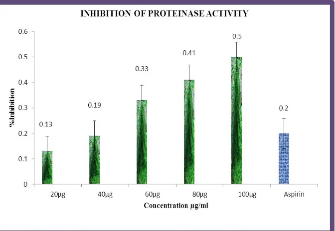
Figure 2 shows the in-vitro anti-inflammatory activity of inhibition of protease activity.
Figure 3 shows the in-vitro anti-inflammatory activity of protease denaturation activity.
Figure 4 shows the in-vitro anti-inflammatory activity of RBC membrane stabilization.
Figure 5 shows the ethanolic extract of GC-MS/MS analysis of Spirulina.
Table 1: Effect of Preliminay Phytochemical Analysis on anti-inflammatory activity of Spirulina plantensis.
S. No. Constituents Results
1 Carbohydrate +
2 Tannins +
3 Saponins + 4 Flavonoids + 5 Alkaloids + 6 Quinones + 7 Terpenoids +
8 Phenols +
9 Steroids and phytosteroids + + = Presence
Table: 2 Effect of Inhibition of proteinase activty on invitro anti-inflammatory Activity of Spirulina plantensis.
Inhibition of Proteinase Activity S.
No.
Concentration (µg)
Inhibition (%) (Mean ±SD) 1 20 0.13±0.098 2 40 0.19±0.050 3 60 0.33±0.102 4 80 0.41±0.061 5 100 0.50±0.083 6 Aspirin(50) 0.20±0.059 Values are expressed as Mean ± SD
Figure 2: Effect of Inhibition of protease activty on invitro anti-inflammatory activity of Spirulina plantensis.
Table 3: Effect of Proteinase denaturatsion on invitro anti-inflammatory activity of Spirulina plantensis. b. Proteinase Denaturation
S. No. Concentration (µg)
Inhibition (%) (Mean±SD) 1 25 0.26±0.020 2 50 0.34±0.052 3 75 0.39±0.054 4 100 0.51±0.020 5 125 0.67±0.028 6 Aspirin(50) 0.30±0.050
Values are expressed as Mean ± SD Students ‘t’ test followed and *P<0.05.
Table 4: Effect of Membrane stabilization on invitro anti-inflammatory activity of Spirulina plantensis. c. Membrane Stabilization
S. No. Concentration (µg) Inhibition (%) (Mean ± SD) 1 50 0.30±0.040 2 100 0.31±0.070 3 150 0.37±0.041 4 200 0.45±0.049 5 250 0.50±0.025 6 Aspirin(50) 0.30±0.065 Values are expressed as Mean ± SD
Students ‘t’ test followed and *P<0.05
Figure 4: Effect of Membrane stabilization on invitro anti-inflammatory activity of Spirulina plantensis.
Table 5: Effect of GCMS/MS analysis on invitro anti-inflammatory activity of Spirulina plantensis. Compounds identified in the Spirulina plantensis sample (Code No. 564).
No. RT Name of the compound Molecular
Formulae
Molecular Weight
Peak Area% 1 9.12 Edulan II C13H20O 192 0.53 2 9.33 Hydroxylamine, O-decyl- C10H23NO 173 0.96
3 9.87 2(4H)-Benzofuranone,
5,6,7,7a-tetrahydro-4,4,7a-trimethyl-, (R)- C11H16O2 180 1.64
4 10.64 Dodecane, 2,6,10-trimethyl- C15H32 212 1.77 5 11.58 4-Trifluoroacetoxypentadecane C17H31F3O2 324 0.69
6 11.86 Nonadecane C19H40 268 41.23
7 13.41 3-Hexadecyne C16H30 222 2.10
8 13.94 9,12-Octadecadienoyl chloride, (Z,Z)- C18H31ClO 298 0.63 9 14.51 Pentadecanoic acid, 14-methyl-, methyl ester C17H34O2 270 1.14 10 15.39 Hexadecanoic acid, ethyl ester C18H36O2 284 3.03 11 16.50 5,8,11,14-Eicosatetraenoic acid, methyl ester, (all-Z)- C21H34O2 318 0.63
12 16.75 9,12-Octadecadienoic acid (Z,Z)-, 2-hydroxy-1- (hydroxymethyl)ethyl ester
C21H38O4 354 1.40
13 17.02 9-Eicosyne C20H38 278 38.68
14 17.69 Linoleic acid ethyl ester C20H36O2 308 0.66
15 20.29 9,12,15-Octadecatrienoic acid, 2,3-dihydroxypropyl ester, (Z,Z,Z)-
C21H36O4
352 0.12
16 20.56 6,9,12,15-Docosatetraenoic acid, methyl ester C23H38O2 346 0.11 17 22.85 Dasycarpidan-1-methanol, acetate (ester) C20H26N2O2 326 2.07 18 24.72 Ursodeoxycholic acid C24H40O4 392 0.35
19 25.53 9,10-Secocholesta-5,7,10(19)-triene-3,24,25-triol, (3β,5Z,7E)- C27H44O3 416 1.19
20 31.71 α-Tocopheryl acetate C31H52O3 472 0.96
21 35.67 Stigmasta-5,22-dien-3-ol, acetate, (3β)- C31H50O2 454 0.13 Chromatogram Plot File: c:\brukerws\data\feb 2017\03.02.2017\564.xms
DISCUSSION
Proteinase inhibitory activity
Proteinases have been implicated in arthritic reactions. Neutrophis are known to be a rich source of proteinase which carries in their lysosomal granules many serine proteinases. It was previously reported that leukocytes proteinase play an important role in the development of tissue damage during inflammatory reactions and significant level of protection was provided by proteinase inhibitors Spirulina ethanolic extract exhibited significant antiproteinase activity at different concentrations. It showed maximum inhibition 86.7±0.098% at 20µg/ml. Aspirin showed the maximum inhibition 60.5±0.059% at 50µg/ml.[21]
Inhibition of albumin denaturation
Denaturation of proteins is a well-documented cause of inflammation. Phenylbutazone, salicylic acid, flufenamic acid (anti-inflammatory drugs) etc, have shown dose dependent ability to thermally induced protein protein denaturation. As a part of the investigation on the mechanism of the anti-inflammatory activity, ability of extract to inhibit protein denaturation was studied. It was effective in inhibiting heat induced albumin denaturation at different concentrations as shown in Table 3.Maximum inhibition, 92.8±0.052% was observed at 50µg/ml. Aspirin, a standard anti-inflammatory drug
showed the maximum inhibition, 64.2±0.050% at the concentration of 50µg/ml.[22]
Membrane stabilization test
GC-MS/MS Analysis
This study revealed a high level of chemical composition characteristic of ethanolic extracted from Spirulina plantensis and analysis by GC-MS and isolated compounds Nonadecane, 9-Eicosyne. From GC MS data, identification of more compounds in their extract and it reported that these compounds has anti-inflammatory activity.[24]
SUMMARY AND CONCLUSION
Spirulina plantensis is highly nutritious and shows great diversity and higher concentrations of nutrients compared to other food sources. In fact, it is among the most nutritious, concentrated whole food sources found in nature, contributing to its being known as a superfood. In this review, we have examined several areas of research showing the potential of Spirulina plantensis as a food supplement. Research on this uni-cellular, blue-green microalgae began in the 1970’s, and has increased in the last ten years. We have examined a series of published studies, most of which were published since the year 2000. Many studies investigated benefits from pure Spirulina plantensis biomass, but some also researched extracts of Spirulina plantensis or isolated compounds from Spirulina plantensis (primarily Nonadecane, 9-Eicosyne found only in Spirulina plantensis and other species of blue-green microalgae). From this review it may be concluded that:
Spirulina plantensis shows in the present study preliminary phytochemicals reveals the presence of Carbohydrate, Tannins, Saponin, Flavonoids, Alkaloids, Quinones, Terpenoids, Phenols,Steroids and phytosteroids.
Spirulina plantensis shows anti-inflammatory activity studies on inhibition of Albumin (protein) activity, inhibition of protease denaturation, Membrane stabilization activity.
From shows GC-MS/MS results it shows that the plant has Nonadecane, 9-Eicosyne compounds were identified in spirulina plantensis.
The alga is also generally considered safe for human consumption on basis of its long historical use.
ACKNOWLEDGEMENT
The Authors are thankful to The Correspondent Sir, Dr. V. Dhivaharan, S.T.E.T. Women’s College, Mannargudi for the Support for the Successful completion of the Project Work.
REFERENCES
1. Gerard J Tortora, Sandra Reynolds, eds. Principles of Anatomy and Physiology. Harper Collins College Publishers, 1993; 7: 695.
2. Sasson, A., Micro Biotechnologies: Recent Developments and Prospects for Developing Countries. BIOTEC Publication, 1997; 1/2542: 11–31.
3. Ciferri, O. Spirulina, the edible organism. Microbiol, 1983; 47: 551-578.
4. Van eykelenburg, C., FUCHS, A. and SCHMIDT, G. H. Shape Biol. Some theoretical considerations on the in vitro of the cross walls in Spirulina spp. J-.-Theor, 1980; 271-282.
5. Tomaselli L. Morphology, ultrastructure and taxonomy, In: Spirulina platensis (Arthrospira): Physiology, Cell-Biology and Biotechnology, 1997, 1-15.
6. Hedenskog G and Hofsten AV,The ultrastrure of Spirulina platensis- a new source of microbial protein. Physiol Plant, 1970; 23:209-216.
7. Gatugel A, Barsanti L and Tredici M. R., Harvest of Arthrospira Platensis from Lake Kossorom (Chad) and its household usage among the Kanembu, Journal of applied Phycology, 2000; 12(3-5): 493-498.
8. Desmorieux, H., Hernandez, F., Biochemical and physical criteria of Spirulina after different drying processes. In: Proceedings of the 14th International Drying Symposium (IDS), B, 2004; 900–907. 9. Burja, A. M., Banaigs,E. B., Abou-Mansour, Marine
cyanobacteria- a profile source of natural products. Tetrahedron, 2001; 57: 9347-9377.
10. Anusuya, D.M.; Subbulakshimi, G.; Madhavi Devi, K.; and Venkataram L. V., Studies on the proteins of mass-cultivated, blue-green alga (Spirulina platensis). 1,981J. Agric. Food Chem., 29: 522-525. 11. Ross, E. and Dominy, W. The nutritional value of
dehydrated, blue-green algae (Spirulina platensis) for poultry. Poultry Science, 1990; 69: 794-800. 12. Ross, E. and Dominy, W. The effect of dehydrated
Spirulina platensis on poultry. Poultry Sci., 1985; 64(S.1): 173.
13. Chatterjee. S Dass. S. N, Anti- arthritic and anti-inflammatory effect of a poly herbal drug. Indian J Pharmacol, 1996; 28: 116-119.
14. Umapathy E, Ndebia EJ, Meeme A, Adam B, Menziura P, Nkeh-Chungag BN and Iputo JE,An experimental evaluation of Albuca setosa aqueous extract on membrane stabilization, protein denaturation and white blood cell migration during acute inflammation. Journal of Medicinal Plant Research, 2010; 4(5): 789-795.
15. Gandhidasan R, Thamaraichelvan A, Baburaj S. Anti-inflammatory action of Lannea coromandelica by HRBC membrane stabilization. Fitoterapia 199; LXII (1): 81-83.
16. Davies W. Gas chromatographic retention indices of mono terpenes and seisqiterpenes on methyl silicone and carbowax 20 M phases. J Chromatogr. 1990; 503: 1–24.
doi: 10.1016/S0021-9673(01)81487-4. [Cross Ref] 17. Adams RP. Identification of essential oils by ion
18. Gilham B, Papachristodoulou K, Thomas JH In: Wills’ Biochemical Basis of Medicine, Oxford: Butterworth‐Heinemenn, 1997.
19. Lewis DA. In: Anti‐inflammatory Drugs from Plants and Marine Sources. Basel: Bikhauser Verlag, 1989. 20. Cotran RS, Kumar V and Robbins SL. In: Robbins,
Pathologic Basis of Disease. Philadelphia: W. B. Saunders Company, 1994.
21. Das SN and Chatterjee S. Long term toxicity study of ART-400.Indian Indg Med., 1995; 16(2): 117-123.
22. Mizushima Y and Kobayashi M. Interaction of anti-inflammatory drugs with serum proteins, especially with some biologically active proteins. J of pharma pharmacol, 1968; 20: 169-173.
23. Chou CT. The anti-inflammatory effect of Tripterygium wifordii Hook F on adjuvantinduced paw edema in rats and inflammatory mediators release. Phytother Res., 1997; 11: 152-154.