REVIEW
Bench to bedside review of myositis
autoantibodies
Boaz Palterer
*, Gianfranco Vitiello, Alessia Carraresi, Maria Grazia Giudizi, Daniele Cammelli
and Paola Parronchi
Abstract
Idiopathic inflammatory myopathies represent a heterogeneous group of autoimmune diseases with systemic
involvement. Even though numerous specific autoantibodies have been recognized, they have not been included,
with the only exception of anti-Jo-1, into the 2017 Classification Criteria, thus perpetuating a clinical-serologic gap.
The lack of homogeneous grouping based on the antibody profile deeply impacts the diagnostic approach,
thera-peutic choices and prognostic stratification of these patients. This review is intended to highlight the comprehensive
scenario regarding myositis-related autoantibodies, from the molecular characterization and biological significance
to target antigens, from the detection tools, with a special focus on immunofluorescence patterns on HEp-2 cells, to
their relative prevalence and ethnic diversity, from the clinical presentation to prognosis. If, on the one hand, a notable
body of literature is present, on the other data are fragmented, retrospectively based and collected from small case
series, so that they do not sufficiently support the decision-making process (i.e. therapeutic approach) into the clinics.
Keywords:
Myositis, Autoantibodies, Immunofluorescence, Anti-nuclear antibodies, Dermatomyositis, Polymyositis,
Immune-mediated necrotizing myopathy, Inclusion body myositis, MDA5, HMGCR
© The Author(s) 2018. This article is distributed under the terms of the Creative Commons Attribution 4.0 International License (http://creativecommons.org/licenses/by/4.0/), which permits unrestricted use, distribution, and reproduction in any medium, provided you give appropriate credit to the original author(s) and the source, provide a link to the Creative Commons license, and indicate if changes were made. The Creative Commons Public Domain Dedication waiver (http://creativecommons.org/ publicdomain/zero/1.0/) applies to the data made available in this article, unless otherwise stated.
Background
The detection of autoantibodies in autoimmune diseases,
either systemic or organ specific, can have both
diagnos-tic and prognosdiagnos-tic importance. Some autoantibodies have
a clear pathogenic role, such as anti-erythrocyte
mem-brane proteins antibodies in autoimmune hemolytic
ane-mia and anti-dsDNA in Systemic Lupus Erythematosus
(SLE). However, the presence of autoantibodies is more
frequently considered to be an epiphenomenon, even
though their detection plays a critical role for the
diagno-sis of some connective tissue diseases (CTDs), [i.e.
anti-SSA/Ro in Sjögren syndrome (SjS) and anti-Sm in SLE],
as included into the classification criteria of CTDs [
1
,
2
].
Repeated serum sampling might be informative about
the clinical course of the disease and response to
immu-nosuppressive therapy, as in the case of anti-dsDNA
antibodies in SLE. Furthermore, the presence of some
autoantibodies could help to discriminate specific clinical
patterns within the same disease, as with diffuse and
lim-ited cutaneous systemic sclerosis (SSc) and their
relation-ship with anti-Scl-70 and anti-centromere, respectively.
In idiopathic inflammatory myopathies (IIMs), albeit
anti-Jo-1 autoantibody was discovered more than thirty
years ago, the percentage of patients in whom an
autoan-tibody could not be recognized (the so called “serologic
gap”) was still high until recently. IIMs have been
his-torically divided in polymyositis (PM) and
dermatomy-ositis (DM) on a purely clinical basis despite phenotypic
variability.
Due to this heterogeneity, the numerous
autoantibod-ies and unavailability of reliable assays in all the
labo-ratories, the clinical use of serology lagged behind and
autoantibodies are not part of the most recent IIM
Clas-sification Criteria [
3
].
A comprehensive review regarding the clinical features,
diagnostic work-up and relationship to some peculiar
autoantibodies has been recently published by Milone
[
4
].
Open Access
*Correspondence: boaz.palterer@unifi.it
Myositis‑specific and –associated autoantibodies:
definitions
Autoantibodies found in IIM patients have been
classi-fied into two main categories: myositis-specific
autoanti-bodies (MSAs), which can be found in IIMs exclusively,
and myositis-associated autoantibodies (MAAs), which
can also be found in other CTDs [
5
,
6
]. MSAs and MAAs
are summarized in Table
1
.
There is no agreement about the attribution of rare
and newly discovered autoantibodies to either MSAs or
MAAs group [
7
]. Anti-synthetase autoantibodies (ARS)
themselves, especially anti-PL-7, PL-12 and KS, often
detected in interstitial pneumonia with autoimmune
features (IPAF) patients, independently from muscular
involvement, are still discussed as MSAs [
8
].
The MAAs group contains Anti-Pm-Scl, U1/U2RNP
and Ku, which are associated with overlap syndromes
with muscular involvement [
9
]. Anti-fibrillarin and
anti-U1-snRNP are sometimes considered as MAAs, even
though they are more specific for the diagnosis of SSc
and mixed connective tissue disease (MCTD),
respec-tively [
10
,
11
]. Anti-Ro52 are usually considered a MAA,
even though they are more frequently found in
associa-tion with other MSA (ARS, anti-MDA5 and anti-SRP, in
particular) [
12
], and define a peculiar clinical spectrum
in which the lung involvement is more common than the
muscular one [
13
].
Detection methods
There are several methods to test for MSAs and MAAs,
with variable sensibility, specificity, costs, complexity
and feasibility in clinical and research settings. Indirect
immuno-fluorescence (IIF) on HEp-2 cells,
counter-immuno-electrophoresis (CIE), immuno-diffusion (ID)
and immuno-enzymatic assays such as enzyme-linked
immunosorbent assay (ELISA), fluorescent
enzyme-linked assay (FEIA) and chemiluminescent
immuno-assay (CLIA) are the most commonly adopted systems
in diagnostic laboratories. However,
immuno-precipita-tion (IP) of RNAs with silver staining and/or protein IP
of cellular lysates (usually K562 cells) radiolabeled with
35S-methionin, is the gold standard for most antibodies.
In order to streamline the detection of many
autoanti-bodies at the same time in a cost/effective manner, recent
multiplex assays, like immunoblots (IB) and Addressable
Laser Beads Immuno Assay (ALBIA) have been
devel-oped [
14
].
Anti-nuclear autoantibodies (ANA) determination by
IIF is virtually universally available and it can be
consid-ered an accessible screening method for many MSAs and
MAAs [
15
]. Furthermore, the recognition of particular
IIF patterns can hint to some specific autoantibodies.
However, using ANA IIF as the sole screening method
for MSAs/MAAs, is not recommended because of low
sensitivity, very low specificity and/or lack of antigen
expression by HEp-2 cells [
16
]. In addition to this, ANA
IIF is burdened by reproducibility issues due to the
oper-ator-dependent recognition of rare patterns and variation
among different commercial HEp-2 substrates [
17
].
CIE and ID have historically been the first methods
to detect specific MSAs/MAAs. Even though they
iden-tify numerous specificities within a single assay, they are
semi-quantitative, work-intensive and scarcely sensitive
[
14
]. For all those reasons, they have been largely
substi-tuted by immunoenzymatic tests.
The main advantages of ELISAs are standardization,
large-scale reproducibility and quantitative results. The
disadvantage of the conjugation of antigens to a substrate
resides on the possible loss of conformational epitopes
and/or the formation of neo-epitopes, which may in turn
impact the test performance [
14
].
Immunoblot (IB) assays can simultaneously test for
many autoantibodies, albeit the denaturation of proteins
during gel preparation implicates the recognition of
lin-ear epitopes only [
14
].
Commercial multiplex IBs, like dot blots or line blot
assays (LIA), based on recombinant or synthetic peptides
have been increasingly available, benefitting from pure
antigens not requiring a gel passage [
18
,
19
].
IP is the gold standard as it evaluates the binding of
the autoantibodies to the RNA and protein complexes
in their native conformation, yielding the best
sensitiv-ity and specificsensitiv-ity [
20
]. The major limitations of IP are
due to technical difficulties, costs and use of radioactive
reagents [
20
]. In addition, interpretation may be
com-plex because the target antigen can co-precipitate with
non-target complexed proteins, with the consequence
of multiple IP bands [
20
]. Thus, a comparison with
ref-erence sera or further purification and characterization
with other methods (i.e. mass spectrometry) is necessary
[
14
]. Quantitative PCR of reverse transcribed RNA
com-ponents extracted from standard IP can be also used as a
detection method for autoantibodies binding
ribonucleo-proteic complexes [
21
].
Myositis‑specific autoantibodies
Anti‑synthetases autoantibodies
ARS are a group of autoantibodies directed against the
aminoacyl transfer RNA (tRNA) synthetases, which are
amino acid-charging enzymes. Autoantibodies to eight
tRNA synthetases have been discovered so far: histidyl
(Jo-1), threonyl (PL-7), alanyl (PL-12), glycyl (EJ),
iso-leucyl (OJ), asparaginyl (KS), phenylalanyl (ZO), and
tyrosyl (YRS/HA) tRNA synthetases [
22
].
Table 1 Summary of the main features of MSAs and MAAs
Antibody Antigen IP IIF Clinical association Proteins (kDa) RNA HEp‑2
Myositis-specific autoantibodies (MSAs) Anti-Jo-1 Histidyl-tRNA
syn-thetase 50 tRNA
His Cytoplasmic fine
speckled Classic anti-synthetase syndrome with more frequent muscle involvement Anti-PL-7 Threonyl-tRNA
syn-thetase 80 tRNA
Thr Cytoplasmic dense fine
speckled Anti-synthetase syn-drome with prevalent ILD
Anti-PL-12 Alanyl-tRNA synthetase 110 tRNAAla Cytoplasmic dense fine
speckled Anti-synthetase syn-drome with prevalent ILD
Anti-EJ Glycyl-tRNA synthetase 75 tRNAGly Cytoplasmic speckled Anti-synthetase
syn-drome Anti-OJ Isoleucyl-tRNA
syn-thetase 150 + 170/130/75 tRNA
Iso Cytoplasmic speckled ILD alone or
anti-syn-thetase syndrome Anti-KS Asparaginyl-tRNA
synthetase 65 tRNA
Asp Cytoplasmic speckled ILD alone or
anti-syn-thetase syndrome Anti-Zo Phenylalanyl-tRNA
synthetase 60/70 tRNA
Phe Cytoplasmic speckled Myositis
Anti-YRS/HA Tyrosyl-tRNA synthetase 59 tRNATyr Cytoplasmic speckled Myositis
Anti-Mi-2 Nucleosome Remod-elling Deacetylase (NuRD) (Mi-2α/β)
240 + 200/150/75/65/63/50/34 Fine speckled Classical DM
Anti-SAE Small ubiquitin-like modifier activating enzyme (SAE1/2)
40/90 Fine speckled Severe cutaneous disease
that classically precede DM with severe dys-phagia and systemic symptoms Anti-MDA5
(anti-CADM140) Melanoma Differentia-tion-Associated gene 5 (MDA5)
140 Negative or
Cytoplas-mic speckled Hypo-amyopathic, ILD with possible RP-ILD and severe and peculiar skin involvement Anti-TIF1γ/α
(anti-p155/p140) Transcription intermedi-ary factor 1 (TIF1γ/α) 155/140 Fine speckled Juvenile DM. Cancer-associated hypo-myo-pathic DM
Anti-TIF1β Transcription
intermedi-ary factor 1β 120 Fine speckled DM
Anti-NXP2 (anti-MJ) Nuclear matrix protein
(NXP-2) 140 Fine speckled and/or multiple nuclear dots Juvenile DM, diffused calcinosis. Cancer-associated DM Anti-SRP Signal recognition
particle 72/68/54/19/14/9 7SL Cytoplasmic dense fine speckled IMNM with frequent esophageal involve-ment. Possible ILD
Anti-HMGCR HMG-CoA reductase 200/100 Negative or
Cytoplas-mic speckled IMNM with or with-out history of statin exposure
Myositis-associated autoantibodies(MAAs) Anti-PM-Scl Exosome protein
com-plex (PM/Scl75/100) 75/100 Nucleolar homogene-ous Overlap PM/SSc
Anti-C1D Exosome associated
protein Overlap PM/SSc
Anti-U1-RNP U1 small nuclear RNP 11–70 U1 Coarse speckled MCTD
Anti-fibrillarin
(anti-U3-snRNP) Fibrillarin 34 U3 Nucleolar clumpy SSc
Anti-Ku DNA-PK regulatory
and were the first MSAs described [
23
]. IP represents the
gold standard for their identification with the following
protein bands: Jo-1 50 kDa, PL-7 80 kDa, PL-12 110 kDa,
EJ 75 kDa, OJ 150 kDa and a multi-enzyme complex of
170, 130, and 75 kDa, KS 65 kDa, ZO 60/70 kDa, YRS/
HA 59 kDa [
22
].
IIF on HEp-2 cells usually demonstrates a cytoplasmic
pattern, ranging from fine (Jo-1) to dense fine speckled
or homogeneous (PL-7, PL-12) whereas the
nucleo-plasm is usually negative (Fig.
1
a–e) [
24
]. In these cases,
also patients without muscular involvement should be
assessed for ARS especially when interstitial lung
dis-ease (ILD), arthritis or scleroderma features are present
[
25
].
Anti-Jo-1 is the only autoantibody routinely tested
as widely available in most commercial ENA screening
assays. An ELISA screening test has been recently
devel-oped to identify ARS, with high sensibility and specificity
if compared to IP [
26
] and some commercially available
IBs can identify some non-Jo-1 anti-synthetase
antibod-ies [
20
,
25
].
Anti-Jo-1 was first discovered in the ‘80ies in patients
with PM [
23
]. Larger cohorts later demonstrated that
its presence was associated with the classical triad of
arthritis, myositis and ILD in the majority of patients,
in addition to Raynaud’s phenomenon, mechanic’s
hands and fever. This clinical presentation together with
anti-Jo-1 autoantibodies, led to the description of the
Table 1 continued
Antibody Antigen IP IIF Clinical association Proteins (kDa) RNA HEp‑2
Anti-Ro52 Ro-52/TRIM21 52 Negative, fine speckled
or cytoplasmic speckled
ILD. Frequently coupled with other MSA
Anti-Ro60/SSA Ro-60/SS-A 60 Fine speckled SjS, SLE
Anti-La/SSB SS-B 48 Fine speckled SjS, SLE
Anti-cN-1A
(anti-Mup44) Cytosolic 5dase 1A′nucleoti- sIBM
Miscellaneous
Anti-RuvBL1/2 RuvBL1/2 complex 48/49 Speckled SSc, PM, Morphea
Anti-Su/Ago2 Argonaute 2 100/102 and 200 Cytoplasmic discrete
dots ILD in absence of cancer. Frequently coupled with MSA, Ro-52 and other antibodies Anti-SMN Survival of Motor
Neuron 38 + 130/120/33 Few nuclear dots PM/SSc
Anti-NUP Nup358/RanBP2, gp210, Nup90, p200/p130, Nup62
Punctate nuclear
envelope Subgroup of PM/SSc patients (so called NUP-syndrome). PBC Anti-mitochondrial
(AMA-M2) Branched-chain α-ketoacid dehydro-genase complex
Cytoplasmic reticular/
AMA Long-lasting myositis with muscle atrophy and cardiac involve-ment. PBC
Anti-KJ Translocation factor 30/43 Cytoplasmic speckled Anti-synthetase-like
syndrome Anti-Fer (anti-eEF1) Eukaryotic elongation
factor 1 Anti-synthetase-like syndrome
Anti-Wa 48 Cytoplasmic speckled Anti-synthetase-like
syndrome Anti-Mas selenocysteine
seryl-tRNA-protein complex 48 tRNA
[Ser]Sec Cytoplasmic speckled Non-immune mediated
rhabdomyolysis. Auto-immune hepatitis Anti-PMS DNA repair mismatch
enzyme (PMS1, PMS2, MLH1)
Mild myositis
Anti-cortactin Cortactin 68 PM. Myasthenia gravis
Anti-FHL1 Four-and-a-Half LIM
domain 1 Myositis and muscular atrophy with severe
antisynthetase syndrome (ASSD), the first attempt to
phenotype IIMs in clinical-serologic syndromes [
27
].
Anti-Jo-1 is detected in 15-25% of patients with
poly-myositis/dermatomyositis (PM/DM), whereas the other
ARS are rarer (anti-PL-7 4-12%, PL-12 < 5%, EJ < 5%,
OJ < 5% and only few cases reported with anti-KS, ZO
e HA/YRS) [
22
]. In two-third of the cases, high titers of
anti-Ro52 antibodies can be also detected and have been
associated with an higher risk of ILD [
28
].
Clinically, anti-PL-7 patients more frequently present
hypo-myopathic features [
29
,
30
], whereas in anti-PL-12
and anti-KS patients the disease can be limited to the
lung [
31
–
33
]. In a quarter of anti-Jo-1 patients, a
sym-metrical polyarthritis mimicking rheumatoid arthritis is
the main presenting finding and, in some of them,
anti-cyclic citrullinated peptide antibodies and rheumatoid
factors can be also detected [
34
].
Regardless of clinical characteristics at
presenta-tion (arthritis, myositis and ILD) every patient tends to
develop the other features of ASSD when not properly
treated [
34
]. Of note, the lower esophageal sphincter is
involved more frequently if compared to the other IIMs
[
35
]. ASSD is histopathologically classified within
per-imysial immune-myopathies in which perimysium
frag-mentation and muscle fiber necrosis are the main feature,
differently from other DM biopsically characterized by
atrophy and vasculopathy [
36
]. Type I
interferon-signa-ture in ASSD is responsible for MHC class I upregulation
[
37
] and MHC class II perifascicular expression [
38
].
Anti‑Mi‑2
Anti-Mi-2 antibodies were the first autoantibodies
spe-cific for DM recognized by DID using calf thymus extract
[
39
]. Mi-2 is a helicase of the Nucleosome Remodeling
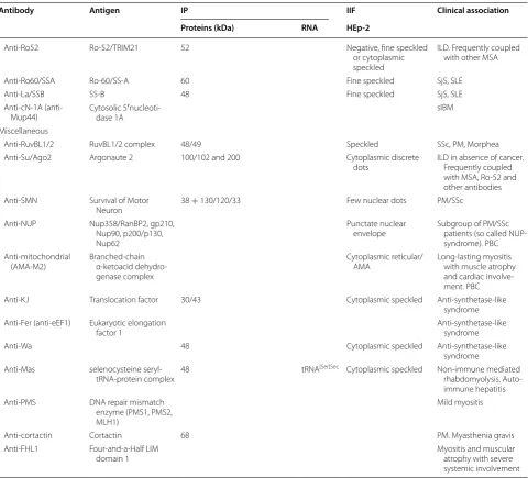
Fig. 1 IIF on HEp-2 ANA slides of myositis-specific autoantibodies from patients with IIMs (EUROIMMUN, Lübeck, Germany). a Anti-Jo-1 and b
anti-Jo-1 and anti-Ro52, c anti-PL-7, d anti-PL-12, e anti-KS, f anti-Mi-2, g anti-SAE-1, h anti-MDA5, i anti-NXP-2 and j anti-NXP-2 with coiled bodies, k
Deacetylase (NuRD) multi-protein complex with
nucleo-some remodeling and histone deacetylase/demethylase
activities [
40
]. Anti-Mi-2 autoantibodies
immunopre-cipitate a major protein of 240 kDa, composed by two
proteins, Mi-2α and Mi-2β of 220 and 218 kDa,
respec-tively. Other NuRD complex proteins co-precipitate at
200, 150, 75, 65, 63, 50 and 34 kDa [
41
,
42
]. IIF on HEp-2
cells reveals a characteristic fine speckled ANA pattern;
during metaphase, chromatin mass is not stained but
the nucleoplasm presents the same fine tiny speckles
(Fig.
1
f). Commercial ELISA and immunoblot kits
iden-tify anti-Mi-2 autoantibodies. Anti-Mi-2 are commonly
detected in DM patients, either in adults (11–59%) or in
children (4–10%), with a great variability among the
stud-ies. Their presence in PM and sporadic inclusion body
myositis (sIBM) is rarer [
40
].
The Mi-2 protein is over-regulated during muscle
regeneration in DM patients and thought to be related
to UV rays exposition, sex and HLA (DRB1*0302 and
DRB1*0701) [
43
–
45
]. Anti-Mi-2 positive DM patients
usually exhibit mild myopathy despite high creatine
kinase (CK) levels, without lung involvement and/or
can-cer [
43
]. Overall, anti-Mi2 positive is associated with a
positive prognosis and a good response to corticosteroids
[
43
].
Anti‑SAE
Small Ubiquitin-like Modifiers (SUMOs) have a key role
in post-transcriptional modification of specific proteins
in a ubiquitin-like fashion. This process is controlled
by the SUMO-Activating Enzyme (SAE), a
heterodi-mer composed of two subunits, SAE-1 and SAE-2 [
46
],
representing the targets of anti-SAE autoantibodies. IP
characteristically shows two bands of 40 and 90 kDa,
respectively [
46
,
47
]. The IIF ANA pattern is coarse
or fine speckled and nucleoli are typically not stained
(Fig.
1
g) [
47
].
Anti-SAE are associated with a typical DM phenotype
with different prevalence in European (4-10%) and Asian
(1–3%) cohorts [
48
–
50
], probably due to the strict
asso-ciation with HLADRB1*04-DQA1*03-DQB1*03
haplo-types [
51
].
The cutaneous involvement is usually severe and
typi-cally precedes the muscular involvement. Other
clini-cal relationships cannot be excluded because of the few
described cases. However, ILD seems to be rare, whereas
severe dysphagia and systemic symptoms have been
reported [
47
]. Only one case series claimed an
associa-tion with cancer [
52
].
Anti‑MDA5
Melanoma Differentiation-Associated gene 5 (MDA5)
or Interferon-induced helicase C domain-containing
protein 1 (IFIH1), is an innate cytosolic sensor, member
of the retinoic acid-inducible gene I (RIG-I)-like
recep-tors family (RLRs). MDA5 is able to recognize
double-stranded RNA and to initiate signaling events leading to
type I interferons production [
53
].
Anti-MDA5 autoantibodies were firstly detected in IP
as a 140 kDa band in a Japanese case series of patients
with clinically amyopathic dermatomyositis (CADM) and
rapidly progressive interstitial lung disease (RP-ILD). For
this reason, they were initially called anti-CADM-140
autoantibodies [
54
]. Nowadays, ELISA and IB tests are
commercially available.
IIF on HEp-2 cells is usually negative. In our
experi-ence, a faint fine speckled cytoplasmic fluorescence may
be detected in scattered cells (unpublished data) (Fig.
1
h).
Clinically, DM anti-MDA5 positive patients present
low grade/absent muscle inflammation and acute or
subacute RP-ILD [
55
,
56
], which is considered the major
negative prognostic factor of this subgroup [
57
].
MDA5 represents the most frequent target antigen in
DM patients of Asian ancestry (10–48% of cases) [
58
]
whereas its prevalence in Europe and USA ranges from 0
to 13%, with great variability among the studies [
59
–
61
]
and a different clinical presentation. A forthcoming
Euro-pean case series is going to be presented at the EuroEuro-pean
League Against Rheumatism 2018 Congress by Cavagna
et al. (unpublished data). A seasonal pattern of CADM
has been proposed by Muro et al. [
62
], suggesting the
influence of environmental factors and HLA-DRB1*04:01
and *12:02 have been proposed as further predisposing
factors [
63
].
In addition to classic DM-related cutaneous
manifesta-tions, skin involvement is usually severe and
character-ized by the so called “inverse Gottron papules”, which
are tender palmar papules that tend to evolve towards
ulcerated-necrotic lesions, with or without digital pulp
ulcers [
64
,
65
]. In addition, polyarthritis, recurrent oral
aphtosis and diffuse alopecia have been described [
66
].
A juvenile DM with anti-MDA5 autoantibodies has been
also described [
67
]. No association with malignancies has
been demonstrated so far. Macrophage activation
syn-drome have been described in CADM associated RP-ILD
patients. Particularly, a ferritin level of above 1500 ng/mL
has been claimed as a predictor of death [
68
,
69
].
Anti-MDA5 autoantibodies titer seems to correlate with
dis-ease activity and response to therapy [
69
].
Anti‑TIF‑1
Targoff et al. and Kaji et al. [
71
,
72
] independently
described two antibodies directed against a 155 and
140 kDa, rapidly identified as TIF-1γ (TRIM33) and
TIF-1α (TRIM24), respectively. Subsequently, a third
120 kDa band, partially overlapping with anti-PL-12, was
identified as TIF1β (TRIM28) [
73
].
IIF on HEp-2 cells demonstrates a fine speckled nuclear
pattern (Fig.
1
i). ELISA and IB, compared to IP, are
reli-able test for the detection of anti-TIF-1γ
autoantibod-ies [
74
]. Two-thirds of the patients present anti-TIF-1γ
and anti-TIF-1α autoantibodies, whereas the
remain-ing one-third is positive for anti-TIF-1γ autoantibodies
exclusively [
70
]. Albeit MSAs are claimed to be mutually
exclusive, double-positive patients for anti-TIF-1α/Mi-2
autoantibodies have been described [
75
].
Hyper-expression of TIF-1γ has been found in tumors
[
76
] and regenerating myofibres of DM patients [
77
]. A
meta-analysis demonstrated that anti-TIF-1γ has a 78%
sensitivity and 89% specificity for the diagnosis of
cancer-associated myositis, with a 58% positive and 95% negative
predictive value [
78
]. The risk of malignancy is higher
in patients with TIF-1γ/α than in those with
anti-TIF-1γ alone [
70
].
Clinically, anti-TIF-1 positive patients can be classified
in two age groups: (1) younger than 40-year-old patients,
with a classical DM at presentation and (2) older than
40-year-old patients, with cancer-associated myositis
[
70
]. Solid tumors, like ovary, lung and breast cancer are
the most commonly associated neoplasia, but
hemato-logic disorders and malignancies have been described as
well [
79
]. In general anti-TIF-1γ patients exhibit a
hypo-myopathic DM with reduced prevalence of systemic
involvement, namely ILD, Raynaud’s phenomenon and
arthritis [
80
]. Conversely, nutcracker esophagus is three
times more frequent in anti-TIF-1γ patients than other
IIMs [
35
]. Widespread cutaneous involvement is
associ-ated with unique features, such as palmar hyper-keratotic
papules, psoriatic-like dermatitis and atrophic
hypo-pig-mented patches with telangiectasias [
80
]. An ovoid
pal-atal patch may be present in about one half of patients,
more frequently females with cancer-associated
amyo-pathic disease [
81
].
Anti‑NXP‑2
Nuclear matrix protein 2 (NXP-2), encoded by the
micro-rchidia 3 gene, is a 140 kDa protein involved in epigenetic
regulation, RNA metabolism and preservation of nuclear
chromatin architecture [
82
]. Anti-NXP-2
autoantibod-ies were found in a cohort of juvenile DM patients as a
140 kDa protein firstly named anti-MJ [
83
].
IIF on HEp-2 cells reveals a fine speckled nuclear
pattern (Fig.
1
k). Moreover, a nuclear dots pattern is
detectable in 60% of sera [
84
], due to co-localization of
NXP-2 with pro-myelocitic leukemia (PML) bodies [
85
]
(Fig.
1
j).
Anti-NXP-2 antibodies have been initially associated
with a severe juvenile DM complicated by calcinosis,
polyarthritis and intestinal vasculitis [
86
]. More recently,
they have been also found in adult patients, with variable
prevalence from 1.6 to 17% [
87
–
89
]. Anti-NXP-2
autoan-tibodies show a bimodal spectrum of clinical association,
with calcinosis being more frequent in younger patients
and cancer more common in the elderly [
90
], especially
in male gender [
88
], even though with a lower prevalence
when compared to anti-TIF-1 [
87
,
88
].
Anti‑SRP
The signal recognition particle (SRP) is a complex of six
proteins (9, 14, 19, 54, 68 and 72 kDa) and a 300
nucleo-tides long RNA (7SL RNA) involved in the recognition
and transportation of proteins to the endoplasmic
reticu-lum [
91
]. Anti-SRP autoantibodies are more frequently
directed against the SRP-54 fragment, albeit anti-SRP-68,
anti-SRP-72 and anti-7SL RNA autoantibodies have been
also described [
91
].
A dense fine speckled cytoplasmic pattern has been
associated with the presence of anti-SRP (Fig.
2
a);
more-over, IIF on stomach–liver–kidney rat sections
dem-onstrates a cytoplasmic staining of gastric chief cells
(Fig.
2
b) and hepatocytes (Fig.
2
c) [
92
].
Anti-SRP-54 autoantibodies ELISA tests are
commer-cially available, but they are less sensitive than IP [
93
].
Anti-SRP antibodies can also be tested on LIA assays,
however careful temperature control is necessary in
order to avoid false positive results [
19
].
autoantibodies level correlates to disease activity, CK
lev-els and response to therapy [
106
].
Anti‑HMGCR
The 3-hydroxy-3-methylglutaryl-coenzyme A reductase
(HMGCR) is the rate-controlling enzyme of the
meva-lonate pathway, bringing to the production of cholesterol.
Of note, HMGCR is the same enzyme targeted by statins.
Autoantibodies towards a complex 200/100 kDa band
were first described in patients with IMNM, and only
after identified as anti-HMGCR autoantibodies [
107
].
IIF pattern is difficult to recognize. In a minority of
cases finely granular cytoplasmic staining with a
peri-nuclear reinforcement is visible on a small number of
scattered cells (3% of the total cellularity) (Fig.
2
g). On
rat liver, a scattered cytoplasmic staining of hepatocytes
around the liver lobules, namely anti-HMGCR Antibody
Associated Liver Immunofluorescence Pattern (HALIP),
can be noted (Fig.
2
i) [
108
,
109
]. Anti-HMGCR
antibod-ies can be identified with different immunoenzymatic
technologies, such as ELISA, CLIA, IB or ALBIA [
110
].
A history of statin exposure is not mandatory to
develop anti-HMGCR positive IMNM, being of some
relevance only in patients older than 50 [
111
,
112
].
Anti-HMGCR antibodies are not found in self-limiting statin
associated myopathy [
113
], albeit they may be associated
with an increased risk of cancer [
114
]. An association
with the DRB1*11:01 haplotype has been demonstrated,
whereas DQA1 and DQB1 seem to have a protective role
[
115
].
Anti-HMGCR positive patients present with a typical
IMNM, responds well to immunosuppressive therapy
Fig. 2 Immunofluorescence patterns from patients with IMNMs with anti-SRP and anti-HMGCR antibodies on HEp-2 ANA slides (EUROIMMUN, Lübeck, Germany) and rat liver and stomach slides (DiaSorin, Italy), compared to AMA-M2 antibodies. a Anti-SRP cytoplasmic dense fine speckled pattern on HEp-2, b chief cells on stomach and c fine granular liver staining; d AMA-M2 cytoplasmic reticular on HEp-2, e granular staining of parietal stomach cells and f diffused fluorescence of hepatocytes; g Anti-HMGCR faint cytoplasmic fluorescence on few numbers of HEp-2 cells, h
and intravenous immunoglobulins [
116
], but tend to
relapse after tapering [
111
]. Younger patients experience
more severe disease with worse prognosis [
117
].
Autoan-tibody titers seem to correlate with CK levels, muscular
weakness and response to therapy [
111
].
Myositis‑associated autoantibodies
Numerous MAAs have been described so far.
Character-istically, they can be found in IIMs, albeit not specific as
found in other CTDs [
118
].
Anti‑PM‑Scl
Anti-PM-Scl autoantibodies are directed against the
exo-some, a macromolecular nucleolar complex composed
by 11–16 proteins (from 20 to 110 kDa) that degrades
mRNA. The two pivotal proteins of the complex are
PM-Scl-75 and PM-Scl-100. IP represents the gold standard
for their determination. Historically, an ID test after
posi-tive nucleolar staining in IIF was used to confirm
anti-PM-Scl reactivity.
PM-Scl-100 and PM-Scl-75 were identified in 1992 and,
in the following years, the immuno-dominant epitope
PM1α was cloned and employed to develop reliable and
specific ELISA tests [
119
]. PM1α ELISA and PM-Scl-100
LIA tests show concordance with IP at high level (> 90
and 98.3%, respectively), whereas PM-Scl-75 LIA has
a lower specificity, especially when considering PM/
SSc overlap syndromes [
120
]. Single positivity against
PM-Scl-75 or -100 can be detected and associates with
different disease phenotypes. HEp-2 IIF typically shows a
mixed homogeneous nucleolar and fine speckled nuclear
pattern when PM-Scl-100 are present, whilst
anti-PM1-α and PM-Scl-75 may show both nucleolar and
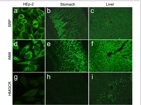
non-nucleolar patterns (Fig.
3
a, b).
PM-Scl autoantibodies are found in 4–12% of adult
patients with myositis [
121
,
122
] with low prevalence in
Asiatic and paediatric cohorts [
123
]. Their presence has
been associated with HLA-DQA1*0501, DQB1*02 and
DRB1*0301 alleles [
122
].
Despite their presence in many connective tissue
dis-eases, these autoantibodies are typically present in
PM/SSc overlap syndromes with an increased risk of
Raynaud’s phenomenon, arthritis, mechanic’s hands and
ILD [
124
]. In detail, isolated anti-PM-Scl-75 have been
more frequently found in patients with joint contractures
and SSc, higher CK levels associate with anti-PM-Scl-100,
whereas the simultaneous presence of anti-PM-Scl-75
and -100 are linked to muscle involvement, digital ulcers
and ILD but lower prevalence of lung hypertension [
125
].
Autoantibodies directed against C1D, an exosome
associated protein, were detected by ELISA and
West-ern blot analysis in 23% of a PM/SSc overlap syndrome
cohort, with frequencies comparable to PM/Scl
anti-bodies [
126
].
Fig. 3 Myositis-associated autoantibodies and some peculiar IIF pattern on HEp-2 ANA slides (EUROIMMUN, Lübeck, Germany). a anti-PM-Scl-100,
Anti‑RNP
The RNP/Sm complex comprises several proteins (70 kD,
A, A
′
, B, B
′
, B
″
, C, D, E, F, G) and five RNA (U1, U2, U4,
U5 and U6). U1 RNA interacts with 70 kD, A and C to
create the U1-snRNP [
127
]. High-titre anti-U1-snRNP
and in particular when targeting the 70kD protein are
considered specific markers of mixed connective
tis-sue disease (MTCD), whereas low titres can be found in
other CTDs [
127
].
Many home-made or commercial assays can detect
anti-RNP autoantibodies, with differences among
immu-noassays depending on the immobilized antigen [
127
].
Usually, anti-U1-snRNP (more often the 70k subunit)
and the anti-Sm (typically the D subunit) are the only
autoantibodies tested in clinical practice. Large speckled
and large coarse speckled are the most frequent HEp-2
IIF patterns observed (Fig.
3
c).
Patients with myositis may exhibit anti-U1-snRNP
positivity, especially those with a mild disease [
128
,
129
]. They are usually steroid-responsive, even though
ILD and/or neurological involvement may be part of the
clinical presentation [
128
,
129
]. Whether the only
pres-ence of anti-U1-snRNP and myositis has to be
consid-ered an incomplete form of MTCD or a true myositis, is
still a matter of debate [
128
,
129
]. In addition,
anti-U2-RNP [
130
], U5-RNP [
131
] and anti-U4/U6-RNP [
132
]
have been described in patients with PM/SSc overlap
syndrome.
Anti‑fibrillarin
Fibrillarin, a highly conserved nucleolar 34 kDa protein
involved in the processing of ribosomal RNA, is part of
the U3-small nucleolar (sno)-RNP complex together
with other proteins and U3 RNA. Fibrillarin is the
pri-mary target of anti-U3-snoRNP autoantibodies [
133
]. IP
is the gold standard for its detection showing good
con-cordance with IB assays that use the recombinant protein
[
134
].
HEp-2 IIF demonstrates a typical “clumpy” nucleolar
pattern, with jagged staining of the nucleoli, coiled bodies
and peri-chromosomal staining at the metaphase plates
[
135
] (Fig.
3
e).
Anti-fibrillarin antibodies are detected in a small
percentage of SSc patients and rarely in SLE, primary
Raynaud’s phenomenon and myositis [
136
]. In detail,
they identify a subset of SSc patients more often of
Afri-can origin, with serious cutaneous and visceral
involve-ment and a higher prevalence of myositis [
137
,
138
].
Anti‑Ku
The Ku protein, involved in the canonical
non-homol-ogous end-joining pathway of the DNA repair, is a
het-erodimer consisting of the two subunits, 70 and 80 kDa
[
139
]. Anti-Ku can be identified with numerous assays,
such as ELISA, CIE or IB. IIF demonstrates a fine
speck-led nuclear pattern with a peculiar ring beam
surround-ing the metaphase on HEp-2 cells and a clumpy speckled
pattern on primate’s liver [
140
] (Fig.
3
d).
Anti-Ku autoantibodies have been identified in 9-19%
of the patients with PM/SSc overlap syndromes and SLE,
being associated with arthralgia, Raynaud’s phenomenon
and ILD [
141
,
142
]. Of note, whilst muscular involvement
seems to be steroid-sensitive, ILD is more frequently
pro-gressive, severe and steroid-resistant [
143
].
Anti‑Ro
Antibodies directed against the ribonucleoproteic
com-plex SSA/Ro and SSB/La have been originally identified
in SjS and SLE. Actually, antigen Ro is made by two
sep-arate complexes of 52 and 60 kDa called Ro52/TRIM21
and SSA/Ro60, respectively. Antigen SSB/La has a
molec-ular weight of 48 kDa [
144
].
ANA may result falsely negative on traditional HEp-2
cells when isolate anti-Ro are present, because Ro52 is
a cytoplasmic antigen and Ro60 may be lost during the
preparation. For this reason, human
SSA/Ro60-trans-fected HEp-2 cells (HEp-2000) are sometimes used [
145
,
146
]. Otherwise, a characteristic pattern defined as
“myr-iad discrete fine speckled” may be observed [
147
].
Anti-SSB autoantibodies show a similar pattern [
148
].
Anti-Ro52 can be found in IIMs [
149
] and are
fre-quently associated with other MSAs, in particular
anti-synthetase [
28
], anti-MDA5 [
61
] and anti-SRP
autoantibodies [
12
].
They are known to be a negative prognostic factor
regarding systemic involvement such as ILD, whereas
their role in the severity of muscular involvement has
not been identified [
13
]. Anti-Ro52 autoantibodies are
known to be associated with atrioventricular congenital
heart block [
150
].
Anti‑cN‑1A
Cytosolic 5
′
nucleotidase 1A (cN-1A o NT5C1A)
is a protein involved in the hydrolysis of adenosine
monophosphate, controlling energy and metabolic cell
balance [
151
]. Anti-cN-1A autoantibodies, first called
anti-Mup44, were simultaneously described by
Sala-jegheh et al. and Pluk et al. [
152
,
153
] as targeting a
44 kDa protein in patients with sIBM.
Anti-cN-1A autoantibodies are demonstrated in
one-third of the patients with sIBM and in less than 5% with
other IIMs or neuromuscular diseases [
151
]. A recent
study demonstrated that positive anti-cN-1A sIBM
patients are included in a more severe sIBM subtype and
represent a homogeneous group as exhibiting higher
mortality risk, less proximal upper limb weakness (not
typical of sIBMs) and a cytochrome oxidase deficiency
in muscular fibers, when compared to negative patients
[
156
].
It is not known whether they have to be considered as
MSAs or MAAs as also demonstrated in other
autoim-mune diseases, such as SjS (30%) and SLE (20%) [
157
].
Furthermore, they have been recently demonstrated in a
cohort of severe juvenile myositis with lung involvement,
juvenile idiopathic arthritis, but also in 12% of healthy
children [
158
].
Despite low sensitivity, anti-cN-1A autoantibodies are
high specific and highly predictive of sIBM [
159
] thus
being of particular importance when bioptic specimens
are not diagnostic.
Miscellaneous autoantibodies in IIM
Several other autoantibodies have been identified as
associated with IIMs, but little is known about their
clini-cal relevance. In fact, they are not routinely determined
because easy-to-perform routine specific immunoassays
still lack and they are rarely found.
Anti‑RuvBL1/2
RuvBL1 (49kD) and RuvBL2 (48kD) constitute a nuclear
complex involved into DNA repair and transcription.
Two distinct bands of approximately 50 kDa are found in
IP [
160
]. By means of ELISA and/or IB techniques,
anti-RuvBL1/2 have been found in several CTDs, but those
involved in SSc and myositis recognize different
confor-mational epitopes identified by IP exclusively [
160
]. On
HEp-2 cells, a fine speckled pattern is associated with
these antibodies, with increased fluorescence in prophase
and decreased in metaphase. Additionally, a fine speckled
pattern can be found in the cytoplasm of about 40%
posi-tive sera [
160
]. Anti-RuvBL1/2 antibodies are highly
spe-cific for SSc, are associated with PM/SSc overlaps with
diffuse cutaneous sclerosis and more frequently found in
older patients of male sex [
160
–
162
] or, less frequently, in
necrotizing polymyositis with morphea [
162
].
Anti‑Su/Ago2
Anti-Su/Argonaute-2 (anti-Su/Ago2) autoantibodies
have been originally identified in SLE patients by means
of immunodiffusion technique in the late ‘80ies [
163
].
Although their high prevalence in CTDs, few studies are
available.
By IP, two distinct 100 and 102 kDa adjacent bands
can be seen in addition to a further 200 kDa band [
164
].
Argonaute-2 protein constitutes the 100 kDa band and
plays a key role in miRNA and interference RNA
matura-tion and metabolism [
163
]. Argonaute-2 colocalized with
GW bodies, a cytoplasmic organelle associated with RNA
metabolism [
164
]. Its location and function is responsible
for the particular cytoplasmic pattern of these
autoanti-bodies also known as “GW-autoanti-bodies-like” or “cytoplasmic
discrete dots” (Fig.
3
f) [
164
].
Anti-Su/Ago2 autoantibodies are frequently associated
with other MSA or MAA antibodies, in particular ARS,
anti-TIF-1γ and anti-MDA5 [
165
]; anti-Ro52 antibodies
are found in almost one half of the patients [
166
]. It has
been reported that anti-Su/Ago2 antibodies can be
dem-onstrated in about 7.5% of the patients of Japanese origin.
Apparently, there is no statistical difference between
anti-Su/Ago2 positive and negative patients; however, a
cor-relation seems to exist with ILD and absence of cancers
[
165
].
Anti‑SMN
The Survival of Motor Neuron (SMN) is a
multi-ribo-nucleoproteic complex able to interact with the
RNP-complex related D–E–F-G proteins. The SMN RNP-complex
is involved into the assembly of snRNPs and
co-local-izes with Cajal bodies. These autoantibodies have been
first described in a small number of PM patients
nega-tive for anti-U1-snRNP and/or anti-Sm but posinega-tive for
RNP D–E–F-G bands by IP. This observation was indeed
responsible for the identification of other
SMN-com-plex components, namely Gemin 2 (33 kDa), Gemin 3
(130 kDa), Gemin 4 (120 kDa) and SMN itself (38 kDa)
[
167
].
Anti-SMN antibodies typically exhibit a few nuclear
dots pattern on HEp-2 cells with well distinguished 2–7
nuclear dots, similarly to anti-p80-coilin, anti-NXP2 and
anti-PML pattern, seldom associated to cytoplasmic or
nuclear speckled patterns (Fig.
3
g).
It is not clear whether positive patients exhibit distinct
clinical features. Anyhow, in the original small group of
positive patients [
167
] and a small Italian cohort [
20
], a
PM/SSc overlap syndrome was present. It is of note that
SMN-complex genetic mutations are frequently found
in neuromuscular degenerative diseases such as
spinal-muscle atrophy. That is why anti-SMN autoantibodies are
of relevance in basic research [
168
].
Anti‑NPC
and, in particular, anti gp210 are typically associated to
Primary Biliary Cholangitis (PBC) and Autoimmune
Hepatitis (AIH) [
170
]. In a cohort study from Canada,
anti-NPC antibodies were found in a PM/SSc overlap
syndrome and called anti-NUP Syndrome, which was
found to be associated with HLA-DQ1*0501. In this case,
a typical nuclear speckled laminar pattern on HEp-2 cells
was observed [
171
] (Fig.
3
h).
AMA‑M2
Among the ten different anti-mitochondrial
antibod-ies (AMA), called M1–M10, anti-M2 antibodantibod-ies
(AMA-M2) are the hallmark of PBC [
172
]. However, they can be
also found in 7–12% of IIM patients without PBC [
173
].
AMA antibodies are readily detectable on HEp-2 cells as
they display a pathognomonic cytoplasmic reticular
pat-tern, and in triple tissue slides (Fig.
2
d–f). In a Japanese
study, the presence of AMA-M2 in the course of IIM was
associated with muscle atrophy, granuloma formation
[
173
] and heart involvement with high risk of
supraven-tricular arrhythmias [
174
]. A distinct inflammatory
phe-notype associated with chronic skeletal muscle disease
and severe cardiac involvement was also found in a North
American cohort [
175
]. These associations have not been
confirmed in an European series [
176
].
Other antibodies
Several cytoplasmic autoantibodies are described in IIM
patients such as anti-KJ towards a 30/43 kDa
transloca-tion factor [
177
], anti-Fer directed against the elongation
factor 1 and anti-Wa recognizing a 48 kDa cytoplasmic
protein with still unknown function [
132
]. All these
antibodies are typically found in anti-synthetase-like
syndromes. Anti-Mas antibodies are directed against a
selenocysteine-containing tRNA complex lacking any
tRNA-synthetase activity but involved in protein
trans-location. The band of precipitation is found at 48 kDa
[
178
]. These antibodies have been described in AIH and
in a single patient with non-immune mediated
rhabdo-myolysis [
178
].
DNA-repairing complexes, especially
mismatch-repair complexes such as PMS1, PMS2 and MLH1, are
frequently recognized as target antigens in IIMs [
179
].
Initially defined as MSAs, they do indeed frequently
associate with other MSAs, in particular anti-Mi-2, but
they can also be found in other non-muscular diseases,
such as SLE. They generally mark mild disease [
180
].
Anti-cortactin antibodies have been initially found in
IIM patients characterized by the simultaneous
pres-ence of anti-MDA5 or anti-HMGCR antibodies by ELISA
[
181
]. As blot confirming assay identified an unexpected
68 kDa band, it was then found that MDA5 and HMGCR
extracts used in the ELISA tests were contaminated by
cortactin [
181
]. Anti-cortactin antibodies were originally
found in myasthenia gravis [
182
] and later in IIM patients
(about 20%) and other systemic connective tissue
dis-eases [
181
].
Anti-Four-and-a-Half LIM domain 1 (FHL1)
antibod-ies were identified in about 25% of IIM patients. These
antibodies associated with a severe prognosis, muscle
atrophy, vasculitis, dysphagia and advanced muscular
damage. Curiously, FHL1 mutations cause hereditary
X-linked congenital myopathies [
183
].
Conclusions
Although autoantibodies are considered to be
epiphe-nomenon in autoimmunity, their presence frequently
plays a pivotal role for the diagnosis of these diseases.
Indeed, several of them exhibit a pathogenethic role in
IIMs. Despite this, there is still a gap between bench and
bedside because the intense basic research efforts have
not been translated in clinical practice, as already
futur-istically underlined more than 20 years ago [
96
,
184
]. As
a fact, only anti-Jo-1 have been included into the 2017
Classification Criteria for Adult and Juvenile IIMs [
3
].
Remarkably, in the context of heterogeneously grouped
diseases such as myositis, they should be even more
appreciated as able to clinically stratify patients in terms
of diagnostic work-up, histological patterns, peculiar
organ involvement, severity, and, therefore, treatment
intensity and prognosis. This process could be
accom-plished by a laboratory auto-immunologist [
185
]
well-trained in recognition of IIF ANA nuclear and, also,
cytoplasmic patterns, in strict collaboration with the
clinical doctor, as a decision-maker for running
in-depth analysis towards the identification of the culprit
autoantibody.
In addition, multicentric studies with a
multidisci-plinary approach may help bridging the divide of the
selection bias depending on the setting where patients
are initially screened (i.e. pneumologic vs. dermatologic
vs. immuno-rheumatologic vs. neurologic outpatient
clinics).
Abbreviations
SUMO-activating enzyme; MDA5: Melanoma Differentiation-Associated gene 5; RIG-I: retinoic acid-inducible gene I; RLR: RIG-I-like receptors family; RP-ILD: rapidly progressive interstitial lung disease; CADM: clinically amyopathic dermatomyositis; TIF1: transcription intermediary factors-1; TRIM: tripartite motif-containing proteins; NXP-2: nuclear matrix protein 2; PML: pro-myeloc-itic leukaemia bodies; SRP: signal recognition particle; IMNM: immune-medi-ated necrotizing myositis; HMGCR: 3-hydroxy-3-methylglutaryl-coenzime A reductase; HALIP: anti-HMGCR antibody associated liver immunofluorescence pattern; MTCD: mixed connective tissue disease; sno: small nucleolar; cN-1A: cytosolic 5′nucleotidase; SMN: survival of motor neuron; NPC: nuclear pore complex; PBC: primary biliary cholangitis; AIH: autoimmune hepatitis; FHL1: anti-four-and-a-half LIM domain 1; AMA: anti-mitochondrial antibodies.
Authors’ contributions
BP and GV drafted the manuscript. All authors read and approved the final manuscript.
Acknowledgements
We thank the team of Immunoallergology Laboratory, AOU-Careggi, Florence for the great deal of teamwork and constant readiness to our requests.
Competing interests
The authors declare that they have no competing interests.
Availability of data and materials Not applicable.
Consent for publication Not applicable.
Ethics approval and consent to participate Not applicable.
Funding
This research received no specific grant from any funding agency in the pub-lic, commercial, or not-for-profit sectors.
Publisher’s Note
Springer Nature remains neutral with regard to jurisdictional claims in pub-lished maps and institutional affiliations.
Received: 31 January 2018 Accepted: 20 February 2018
References
1. Petri M, Orbai A-M, Alarcón GS, Gordon C, Merrill JT, Fortin PR, et al. Deri-vation and validation of the Systemic Lupus International Collaborating Clinics classification criteria for systemic lupus erythematosus. Arthritis Rheum. 2012;64:2677–86.
2. Shiboski CH, Shiboski SC, Seror R, Criswell LA, Labetoulle M, Lietman TM, et al. 2016 American College of Rheumatology/European League Against Rheumatism classification criteria for primary Sjögren’s syn-drome. Ann Rheum Dis. 2017;76:9–16.
3. Lundberg IE, Tjärnlund A, Bottai M, Werth VP, Pilkington C, de Visser M, et al. 2017 European League Against Rheumatism/American College of Rheumatology classification criteria for adult and juvenile idiopathic inflammatory myopathies and their major subgroups. Ann Rheum Dis. 2017;76:1955–64.
4. Milone M. Diagnosis and Management of Immune-Mediated Myopa-thies. Mayo Clin Proc. 2017;92:826–37.
5. Targoff IN. Autoantibodies in polymyositis. Rheum Dis Clin North Am. 1992;18:455–82.
6. Targoff IN. Idiopathic inflammatory myopathy: autoantibody update. Curr Rheumatol Rep. 2002;4:434–41.
7. Satoh M, Tanaka S, Ceribelli A, Calise SJ, Chan EKL. A comprehensive overview on myositis-specific antibodies: new and old biomarkers
in idiopathic inflammatory myopathy. Clin Rev Allergy Immunol. 2015;52:1–9.
8. Nakashima R, Imura Y, Hosono Y, Seto M, Murakami A, Watanabe K, et al. The multicenter study of a new assay for simultaneous detection of multiple anti-aminoacyl-tRNA synthetases in myositis and interstitial pneumonia. PLoS ONE. 2014;9:e85062.
9. Colafrancesco S, Priori R, Valesini G. Inflammatory myopathies and overlap syndromes: update on histological and serological profile. Best Pract Res Clin Rheumatol. 2015;29:810–25.
10. Gunawardena H, Betteridge ZE, McHugh NJ. Myositis-specific autoanti-bodies: their clinical and pathogenic significance in disease expression. Rheumatology (Oxford). 2009;48:607–12.
11. Chinoy H, Fertig N, Oddis CV, Ollier WER, Cooper RG. The diagnostic utility of myositis autoantibody testing for predicting the risk of cancer-associated myositis. Ann Rheum Dis. 2007;66:1345–9.
12. Frank MB, McCubbin V, Trieu E, Wu Y, Isenberg DA, Targoff IN. The association of anti-Ro52 autoantibodies with myositis and scleroderma autoantibodies. J Autoimmun. 1999;12:137–42.
13. Ferreira JP, Almeida I, Marinho A, Cerveira C, Vasconcelos C. Anti-ro52 antibodies and interstitial lung disease in connective tissue diseases excluding scleroderma. ISRN Rheumatol. 2012;2012:415272. 14. van Dooren SHJ, van Venrooij WJ, Pruijn GJM. Myositis-specific
autoan-tibodies: detection and clinical associations. Autoimmun Highlights. 2011;2:5–20.
15. Chan EKL, Damoiseaux J, Carballo OG, Conrad K, de Melo Cruvinel W, Francescantonio PLC, et al. Report of the first international consensus on standardized nomenclature of antinuclear antibody HEp-2 cell pat-terns 2014–2015. Front Immunol. 2015;6:412.
16. García-DeLaTorre I. Clinical usefulness of autoantibodies in idiopathic inflammatory myositis. Front Immunol. 2015;6:331.
17. Damoiseaux J, von Mühlen CA, Garcia-De La Torre I, Carballo OG, de Melo Cruvinel W, Francescantonio PLC, et al. International consensus on ANA patterns (ICAP): the bumpy road towards a consensus on report-ing ANA results. Autoimmun Highlights. 2016;7:1–8.
18. Ghirardello A, Bendo R, Rampudda ME, Bassi N, Zampieri S, Doria A. Commercial blot assays in the diagnosis of systemic rheumatic dis-eases. Autoimmun Rev. 2009;8:645–9.
19. Rönnelid J, Barbasso Helmers S, Storfors H, Grip K, Rönnblom L, Franck-Larsson K, et al. Use of a commercial line blot assay as a screening test for autoantibodies in inflammatory myopathies. Autoimmun Rev. 2009;9:58–61.
20. Cavazzana I, Fredi M, Ceribelli A, Mordenti C, Ferrari F, Carabellese N, et al. Testing for myositis specific autoantibodies: comparison between line blot and immunoprecipitation assays in 57 myositis sera. J Immu-nol Methods. 2016;433:1–5.
21. Ceribelli A, Satoh M, Chan EK. A new immunoprecipitation-real time quantitative PCR assay for anti-Th/To and anti-U3RNP antibody detec-tion in systemic sclerosis. Arthritis Res Ther. 2012;14:R128.
22. Mahler M, Miller FW, Fritzler MJ. Idiopathic inflammatory myopathies and the anti-synthetase syndrome: a comprehensive review. Autoim-mun Rev. 2014;13:367–71.
23. Nishikai M, Reichlin M. Heterogeneity of precipitating antibodies in polymyositis and dermatomyositis. Characterization of the Jo-1 anti-body system. Arthritis Rheum. 1980;23:881–8.
24. Agmon-Levin N, Damoiseaux J, Kallenberg C, Sack U, Witte T, Herold M, et al. International recommendations for the assessment of autoanti-bodies to cellular antigens referred to as anti-nuclear antiautoanti-bodies. Ann Rheum Dis. 2014;73:17–23.
25. Infantino M, Palterer B, Biagiotti R, et al. Reflex testing of speckled cyto-plasmic patterns observed in routine ANA HEp-2 indirect immunofluo-rescence with a multiplex anti-synthetase dot-blot assay: a multicentric pilot study. Immunol Res. 2018;66(1):74–8.
26. Abe T, Tsunoda S, Nishioka A, Azuma K, Tsuboi K, Ogita C, et al. Reli-ability and clinical utility of enzyme-linked immunosorbent assay for detection of anti-aminoacyl-tRNA synthetase antibody. Nihon Rinsho Meneki Gakkai Kaishi. 2016;39:140–4.
28. Yamasaki Y, Satoh M, Mizushima M, Okazaki T, Nagafuchi H, Ooka S, et al. Clinical subsets associated with different anti-aminoacyl transfer RNA synthetase antibodies and their association with coexisting anti-Ro52. Mod Rheumatol. 2016;26:403–9.
29. Yamasaki Y, Yamada H, Nozaki T, Akaogi J, Nichols C, Lyons R, et al. Unu-sually high frequency of autoantibodies to PL-7 associated with milder muscle disease in Japanese patients with polymyositis/dermatomyosi-tis. Arthritis Rheum. 2006;54:2004–9.
30. Marie I, Josse S, Decaux O, Diot E, Landron C, Roblot P, et al. Clinical manifestations and outcome of anti-PL7 positive patients with antisyn-thetase syndrome. Eur J Intern Med. 2013;24:474–9.
31. Targoff IN, Arnett FC. Clinical manifestations in patients with antibody to PL-12 antigen (alanyl-tRNA synthetase). Am J Med. 1990;88:241–51. 32. Hirakata M, Suwa A, Nagai S, Kron MA, Trieu EP, Mimori T, et al. Anti-KS: identification of autoantibodies to asparaginyl-transfer RNA synthetase associated with interstitial lung disease. J Immunol. 1999;162:2315–20. 33. Scirè CA, Gonzalez-Gay MA, O’Callaghan A, Cavagna L,
Selva-O’Callaghan A, Cavagna L. Clinical spectrum time course of interstitial pneumonia with autoimmune features in patients positive for antisyn-thetase antibodies. Respir Med. 2017;132:265–6.
34. Cavagna L, Nuño L, Scirè CA, Govoni M, Longo FJL, Franceschini F, et al. Clinical spectrum time course in anti Jo-1 positive antisynthetase syndrome: results from an international retrospective multicenter study. Medicine (Baltimore). 2015;94:e1144.
35. Casal-Dominguez M, Pinal-Fernandez I, Mego M, Accarino A, Jubany L, Azpiroz F, et al. High-resolution manometry in patients with idiopathic inflammatory myopathy: Elevated prevalence of esophageal involve-ment and differences according to autoantibody status and clinical subset. Muscle Nerve. 2016;45(suppl_4):iv18–21.
36. Pestronk A. Acquired immune and inflammatory myopathies. Curr Opin Rheumatol. 2011;23:595–604.
37. Mescam-Mancini L, Allenbach Y, Hervier B, Devilliers H, Mariampillay K, Dubourg O, et al. Anti-Jo-1 antibody-positive patients show a charac-teristic necrotizing perifascicular myositis. Brain. 2015;138(Pt 9):2485–92. 38. Aouizerate J, De Antonio M, Bassez G, Gherardi RK, Berenbaum F,
Guillevin L, et al. Myofiber HLA-DR expression is a distinctive biomarker for antisynthetase-associated myopathy. Acta Neuropathol Commun. 2014;2:154.
39. Targoff IN, Reichlin M. The association between Mi-2 antibodies and dermatomyositis. Arthritis Rheum. 1985;28:796–803.
40. Ghirardello A, Zampieri S, Iaccarino L, Tarricone E, Bendo R, Gambari PF, et al. Anti-Mi-2 antibodies. Autoimmunity. 2005;38:79–83.
41. Nilasena DS, Trieu EP, Targoff IN. Analysis of the Mi-2 autoantigen of dermatomyositis. Arthritis Rheum. 1995;38:123–8.
42. Zhang Y, LeRoy G, Seelig HP, Lane WS, Reinberg D. The dermatomyosi-tis-specific autoantigen Mi2 is a component of a complex contain-ing histone deacetylase and nucleosome remodelcontain-ing activities. Cell. 1998;95:279–89.
43. Petri MH, Satoh M, Martin-Marquez BT, Vargas-Ramírez R, Jara LJ, Saave-dra MA, et al. Implications in the difference of anti-Mi-2 and -p155/140 autoantibody prevalence in two dermatomyositis cohorts from Mexico City and Guadalajara. Arthritis Res Ther. 2013;15:R48.
44. Love LA, Weinberg CR, McConnaughey DR, Oddis CV, Medsger TA, Reveille JD, et al. Ultraviolet radiation intensity predicts the relative dis-tribution of dermatomyositis and anti-Mi-2 autoantibodies in women. Arthritis Rheum. 2009;60:2499–504.
45. Prieto S, Grau JM. The geoepidemiology of autoimmune muscle dis-ease. Autoimmun Rev. 2010;9:A330–4.
46. Tarricone E, Ghirardello A, Rampudda M, Bassi N, Punzi L, Doria A. Anti-SAE antibodies in autoimmune myositis: identification by unlabelled protein immunoprecipitation in an Italian patient cohort. J Immunol Methods. 2012;384:128–34.
47. Betteridge Z, Gunawardena H, North J, Slinn J, McHugh N. Identifica-tion of a novel autoantibody directed against small ubiquitin-like modifier activating enzyme in dermatomyositis. Arthritis Rheum. 2007;56:3132–7.
48. Ge Y, Lu X, Shu X, Peng Q, Wang G. Clinical characteristics of anti-SAE antibodies in Chinese patients with dermatomyositis in comparison with different patient cohorts. Sci Rep. 2017;7:188.
49. Muro Y, Sugiura K, Akiyama M. Low prevalence of anti-small ubiquitin-like modifier activating enzyme antibodies in dermatomyositis patients. Autoimmunity. 2013;46:279–84.
50. Fujimoto M, Matsushita T, Hamaguchi Y, Kaji K, Asano Y, Ogawa F, et al. Autoantibodies to small ubiquitin-like modifier activating enzymes in Japanese patients with dermatomyositis: comparison with a UK Cauca-sian cohort. Ann Rheum Dis. 2013;72:151–3.
51. Betteridge ZE, Gunawardena H, Chinoy H, North J, Ollier WER, Cooper RG, et al. Clinical and human leucocyte antigen class II haplotype associations of autoantibodies to small ubiquitin-like modifier enzyme, a dermatomyositis-specific autoantigen target, in UK Caucasian adult-onset myositis. Ann Rheum Dis. 2009;68:1621–5.
52. Muro Y, Sugiura K, Nara M, Sakamoto I, Suzuki N, Akiyama M. High incidence of cancer in anti-small ubiquitin-like modifier activating enzyme antibody-positive dermatomyositis. Rheumatology (Oxford). 2015;54:1745–7.
53. Nakashima R, Imura Y, Kobayashi S, Yukawa N, Yoshifuji H, Nojima T, et al. The RIG-I-like receptor IFIH1/MDA5 is a dermatomyositis-specific autoantigen identified by the anti-CADM-140 antibody. Rheumatology (Oxford). 2010;49:433–40.
54. Sato S, Hirakata M, Kuwana M, Suwa A, Inada S, Mimori T, et al. Autoantibodies to a 140-kd polypeptide, CADM-140, in Japanese patients with clinically amyopathic dermatomyositis. Arthritis Rheum. 2005;52:1571–6.
55. Sato S, Hoshino K, Satoh T, Fujita T, Kawakami Y, Fujita T, et al. RNA helicase encoded by melanoma differentiation-associated gene 5 is a major autoantigen in patients with clinically amyopathic dermato-myositis: association with rapidly progressive interstitial lung disease. Arthritis Rheum. 2009;60:2193–200.
56. Parronchi P, Radice A, Palterer B, Liotta F, Scaletti C. MDA5-positive dermatomyositis: an uncommon entity in Europe with variable clinical presentations. Clin Mol Allergy. 2015;13:22.
57. Zhang L, Wu G, Gao D, Liu G, Pan L, Ni L, et al. Factors Associated with interstitial lung disease in patients with polymyositis and dermatomyositis: a systematic review and meta-analysis. PLoS ONE. 2016;11:e0155381.
58. Chen Z, Hu W, Wang Y, Guo Z, Sun L, Kuwana M. Distinct profiles of myositis-specific autoantibodies in Chinese and Japanese patients with polymyositis/dermatomyositis. Clin Rheumatol. 2015;34:1627–31. 59. Ceribelli A, Fredi M, Taraborelli M, Cavazzana I, Tincani A, Selmi C, et al.
Prevalence and clinical significance of anti-MDA5 antibodies in Euro-pean patients with polymyositis/dermatomyositis. Clin Exp Rheumatol. 2014;32:891–7.
60. Labrador-Horrillo M, Martinez MA, Selva-O’Callaghan A, Trallero-Araguas E, Balada E, Vilardell-Tarres M, et al. Anti-MDA5 antibodies in a large Mediterranean population of adults with dermatomyositis. J Immunol Res. 2014;2014:290797.
61. Hall JC, Casciola-Rosen L, Samedy L-A, Werner J, Owoyemi K, Danoff SK, et al. Anti-melanoma differentiation-associated protein 5-associated dermatomyositis: expanding the clinical spectrum. Arthritis Care Res (Hoboken). 2013;65:1307–15.
62. Muro Y, Sugiura K, Hoshino K, Akiyama M, Tamakoshi K. Epidemiologic study of clinically amyopathic dermatomyositis and anti-melanoma differentiation-associated gene 5 antibodies in central Japan. Arthritis Res Ther. 2011;13:R214.
63. Chen Z, Wang Y, Kuwana M, Xu X, Hu W, Feng X, et al. HLA-DRB1 alleles as genetic risk factors for the development of anti-MDA5 antibodies in patients with dermatomyositis. J Rheumatol. 2017;44:1389–93. 64. Ward I, Hiles P, Arroyo R, Downs W, Bell D. Digital pulp ulcerations and
inverse gottron papules in melanoma differentiation-associated gene 5-related dermatomyositis. J Clin Rheumatol. 2016;22:274–5. 65. Cao H, Xia Q, Pan M, Zhao X, Li X, Shi R, et al. Gottron papules and
gottron sign with ulceration: a distinctive cutaneous feature in a subset of patients with classic dermatomyositis and clinically amyopathic dermatomyositis. J Rheumatol. 2016;43:1735–42.
66. Fiorentino D, Chung L, Zwerner J, Rosen A, Casciola-Rosen L. The mucocutaneous and systemic phenotype of dermatomyositis patients with antibodies to MDA5 (CADM-140): a retrospective study. J Am Acad Dermatol. 2011;65:25–34.
diagnostic value and associated clinical phenotype in a large UK cohort. J Autoimmun. 2017;84:55–64.
68. Gono T, Kawaguchi Y, Hara M, Masuda I, Katsumata Y, Shinozaki M, et al. Increased ferritin predicts development and severity of acute interstitial lung disease as a complication of dermatomyositis. Rheumatology (Oxford). 2010;49:1354–60.
69. Muro Y, Sugiura K, Akiyama M. Limitations of a single-point evaluation of anti-MDA5 antibody, ferritin, and IL-18 in predicting the prognosis of interstitial lung disease with anti-MDA5 antibody-positive dermatomy-ositis. Clin Rheumatol. 2013;32:395–8.
70. Fujimoto M, Hamaguchi Y, Kaji K, Matsushita T, Ichimura Y, Kodera M, et al. Myositis-specific anti-155/140 autoantibodies target transcription intermediary factor 1 family proteins. Arthritis Rheum. 2012;64:513–22. 71. Targoff IN, Mamyrova G, Trieu EP, Perurena O, Koneru B, O’Hanlon TP,
et al. A novel autoantibody to a 155-kd protein is associated with dermatomyositis. Arthritis Rheum. 2006;54:3682–9.
72. Kaji K, Fujimoto M, Hasegawa M, Kondo M, Saito Y, Komura K, et al. Identification of a novel autoantibody reactive with 155 and 140 kDa nuclear proteins in patients with dermatomyositis: an association with malignancy. Rheumatology (Oxford). 2007;46:25–8.
73. Satoh M, Chan JYF, Ross SJ, Li Y, Yamasaki Y, Yamada H, et al. Autoan-tibodies to transcription intermediary factor TIF1β associated with dermatomyositis. Arthritis Res Ther. 2012;14:R79.
74. Labrador-Horrillo M, Martínez MA, Selva-O’Callaghan A, Trallero-Araguás E, Balada E, Vilardell-Tarrés M, et al. Anti-TIF1γ antibodies (anti-p155) in adult patients with dermatomyositis: comparison of different diagnos-tic assays. Ann Rheum Dis. 2012;71:993–6.
75. Muro Y, Ishikawa A, Sugiura K, Akiyama M. Clinical features of anti-TIF1-α antibody-positive dermatomyositis patients are closely associated with coexistent dermatomyositis-specific autoantibodies and anti-TIF1-γ or anti-Mi-2 autoantibodies. Rheumatology (Oxford). 2012;51:1508–13. 76. Kasuya A, Hamaguchi Y, Fujimoto M, Tokura Y. TIF1γ-overexpressing, highly progressive endometrial carcinoma in a patient with dermato-myositis positive for malignancy-associated anti-p155/140 autoanti-body. Acta Derm Venereol. 2013;93:715–6.
77. Mohassel P, Rosen P, Casciola-Rosen L, Pak K, Mammen AL. Expression of the dermatomyositis autoantigen transcription intermediary factor 1γ in regenerating muscle. Arthritis Rheumatol. 2015;67:266–72. 78. Trallero-Araguás E, Rodrigo-Pendás JÁ, Selva-O’Callaghan A,
Martínez-Gõmez X, Bosch X, Labrador-Horrillo M, et al. Usefulness of anti-p155 autoantibody for diagnosing cancer-associated dermatomyositis: a systematic review and meta-analysis. Arthritis Rheum. 2012;64:523–32. 79. Palterer B, Vitiello G, Cammelli D. First report of anti-TIF1γ
dermato-myositis in a patient with myelodysplastic syndrome. Reumatismo. 2017;69:75–7.
80. Fiorentino DF, Kuo K, Chung L, Zaba L, Li S, Casciola-Rosen L. Distinctive cutaneous and systemic features associated with antitranscriptional intermediary factor-1γ antibodies in adults with dermatomyositis. J Am Acad Dermatol. 2015;72:449–55.
81. Bernet LL, Lewis MA, Rieger KE, Casciola-Rosen L, Fiorentino DF. Ovoid palatal patch in dermatomyositis: a novel finding associated with anti-TIF1γ (p155) antibodies. JAMA Dermatol. 2016;152:1049–51.
82. Kimura Y, Sakai F, Nakano O, Kisaki O, Sugimoto H, Sawamura T, et al. The newly identified human nuclear protein NXP-2 possesses three distinct domains, the nuclear matrix-binding, RNA-binding, and coiled-coil domains. J Biol Chem. 2002;277:20611–7.
83. Targoff IN, Trieu EP, Levy-Neto M. Sera with autoantibodies to the MJ antigen react with NXP2. Arthritis Rheum. 2007;56:S787.
84. Fredi M, Bartoli F, Cavazzana I, Ceribelli A, Carabellese N, Tincani A, et al. Calcinosis in poly-dermatomyositis: clinical and laboratory predictors and treatment options. Clin Exp Rheumatol. 2017;35:303–8. 85. Mimura Y, Takahashi K, Kawata K, Akazawa T, Inoue N. Two-step
colo-calization of MORC3 with PML nuclear bodies. J Cell Sci. 2010;123(Pt 12):2014–24.
86. Espada G, Maldonado Cocco JA, Fertig N, Oddis CV. Clinical and serologic characterization of an Argentine pediatric myositis cohort: identification of a novel autoantibody (anti-MJ) to a 142-kDa protein. J Rheumatol. 2009;36:2547–51.
87. Ichimura Y, Matsushita T, Hamaguchi Y, Kaji K, Hasegawa M, Tanino Y, et al. Anti-NXP2 autoantibodies in adult patients with idiopathic
inflammatory myopathies: possible association with malignancy. Ann Rheum Dis. 2012;71:710–3.
88. Fiorentino DF, Chung LS, Christopher-Stine L, Zaba L, Li S, Mammen AL, et al. Most patients with cancer-associated dermatomyositis have antibodies to nuclear matrix protein NXP-2 or transcription intermedi-ary factor 1γ. Arthritis Rheum. 2013;65:2954–62.
89. Ceribelli A, Fredi M, Taraborelli M, Cavazzana I, Franceschini F, Quinzanini M, et al. Anti-MJ/NXP-2 autoantibody specificity in a cohort of adult Italian patients with polymyositis/dermatomyositis. Arthritis Res Ther. 2012;14:R97.
90. Tansley SL, Betteridge ZE, Shaddick G, Gunawardena H, Arnold K,