R E S E A R C H A R T I C L E
Open Access
Endoscopic gallbladder stenting for acute
cholecystitis: a retrospective study of 46 elderly
patients aged 65 years or older
Satoshi Maekawa
1*, Ryosuke Nomura
1, Takayuki Murase
1, Yasuyoshi Ann
1, Masayuki Oeholm
1and Masaru Harada
2Abstract
Background:Endoscopic transpapillary pernasal gallbladder drainage and endoscopic gallbladder stenting (EGS) have recently been reported to be useful in patients with acute cholecystitis for whom a percutaneous approach is contraindicated. The aim of this study was to evaluate the efficacy of permanent EGS for management of acute cholecystitis in elderly patients who were poor surgical candidates.
Methods:We retrospectively studied 46 elderly patients aged 65 years or older with acute cholecystitis who were treated at Japan Labour Health and Welfare Organization Niigata Rosai Hospital. In 40 patients, acute cholecystitis was diagnosed by transabdominal ultrasonography and computed tomography, while 6 patients were transferred from other hospitals after primary management of acute cholecystitis. All patients underwent EGS, with a 7Fr double pig-tail stent being inserted into the gallbladder. If EGS failed, percutaneous transhepatic gallbladder drainage or percutaneous transhepatic gallbladder aspiration was subsequently performed. The main outcome measure of this study was the efficacy of EGS.
Results:Permanent EGS was successful in 31 patients (77.5%) with acute cholecystitis, without any immediate postprocedural complications such as pancreatitis, bleeding, perforation, or cholangitis. The most common comorbidities of these patients were cerebral infarction (n=14) and dementia (n=13). In 30 of these 31 patients (96.7%), there was no recurrence of cholecystitis and 29 patients (93.5%) remained asymptomatic until death or the end of the study period (after 1 month to 5 years).
Conclusions:EGS can be effective for elderly patients with acute cholecystitis who are poor surgical candidates and can provide a solution for several years.
Keywords:Endoscopic gallbladder stenting, Cholecystitis, Percutaneous transhepatic gallbladder drainage, percutaneous transhepatic gallbladder aspiration, Endoscopic transpapillary naso-gallbladder drainage, Elderly patients
Background
Acute calculous cholecystitis appears to arise from ob-struction of the cystic duct or the junction between the gallbladder and cystic duct by a stone or edema resulting from local mucosal erosion and inflammation caused by a stone [1]. Early cholecystectomy is the standard therapy for acute cholecystitis [1,2]. Although cholecystectomy is generally safe, its mortality rate increases markedly in
high-risk patients with comorbidities [3,4]. In critically ill elderly patients, the mortality rate of emergency chole-cystectomy can be as high as 30% [3,4]. Therefore, as a temporary measure, high-risk patients are treated by de-compression of the gallbladder using percutaneous transhepatic gallbladder drainage (PTGBD) or percutan-eous transhepatic gallbladder aspiration (PTGBA) [5-8]. In addition, several authors have reported that endo-scopic transpapillary naso-gallbladder drainage (ETGBD) is a safe and effective procedure for acute cholecystitis [9-12]. Endoscopic gallbladder stenting (EGS) may not only be effective for acute cholecystitis, but could also be * Correspondence:s5083m@niirou.jp
1Department of Gastroenterological Medicine, Japan Labour Health and
Welfare Organization Niigata Rosai Hospital, 1-7-12 Touncho, Joetsu, Niigata 942-8502, Japan
Full list of author information is available at the end of the article
a viable strategy for long-term management of symptom-atic cholelithiasis in patients who are poor surgical candi-dates [13-24]. However, very few studies have provided detailed long-term follow up analysis of EGS.
Here, we report our experience with various EGS tech-niques for the management of acute cholecystitis and evaluate the efficacy of permanent stenting as an option in elderly patients who are poor surgical candidates.
Methods
The study population consisted of 46 patients with acute cholecystitis who were 65 years of age or older. They were admitted to Japan Labour Health and Welfare Organization Niigata Rosai Hospital between 2007 and 2012. In 40 pa-tients, acute cholecystitis was diagnosed by transabdominal ultrasonography (US) and computed tomography (CT) at our hospital, while 6 patients were transferred from other hospitals after primary management of acute cholecystitis. After endoscopic retrograde cholangiography (ERC), EGS was attempted in all 46 patients.
ERC was performed with a video duodenoscope (JF-260V; Olympus Medical Systems, Tokyo, Japan). Cannula-tion of the bile duct and cystic duct was done with an over-the-wire endoscopic retrograde cholangiopancreatography (ERCP) cannula (Tandem XL; Boston Scientific, Tokyo, Japan) and an 0.018, 0.025, or 0.035 inch guidewire (Radifocus; Terumo Tokyo, Japan or Chiarida; Century Medical Ink, Tokyo, Japan). The guidewire was advanced retrogradely through the cannula and coiled within the gallbladder. Over this wire, a 7Fr double pig-tail stent (10 or 15 cm, Olympus) was advanced into the gall-bladder (Additional file 1).
Clinical success with EGS was defined as complete resolution of symptoms, improvement of laboratory data (white blood cell count and C-reactive protein), and im-provement of US findings. Follow-up of permanent EGS patients was performed until death or the end of the study period, with review of symptoms and laboratory data every 3 months plus US every 6 months.
Written informed consent was obtained from the pa-tients (or from a family member if the patient had cere-bral infarction or dementia) and the ethics committee of Japan Labour Health and Welfare Organization Niigata Rosai Hospital approved this study. We obtained con-sent for publication of all material in this article, includ-ing the individual information contained in the tables.
Results
The characteristics of the 46 subjects are depicted in Table 1. Their average age was 79.70±7.96 years (mean ± standard deviation [SD]). Twenty-four patients also had
choledocholithiasis, and we performed endoscopic
sphincterotomy (EST) in 34 patients, including 10 with-out choledocholithiasis to obtain adequate post-EGS
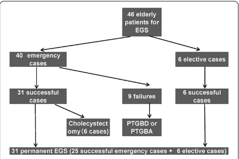
flow in the common bile duct. Emergency EGS was suc-cessful in 31 out of 40 patients (77.5%) with acute chole-cystitis. We also performed elective EGS for the purpose of permanent placement in 6 patients. We inserted a 10 cm double pig-tail stent in 7 patients and a 15 cm double pigtail stent in 30 patients. There were no imme-diate postprocedural complications, such as pancreatitis, bleeding, perforation, or cholangitis.
We performed emergency EGS in 40 patients who presented with acute severe cholecystitis and were poor candidates for cholecystectomy. In 31 patients (77.5%), emergency EGS was successful and the procedural time was 27.6 ± 15.1 min (mean ± SD). Clinical success was achieved within 3 days in all 31 patients. Their symptoms (abdominal pain, fever, and vomiting) resolved and they started to eat from the day after emergency EGS, and all 31 showed normalization of WBC and a decrease of C-reactive protein after 3 days. All were discharged from hospital within 1 week. We administered antibiotics to all 31patients for 3 days after emergency EGS, but they did not need ICU support, intubation, or inotropes. Among the 31 successful patients, we used a microcatheter for EGS in 4 difficult cases. The reasons for difficulty were se-vere stricture and kinking of the cystic duct and we ac-complished EGS in these cases by using an 0.018 inch guidewire and a microcatheter (Additional file 2). The microcatheter had an inner diameter of 0.59 mm, outer diameter of 0.89 mm, and an effective length of 2,600 mm (Figure 1). Of the 31 patients, 6 patients underwent chole-cystectomy within 2 months of EGS, while 25 patients were followed up without surgery due to their poor gen-eral condition (Figure 2).
Among the 40 patients in whom emergency EGS was attempted, it failed in 9 patients because the cystic duct branched caudally from the common bile duct (n=3), because there was severe kinking of the cystic duct (n=3), because the cystic duct was completely blocked by stones (n=2), or because of cystic duct adhe-sions (n=1). Among the 9 patients in whom EGS failed, we performed PTGBD in 6 patients and repeated PTGBA in 3 patients. Of the 6 patients receiving PTGBD, 5 underwent cholecystectomy within 2 months, while 1 patient died of aspiration pneumonia after 1 Table 1 Characteristics of the patients receiving EGS
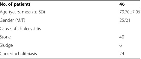
No. of patients 46
Age (years, mean ± SD) 79.70±7.96
Gender (M/F) 25/21
Cause of cholecystitis
Stone 40
Sludge 6
Choledocholithiasis 24
month. The 3 patients receiving repeated PTGBA were discharged from hospital after 2 or 3 weeks.
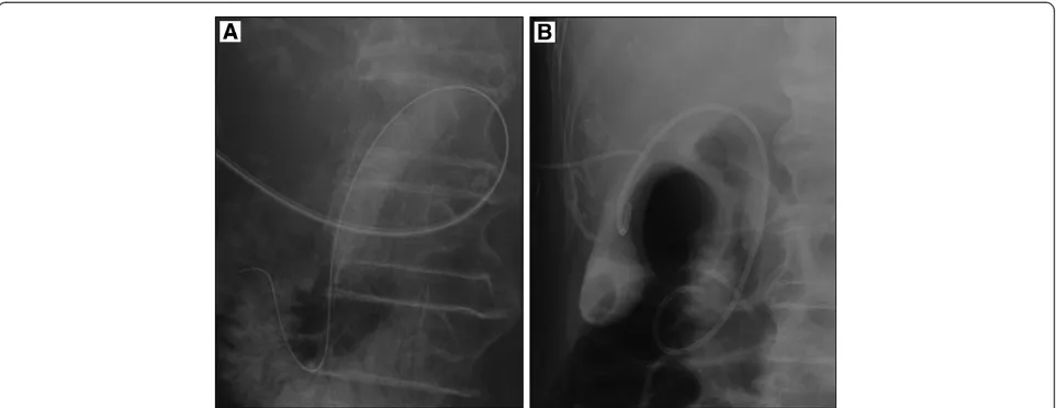
We also performed elective EGS for the purpose of per-manent placement in 6 patients (5 after PTGBD and 1 after conservative treatment) who were transferred from other hospitals within 1 week after the onset of acute cholecystitis, and the procedurewas successful in all 6 patients. In 5 patients, EGS was done by passing the guidewire antegradely into the duodenum via the PTGBD route and then the guidewire was used retrogradely (Figure 3). In 1 patient, EGS was done via peroral cholangioscopy (POCS) with a GIF-XP260 (Olympus) because the cystic duct branched caudally from the common bile duct and this was considered to be a very difficult case for EGS. The procedure was started by inserting the GIF-XP260 into the common bile duct
after endoscopic sphincterotomy. We endoscopically explored the cystic duct and then easily inserted the guidewire and the cannula into the duct. Then the guidewire was advanced retrogradely through the can-nula and coiled within the gallbladder. Afterwards, we exchanged the GIF-XP260 for the GIF-Q260, and a double pigtail stent was advanced into the gallbladder over the wire (Additional file 3).
Permanent EGS was useful for prevention of cholecyst-itis in 31 elderly patients who were poor surgical candi-dates. Their clinical characteristics and outcomes are listed in Table 2. The average age of the patients was 82.19 ± 7.15 years (mean ± SD). Sequelae of cerebral infarction were present in 14 patients, dementia in 13 patients, se-vere heart disease in 1 patient, cerebral hemorrhage in 1 patient, malignant mesothelioma in 1 patient, and polymyalgia rheumatica in 1 patient. The follow-up period ranged from 1 month to 5 years. In 30 of 31patients (96.7%), there was no recurrence of cholecystitis and 29 patients (93.5%) remained asymptomatic for 1 month to 5 years (until death or the end of the study period). Sixteen patients (51.6%) died from 1 month to 5 years after the procedure due to aspiration pneumonia, heart failure, cerebral infarction, gastric cancer, or malignant mesotheli-oma. Two patients developed late complications. Patient no. 3 developed vomiting at 2 months and 8 months after EGS. We diagnosed her as acute cholecystitis from the US and CT findings (occlusion at 2 months and migration at 8 months). After the stent was exchanged with ERC, the symptoms soon resolved. Following the second exchange, there have been no further complications. Patient no. 5 underwent removal of the gallbladder stent at 3.5 years after EGS because he developed a liver abscess (without cholecystitis). He was treated with antibiotics and the ab-scess resolved after 2 months. We did not reinsert a
Figure 1The structure of the microcatheter used for endoscopic gallbladder stenting.
Figure 2Outcome of 46 elderly patients with acute
gallbladder stent because the patient refused it. Although scheduled stent exchange was not done, the other 29 patients (93.5%) did not have any late complications.
Discussion
The definitive treatment for acute cholecystitis is chole-cystectomy [1,2], but in critically ill elderly patients, the mortality rate of emergency cholecystectomy can reach 30% [3,4]. Several authors have described the usefulness of PTGBD or PTGBA for such poor surgical candidates [5-8]. However, we sometimes encounter patients who cannot tolerate the percutaneous transhepatic approach (e.g., because of anticoagulant/antiplatelet therapy, dis-seminated intravascular coagulation, gallbladder malpo-sition, or severe contracture). Such patients are likely to develop intra-abdominal bleeding, biloma or even biliary peritonitis, if these procedures are performed. Recently, ETGBD has been reported as an alternative to PTGBD or PTGBA [9-12]. Although it is possible to wash the gallbladder via an ETGBD tube, there is a danger of the tube being pulled out by elderly patients and it cannot be left in the gallbladder over the long-term.
Otherwise EGS could be a viable strategy for long-term management of symptomatic cholelithiasis in pa-tients who are poor surgical candidates. EGS has been shown to be an effective long-term option in cirrhosis patients with cholelithiasis as a bridge to liver trans-plantation [13-17]. Studies on EGS have demonstrated a favorable clinical outcome up to 3 years after stenting without any need for routine replacement of the stent [14,15]. In a prospective follow-up study, Lee et al. [18] also showed that at least 80% of 20 patients undergoing EGS maintain stent patency without requiring stent ex-change for at least 2 years. In the present series, we performed permanent EGS in 31 elderly patients. As a
result, 30 patients (96.7%) had no recurrence of chole-cystitis and 29 patients (93.5%) remained asymptomatic for the duration of their survival. There were no late complications in 14 patients after follow up for more than 3 years, 9 patients after follow up for more than 4 years, and 1 patient after follow up for more than 5 years. Accordingly, this study has demonstrated the lon-gest good clinical outcome for up to 5 years after EGS in comparison with earlier studies without routine stent re-placement [10,12]. Stents are thought to protect against recurrent cholecystitis by occupying the lumen of the cystic duct, thereby preventing stone impaction [24]. It has also been postulated that after probable occlusion of the stent, adequate flow of bile from the gallbladder to the duodenum still occurs along the outer surface of the stent through capillary action, a phenomenon known as “wicking”[24]. Moreover, the stent may prevent bile flow from the common hepatic duct into the cystic duct. In our series, we found that the gallbladders of 12 patients became very small.
Only 2 patients developed late complications. One pa-tient (Papa-tient no. 3) had 2 episodes of recurrent chole-cystitis at 2 months and 8 months after EGS (stent occlusion at 2 months and stent migration at 8 months). When occlusion occurred at 2 months, we thought that the probable cause was placing the stent without wash-ing sludge out of the gallbladder with saline. After that, we had no recurrent cholecystitis due to occlusion of stents because we washed the gallbladder several times with 20 mL of sterile saline when EGS was performed. Stent migration at 8 months was caused by strong intes-tinal peristalsis, so we inserted the next stent as far as the fundus of the gallbladder because the previous stent had only reached the neck of the gallbladder. Subse-quently, none of the stents have fallen out. We think
that it is important to insert the stent as far as the fun-dus of the gallbladder and wash the gallbladder several times with sterile saline when EGS is performed to pre-vent stent migration and occlusion. In 1 patient (Patient no. 5), the stent was removed at 3.5 years after EGS be-cause of a liver abscess without cholecystitis. The major cause of liver abscess may have been inadequate flow in the common bile duct because EST was not performed since that patient was on antiplatelet therapy. We rec-ommend performing EST if possible and opening the stent side holes if that is impossible to obtain adequate flow in the common bile duct after EGS.
In 1984, Kozarek [25] introduced endoscopic transpapillary cannulation of the gallbladder. Subsequently, new cath-eters and guidewires have been developed to facilitate selective cannulation of the cystic duct and gallbladder [10,12,20,25]. Several other authors have attempted EGS and ETGBD in patients with acute cholecystitis [9-25]. The most recent evidenced-based report on ETGBD by Hirota et al. recommends it as an option for gallbladder drainage [2]. Clinical success was achieved in all 31 patients (100%) with acute cholecystitis by performing EGS within 3 days. Moreover, after successful emergency EGS, patients were discharged from hospital Table 2 Outcome of stenting
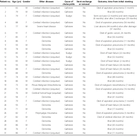
Patient no. Age (yrs) Gender Other diseases Cause of cholecystitis
Stent exchange or removal
Outcome, time from initial stenting
1 65 M Cerebral infarction (sequelae) Gallstone No Died of aspiration pneumonia (1 month)
2 79 M Cerebral infarction (sequelae) Gallstone No Alive (60 months)
3 79 F Cerebral infarction (sequelae) Sludge Yes Stent occlusion (2 months) and stent migration (8 months): alive after 2 exchanges (59 months)
4 88 F Cerebral infarction (sequelae) Gallstone No Died of aspiration pneumonia (56 months)
5 77 M Severe heart disease Gallstone Yes Liver abscess (44 months): alive after drainage
(57 months)
6 91 F Cerebral infarction (sequelae) Gallstone No Died of gastric cancer, 24 months
7 85 F Dementia Gallstone No Alive (56 months)
8 86 F Dementia Gallstone No Died of aspiration pneumonia (11 months)
9 93 M Dementia Gallstone No Died of aspiration pneumonia (51 months)
10 78 M Dementia Gallstone No Alive (52 months)
11 85 M Cerebral infarction (sequelae) Sludge No Died of heart failure (22 months)
12 76 F Cerebral infarction (sequelae) Sludge No Alive (52 months)
13 83 M Cerebral infarction (sequelae) Sludge No Died of heart failure (2 months)
14 93 M Dementia Gallstone No Died of heart failure (26 months)
15 88 F Cerebral infarction (sequelae) Gallstone No Died of heart failure (35 months)
16 84 M Dementia Gallstone No Died of aspiration pneumonia (2 months)
17 74 M Cerebral infarction (sequelae) Gallstone No Alive (48 months)
18 86 M Cerebral infarction (sequelae) Gallstone No Alive (46 months)
19 73 M Pleura Malignant Mesothelioma Gallstone No Died of malignant mesothelioma (7 months)
20 75 M Cerebral infarction (sequelae) Gallstone No Died of aspiration pneumonia (16 months)
21 76 M Cerebral hemorrhage (sequelae) Gallstone No Alive (44 months)
22 94 F Dementia Gallstone No Alive (41 months)
23 79 F Cerebral infarction (sequelae) Gallstone No Died of aspiration pneumonia (1 month)
24 93 F Dementia Gallstone No Died of heart failure (26 months)
25 71 M Dementia Gallstone No Alive (17 months)
26 82 F Polymyalgia rheumatica Gallstone No Died of aspiration pneumonia (5 months)
27 82 M Dementia Gallstone No Died of cerebral infarction (29 months)
28 85 F Dementia Gallstone No Alive (48 months)
29 86 M Dementia Gallstone No Alive (43 months)
30 76 F Dementia Gallstone No Alive (26 months)
within 1 week. Thus, EGS may shorten the duration of hospitalization compared with PTGBD or ETGBD.
We consider that EGS and ETGBD are best indicated for patients with acute cholecystitis in whom transhepatic route cannot be used. Ferritis et al. [9], Toyota et al. [10], Kjaer et al. [11], and Itoi et al. [12] reported ETGBD suc-cess rates of 89% (16/18), 82% (18/22), 70.6% (24/34), and 83.7% (38/43) in smaller series. Our success rate for emer-gency EGS was 77.5% (31/40), which is similar to the suc-cess rate for ETGBD. We believe that EGS is slightly more difficult than ETGBD because the stent is harder to ma-nipulate compared with an ETGBD tube (a 5Fr soft drain tube is often used for ETGBD). We encountered 3 cases in which we could not place a stent in the gallbladder des-pite inserting the guidewire, because of cystic duct occlu-sion by stones in 2 cases and severe kinking of the cystic duct in 1 case. Moreover, emergency EGS for acute chole-cystitis is more difficult than elective EGS because the cys-tic duct often shows stricture or obstruction in acute patients. In our series, the success rate of emergency EGS was 77.5% (31/40) and this was lower than that of elective EGS (100%, 6/6).
EGS has not become very popular so far. The main reason is difficulty in negotiating the cystic duct and gallbladder with the guide wire. We used microcatheters for 4 difficult patients, and used POCS for 1 patient. The microcatheters used in 4 cases were designed for abdominal angiography and were adapted to the EGS procedure. After the microcatheter and 0.018 inch guidewire were advanced through the ERCP cannula, we could pass severe strictures and kinks of the cystic duct in all 4 cases. Thus, a microcatheter is useful for contrast studies of the cystic duct and for crossing strictures and kinks, but more highly visible and softer microcatheters should be de-veloped. Barkay et al. [26] reported successful cannula-tion of the cystic duct by visualizacannula-tion with the SpyGlass cholangiopancreatography system (SpyGlass Direct Visualization System; Microvasive Endoscopy, Boston Scientific, Natick, MA) with EGS in a patient with cystic duct obstruction and acute cholecystitis. In 1 of our patients, a novel EGS technique using POCS was tried. POCS enabled us to endoscopically explore the cystic duct and easily insert the guidewire into the duct by using the angulation and rotation of the GIF-XP260. In our opinion, use of a microcatheter and POCS could increase the technical success rate of EGS.
Our study showed that EGS can be effective for eld-erly patients with acute cholecystitis who are poor sur-gical candidates. Generally, EGS is associated with a higher risk of aspiration pneumonia in comparison with PTGBA because EGS is performed by peroral en-doscopy. EGS should be performed as rapidly as pos-sible and improvement of EGS technology is needed in the future.
Conclusions
We conclude that EGS can be effective for elderly pa-tients with acute cholecystitis who are poor surgical can-didates and the stent can be left for many months to several years. However, further investigations will be needed to establish the appropriate role of EGS.
Additional files
Additional file 1:Standard EGS procedure.Cannulating the bile duct and cystic duct with an over-the-wire ERCP cannula. The guide wire was advanced retrogradely through the cannula into the gallbladder and a 7Fr double pig-tail stent was then advanced.
Additional file 2:Difficult EGS case using a microcatheter.We accomplished EGS by using an 0.018 inch guide wire and microcatheter to pass through a severe stricture of the cystic duct.
Additional file 3:EGS via peroral cholangioscopy using a GIF-XP260.
We inserted the GIF-XP260 into the common duct, explored the cystic duct, and advanced the guide wire into the gallbladder. Then we exchanged GIF-XP260 for GIF-Q260, and advanced a double pigtail stent.
Abbreviations
CT:Computed tomography; EGS: Endoscopic gallbladder stenting; ERC: Endoscopic retrograde cholangiography; ERCP: Endoscopic retrograde cholangiopancreatography; EST: Endoscopic sphincterotomy;
ETGBD: Endoscopic transpapillary naso-gallbladder drainage; POCS: Peroral cholangioscopy; PTGBD: Percutaneous transhepatic gallbladder drainage; PTGBA: Percutaneous transhepatic gallbladder aspiration; SD: Standard deviation; US: Ultrasonography.
Competing interests
The authors declare that there are no competing interests.
Authors’contributions
SM, YA, and MO contributed to research design. SM, RN, TM, MO, and YA collected the data. SM drafted the manuscript. MH, RN, TM, YA, and MO contributed to revising the manuscript. All authors approved the final version.
Acknowledgements
The authors declared that they haved no competing interests.
Author details
1Department of Gastroenterological Medicine, Japan Labour Health and
Welfare Organization Niigata Rosai Hospital, 1-7-12 Touncho, Joetsu, Niigata 942-8502, Japan.2Third Department of Internal Medicine, University of Occupational and Environmental Health, 1-1 Iseigaoka, Yahata-nishi-ku, Kitakyushu, Fukuoka 807-8555, Japan.
Received: 6 December 2012 Accepted: 10 April 2013 Published: 12 April 2013
References
1. Nahrwold DL:Acute cholecystitis. InTextbook of Surgery.15th edition. Edited by Sabiston D Jr. Philadelphia: WB Saunders; 1997:1126–1131. 2. Hirota M, Takada T, Kawarada Y, Nimura Y, Miura F, Hirata K, Mayumi T,
Yoshida M, Strasberg S, Pitt H, Gadacz TR, de Santibanes E, Gouma DJ, Solomkin JS, Belghiti J, Neuhaus H, Büchler MW, Fan ST, Ker CG, Padbury RT, Liau KH, Hilvano SC, Belli G, Windsor JA, Dervenis C:Diagnostic criteria and severity assessment of acute cholecystitis: Tokyo guidelines.
J Hepatobiliary Pancreat Surg2007,14:78–82.
3. Houghton PWG, Jenkinson LR, Donaldson LA:Cholecystectomy in the elderly: a prospective study.Br J Surg1985,72:220–222.
4. Frazee RC, Nagorney DM, Mucha P Jr:Acute calculus cholecystitis.Mayo Clin Proc1989,64:163–167.
cholecystostomy and interval laparoscopic cholecystectomy.Surg Endosc
1996,10:1185–1188.
6. McGahan JP, Walter JP:Diagnostic perctaneous aspiration of the gallbladder.Radiology1985,155:619–622.
7. Chopra S, Dodd GD 3rd, Mumbower AL, Chintapalli KN, Schwesinger WH, Sirinek KR, Dorman JP, Rhim H:Treatment of acute cholecystitis in non-critically ill patients at high surgical risk: comparison of clinical outcomes after gallbladder aspiration and after perctaneous cholecyststomy.AJR Am J Roentgenol2001,176:1025–1031.
8. Ito K, Fujita N, Noda Y, Kobayashi G, Kimura K, Sugawara T, Horaguchi J: Percutaneous cholecystostomy versus gallbladder aspiration for acute cholecystitis: a prospective randomized controlled trial.AJR Am J Roentgenol2004,183:193–196.
9. Feretis C, Apostolidis N, Mallas E, Manouras A, Papadimitriou J:Endoscopic drainage of acute obstructive cholecystitis in patients with increased operative risk.Endocopy1993,25:392–395.
10. Toyota N, Takada T, Amano H, Yoshida M, Miura F, Wada K:Endoscopic naso-gallbladder drainage in the treatment of acute cholecystitis: alleviates inflammation and fixes operator’s aim during early laparoscopic cholecystectomy.J Hepatobiliary Pancreat Surg2006, 13:80–85.
11. Kjaer DW, Kruse A, Funch-Jensen P:Endoscopic gallbladder drainage of patients with acute cholecystitis.Endoscopy2007,39:304–308. 12. Itoi T, Sofuni A, Itokawa F, Tsuchiya T, Kurihara T, Ishii K, Tsuji S, Ikeuchi N,
Tsukamoto S, Takeuchi M, Kawai T, Moriyasu F:Endoscopic transpapollary gallbladder drainage in patients with acute cholecystitis in whom percutaneous transhepatic approach is contraindicated or anatomically impossible (with video).Gastrointest Endosc2008,68:455–460.
13. Conway JD, Russo MW, Shrestha R:Endoscopic stent insertion into the gallbladder for symptomatic gallbladder disease in patients with end-stage liver disease.Gastrointest Endosc2005,61:32–36.
14. Schlenker C, Trotter JF, Shah RJ, Everson G, Chen YK, Antillon D, Antillon MR: Endoscopic gallbladder stent placement for treatment of symptomatic cholelithiasis in patients with end-stage liver disease.Am J Gastroenterol
2006,101:278–283.
15. Shrestha R, Trouillot TE, Everson GT:Endoscopic stenting of the gallbladder for symptomatic gallbladder disease in patients with end-stage liver disease awaiting orthotopic liver transplantation.Liver Transplant Surg1999,5:275–281.
16. Shrestha R, Bilir BM, Everson GT:Endoscopic stenting of gallbladder for symptomatic cholelithiasis in patients with end-stage liver disease awaiting orthotopic liver transplantation.Am J Gastroenterol1996, 91:595–598.
17. Gaglio PJ, Buniak B, Leevy CB:Primary endoscopic retrograde cholecystoendoprosthesis: a nonsurgical modality for symptomatic cholelithiasis in cirrhotic patients.Gastrointest Endosc1996, 44:339–342.
18. Lee TH, Park DH, Lee SS, Seo DW, Park SH, Lee SK, Kim MH, Kim SJ: Outcome of endoscopic transpapillary gallbladder stenting for symptomatic gallbladder disease: a multicenter prospective follow-up study.Endoscopy2011,43:702–708.
19. Kozarek RA:The future of invasive pancreatobiliary endoscopy.J Clin Gastroenterol1988,10:153–157.
20. McCarthy JH, Miller GL, Laurence BH:Cannulation of the biliary tree, cystic duct and gallbladder using a hydrophilic polymer-coated steerable guidewire.Gastrointest Endosc1990,36:386–389.
21. Tamada K, Seki H, Sato K, Kano T, Sugiyama S, Ichiyama M, Wada S, Ohashi A, Tomiyama G, Ueno A:Efficacy of endoscopic retrograde
cholecystoendoprosthesis (ERCCE) for cholecystitis.Endoscopy1991, 23:2–3.
22. Kalloo AN, Thuluvath PJ, Pasricha PJ:Treatment of high-risk patients with symptomatic cholelithiasis by endoscopic gallbladder stenting.
Gastrointest Endosc1994,40:608–610.
23. Feretis C, Apostolidis N, Mallas E, Manouras A, Papadimitriou J:Endoscopic drainage of acute obstructive cholecystitis in patients with increased operative risk.Endoscopy1993,25:392–395.
24. Elmunzer BJ, Novelli PM, Taylor JR, Piraka CR, Shields JJ:Percutaneous cholecystostomy as a bridge to definitive endoscopic gallbladder stent placement.Gastrointest Endosc2011,9:18–20.
25. Kozarek RA:Selective cannulation of the cystic duct at the time of ERCP.
J Clin Gastroenterol1984,6:37–40.
26. Barkay O, Bucksot L, Sherman S:Endoscopic transpapillary gallbladder drainage with SpyGlass cholangiopancreatoscopy system.Gastrointest Endosc2009,70:1039–1040.
doi:10.1186/1471-230X-13-65
Cite this article as:Maekawaet al.:Endoscopic gallbladder stenting for acute cholecystitis: a retrospective study of 46 elderly patients aged 65 years or older.BMC Gastroenterology201313:65.
Submit your next manuscript to BioMed Central and take full advantage of:
• Convenient online submission
• Thorough peer review
• No space constraints or color figure charges
• Immediate publication on acceptance
• Inclusion in PubMed, CAS, Scopus and Google Scholar
• Research which is freely available for redistribution