R E S E A R C H A R T I C L E
Open Access
The bicipital groove as a landmark for
reconstruction of complex proximal
humeral fractures with hybrid double plate
osteosynthesis
Jan Theopold
1*, Bastian Marquaß
1, Johannes Fakler
1, Hanno Steinke
2, Christoph Josten
1and Pierre Hepp
1Abstract
Background:Complex proximal humerus fractures with metaphyseal comminution remain challenging regarding
reduction and stability. In most fracture patterns the hard bone of the bicipital groove remains intact. In this case series, we describe a novel technique of hybrid double plate osteosynthesis of complex proximal humerus fractures with metaphyseal comminution.
Methods:In randomly chosen shoulder specimens and synthetic bones, pilot studies for evaluation of the
feasibility of the technique were performed. Between 4/2010 and 1/2012 10 patients underwent hybrid double plate osteosynthesis. Seven patients (4 male, 3 female, mean age was 50 years (range 27–73)) were available for retrospective analysis. Based on plain radiographs (anterior-posterior and axial view), the fractures were classified according to the Orthopaedic Trauma Association classification (OTA) and by descriptive means (head-split variant (HS), diaphyseal extension or comminution (DE)).
Results: Follow-up radiographs demonstrated complete fracture healing in six patients and one incomplete
avascular necrosis. None of the patients sustained loss of reduction. Three patients where reoperated. The medium, not adapted, Constant score was 80 Points (58–94). Patients subjective satisfaction was graded mean 3 (range: 0–6) in the visual analog scoring system (VAS).
Conclusion: The technique of hybrid double plate osteosynthesis using the bicipital groove as anatomic landmark may re-establish shoulder function after complex proximal humerus fractures in two dimensions. Firstly the anatomy is restored due to a proper reduction based on intraoperative landmarks. Secondly additional support by the second plate may provide a higher stability in complex fractures with metaphyseal comminution.
Keywords:Proximal humerus fracture, Metaphyseal comminution, Double plate, Locked plate
Background
Complex proximal humerus fractures with metaphyseal extension or comminution are challenging regarding reduction and stability of the osteosynthesis [1, 2]. The metaphysis may be broken into several fragments and anatomical landmarks for reduction may be absent. In addition there will not be a sufficient metaphyseal substance to provide support for fixation. The missing
restoration of the medial cortical support has been identified being responsible for an increased failure rate [1, 2] and nonunions [3] of the proximal Humerus. The initial calcar comminution is an independent prognostic factor for bad clinical outcome [1, 2]. This may lead to secondary varus dislocation with biomechanical alteration due to decreased supraspinatus efficiency [4]. Whereas locking plates with placement of calcar screws [2, 5] and locking plates in combination with fibular bone grafts [6, 7] have been described to increase the medial sup-port, hybrid double-plating has not been considered for improving stability, yet.
* Correspondence:Jan.theopold@medizin.uni-leipzig.de
1Department of Orthopedics, Trauma and Plastic Surgery, University of
Leipzig, Liebigstrasse 20, 04103 Leipzig, Germany
Full list of author information is available at the end of the article
In this report, we describe a technique of double plate osteosynthesis of complex proximal humerus fractures with metaphyseal comminution. In the first step the bicipital groove is used as an anatomical landmark for reduction of the anterior column and a one-third tubular plate is placed into the groove in an inverted fashion. Secondly the reduction is completed and the fracture is fixed by a laterally placed locking plate.
Methods
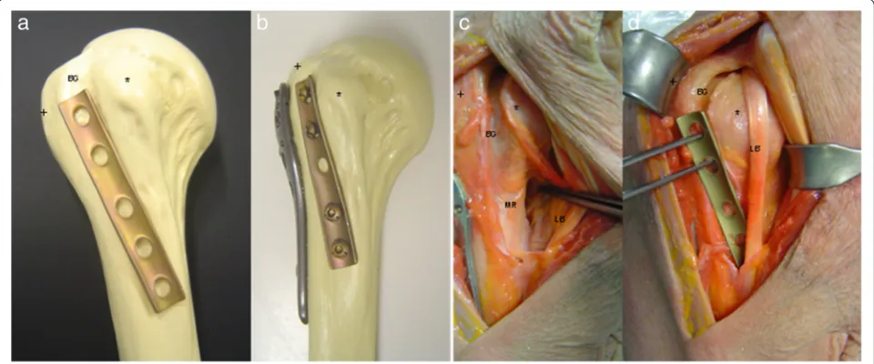
In randomly chosen shoulder specimens (donors gave directed consent that their cadavers were to be used in teaching or for research projects at the Institute of anat-omy) and synthetic bones, pilot studies for evaluation of the feasibility of the technique were performed (Fig. 1) The double plate osteosynthesis involves a one third tubular plate placed into the bicipital groove in an inverted fashion. The locking plate is fixed laterally in classical fashion.
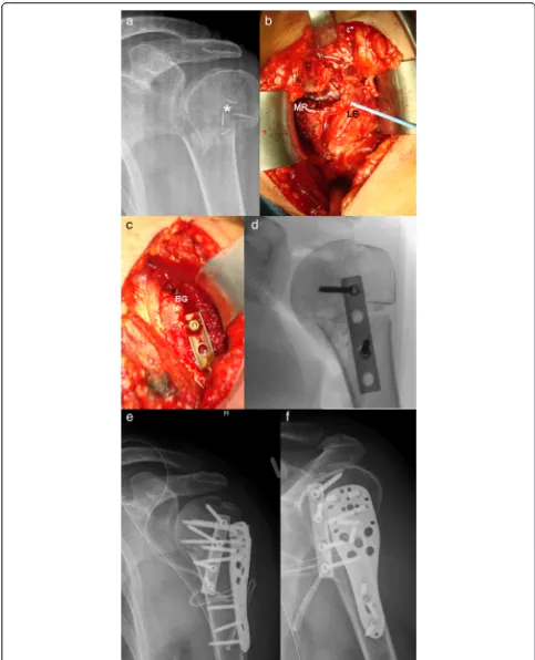
The patient is placed in beachchair position and posi-tioned such that anteroposterior and axillary views of the proximal humerus can be easily obtained using C-arm fluoroscopy. The fracture is exposed through a deltopec-toral approach. The long biceps tendon is identified and followed proximally. The rotator interval is partially opened and a biceps tendon tenotomy is performed. Now the anterior aspect of the fracture including parts of the medial metaphysical region can be visualised and analysed (Fig. 2b). Fracture reduction is performed by standard methods, including Kirschner wire and eleva-tors to control the humeral head fragment. A one third tubular plate is placed into the groove in an inverted
fashion and fixed to the proximal part with 2 screws. Further reduction is achieved by fixing the plate distally (Fig. 2c, d). The pectoralis major tendon may be par-tially released from the humeral shaft to achieve suffi-cient overview. Further reduction of the fracture is accomplished (e.g. greater tuberosity) and the locking plate is applied laterally (Fig. 2e, f ). Special attention should be paid to the position of the implant. Once the fracture has been provisionally reduced and the implant temporarily fixed with Kirschner wires, the reduction and implant placement should be verified on C-arm fluoroscopy. Depending on the patient’s age a soft tissue tenodesis of the biceps tendon is performed beside the tubular plate.
After surgery patients had individual patient-related postoperative management. For better comfort, the patient’s arm was placed in a sling for a maximum of 2 weeks. Passive and active range of motion exercises were started after surgery, depending on pain and ac-tivity level.
Patients
Between 4/2010 and 1/2012 10 patients underwent hybrid double plate osteosynthesis. All patients were treated by one surgeon (P.H.). In the present case series, the indica-tion for double-plating was individual and depending on intraoperative reduction. Retrospective review of patient records and radiographs was approved by the institutional review board of the medical faculty of the University of Leipzig, Germany (no. 062-14-10032014). All included
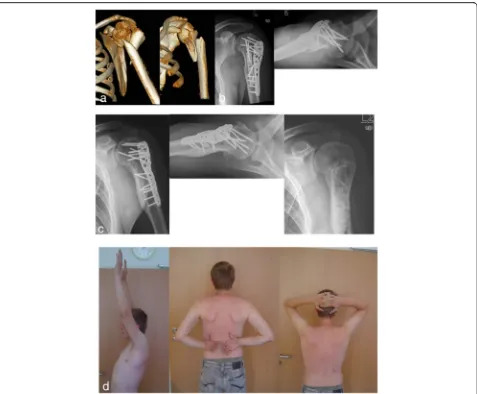
patients gave their informed consent. Consent to publish Fig. 3 and the patient information contained in Table1was also obtained. Three patients were excluded (one died, one was lost to follow-up and one patient sustained a pathological fracture due to a mamma cancer). Thus, seven patients (4 male, 3 female, mean age was 50 years (range 27–73)) were available for retrospective analysis. Based on plain radiographs (anterior-posterior and axial view), the fractures were classified according to the Ortho-paedic Trauma Association classification (OTA) and by descriptive means (head-split variant (HS), diaphyseal extension or comminution (DE)).
Results
Table 1 summarizes the demographic, injury and fol-low up data for each patient in our series. Folfol-low-up
radiographs demonstrated complete fracture healing in six patients and one incomplete avascular necrosis. None of the patients sustained loss of reduction. Three patients where reoperated: Two elective mater-ial removal after one year (Fig. 3) and one removal of perforating screws after 9 months and complete ma-terial removal with arthrolysis of the shoulder joint after 21 months. The medium, not adapted, Constant score is 80 Points (58–94). The medium Constant score of the non-affected side was 91 Points (84–96). Functional results revealed a mean range of motion (flexion/extension/adduction/abduction/internal rota-tion, external rotation) of 150 (110–170)/40 (30–50)/ 40 (30–50)/140 (90–180)/60 (50–90)/and 80 (40–90)°. Patients subjective satisfaction was graded mean 3 (range: 0–6) in the visual analog scoring system (VAS).
Discussion
Taking into account that adequate reduction and stable conditions in osteosynthesis of proximal humerus frac-tures may be crucial for preservation and revascularization of the humeral head [8], the double plate osteosynthesis with consideration of the bicipital groove as anatomical landmark has been developed to restore the anatomy and optimize the stability after complex proximal humerus fractures.
Double plating with two one-third tubular plates in 90° configuration has previously been described in clin-ical studies providing high stability and allowing early mobilization [9]. On the other hand biomechanical studies pointed out superior biomechanical properties of locking plates when compared to double plate with one-third tubular plates [10]. Whereas hybrid plating– the combination of locking and nonlocking screws in the same plate–has been shown to provide an attract-ive alternatattract-ive to an all-locked construct [11], to our knowledge the combination of a locking plate with non-locking tubular plate has not been described, yet. This hybrid configuration of the one-third tubular plate ana-tomically fitting in the bicipital groove and the advantages of the locking plate in osteoporotic bone may lead to higher strength of the construct.
Whereas the bicipital groove is used as an anatomical landmark to restore humeral head retroversion when treating complex proximal humeral fractures with arthro-plasty [12, 13], it has not been taken into account as land-mark for the reconstruction with plate osteosynthesis. In most fracture patterns the hard bone of the bicipital groove remains intact [14]. The inverted one-third tubular plate fits into this sulcus. Nevertheless anatomical varia-tions have to be taken into account [15] and the tubular plate may not always optimally fit. One may criticize the
tenotomy of the biceps tendon for exposure of the sulcus. On the other hand the tendon has recently been identified as potential source for pain in complex proximal humerus fractures [16] and is routinely performed in hemiarthro-plasty. In a series of arthroscopic material removal 30 % tenotomies were performed due to biceps tendon path-ologies after proximal humerus fractures [17]. In our ex-perience the biceps tendon tenotomy provides a greater exposure of the metaphyseal comminution zone, enables a distinct visualisation and palpation of the joint surface through the rotator interval and this may potentially re-veal hidden head-split variants.
Limitations: Our study also has the same inherent weakness that is seen in many other retrospective stud-ies. One concern may be the lack of any controls. In order to achieve a desired test power of 0.95, with an expected effect size d of 0.5 a total sample size of n = 176 (88 per group) would have been required for patients and controls.
In our series, the patients treated with double plate technique sustained complex proximal humerus fracture. Retrospectively, 57 % (n = 4) showed a head-split variant in combination with the metaphyseal comminution. In the literature this kind of fracture are often treated with arthroplasty [18, 19]. Except of one patient with persisting pain and functional impairment, the overall Constant score was predominately excellent and good. The functional results were comparable with current literature[8, 20].
One further limitation of the presented case series is the lack of a reliable algorithm for the use of double plate osteosynthesis. The decision making Table 1Overall characteristics and results of the seven patients with minimum 12 months follow-up
Case no.
Age Sex OTA proximal segment classification Fracture morphology Follow-up (months) Constant score (affected/ unaffected side)
Shoulder range of motion Cause of injury
Complications/ Reoperation Flexion Abduction Internal
rotation External rotation
1 24 M 11-B2.3 DE 13 94/96 170 180 90 90 MVA Material removal after 12 months 2 65 F 11-C2.2 DE, HS 35 72/93 160 90 90 50 F
3 73 M 11-C2.1 DE, HS 34 79/85 160 150 90 50 F
4 39 F 11-C2.1 DE, HS 28 85/93 170 160 90 50 BA AVN, Screw perforation, frozen shoulder, 2 reoperations 5 36 M 11-B2.3 DE 18 84/93 150 150 40 60 FFH
6 63 M 11-C2.1 DE, HS 26 91/94 160 130 90 60 BA
7 51 F 11-B2.3 DE 24 58/84 110 100 90 50 F Material removal after 13 months
was individual in each case and was performed, when reduction or stability was subjectively unsatisfying.
Conclusion
In conclusion, the technique of hybrid double plate osteo-synthesis using the bicipital groove as anatomic landmark may re-establish shoulder function after complex proximal humerus fractures in two dimensions. Firstly the anatomy is restored due to a proper reduction based on intraopera-tive landmarks. Secondly additional support by the second plate may provide a higher stability in complex fractures with metaphyseal comminution. Ultimately, comparative biomechanical evaluations and long term follow-up stud-ies in vivo are required to delineate the advantages.
Competing interests
The authors declare that they have no competing interests.
Authors’contributions
Data collection: JT, BM, HS, PH. Data analysis: JT, CJ, PH. Drafting manuscript: JT, BM, HS, JK, CJ, PH. Approving final version: JT, BM, HS, JK, CJ, PH. PH takes responsibility for the integrity of the data.
Acknowledgements
We acknowledge support from the German Research Foundation (DFG) and Universität Leipzig within the program of Open Access Publishing.
Disclosures
All authors state that they have no conflict of interest and disclose all restrictions on full access for all authors to all raw data.
Author details
1Department of Orthopedics, Trauma and Plastic Surgery, University of
Leipzig, Liebigstrasse 20, 04103 Leipzig, Germany.2Institute of Anatomy,
University of Leipzig, Liebigstrasse 13, 04103 Leipzig, Germany.
Received: 6 August 2015 Accepted: 8 March 2016
References
1. Krappinger D, Bizzotto N, Riedmann S, et al. Predicting failure after surgical fixation of proximal humerus fractures. Injury. 2011;42:1283–8.
2. Gardner MJ, Weil Y, Barker JU, et al. The importance of medial support in locked plating of proximal humerus fractures. J Orthop Trauma. 2007;21:185–91.
3. Court-Brown CM, McQueen MM. Nonunions of the proximal humerus: their prevalence and functional outcome. J Trauma. 2008;64:1517–21. 4. Voigt C, Kreienborg S, Megatli O, et al. How does a varus deformity of the
humeral head affect elevation forces and shoulder function? A biomechanical study with human shoulder specimens. J Orthop Trauma. 2011;25:399–405.
5. Osterhoff G, Ossendorf C, Wanner GA, et al. The calcar screw in angular stable plate fixation of proximal humeral fractures–a case study. J Orthop Surg Res. 2011;6:50.
6. Osterhoff G, Baumgartner D, Favre P, et al. Medial support by fibula bone graft in angular stable plate fixation of proximal humeral fractures: an in vitro study with synthetic bone. J Shoulder Elbow Surg. 2011;20:740–6. 7. Gardner MJ, Boraiah S, Helfet DL, et al. Indirect medial reduction and
strut support of proximal humerus fractures using an endosteal implant. J Orthop Trauma. 2008;22:195–200.
8. Bastian JD, Hertel R. Osteosynthesis and hemiarthroplasty of fractures of the proximal humerus: outcomes in a consecutive case series. J Shoulder Elbow Surg. 2009;18:216–9.
9. Wanner GA, Wanner-Schmid E, Romero J, et al. Internal fixation of displaced proximal humeral fractures with two one-third tubular plates. J Trauma. 2003;54:536–44.
10. Hessmann MH, Korner J, Hofmann A, et al. Angle-fixed plate fixation or double-plate osteosynthesis in fractures of the proximal humerus: a biomechanical study. Biomedizinische Technik Biomedical Engineering. 2008;53:130–7.
11. Doornink J, Fitzpatrick DC, Boldhaus S, et al. Effects of hybrid plating with locked and nonlocked screws on the strength of locked plating constructs in the osteoporotic diaphysis. J Trauma. 2010;69:411–7.
12. Angibaud L, Zuckerman JD, Flurin PH, et al. Reconstructing proximal humeral fractures using the bicipital groove as a landmark. Clin Orthop Relat Res. 2007;458:168–74.
13. Kontakis GM, Damilakis J, Christoforakis J, et al. The bicipital groove as a landmark for orientation of the humeral prosthesis in cases of fracture. J Shoulder Elbow Surg. 2001;10:136–9.
14. Edelson G, Kelly I, Vigder F, et al. A three-dimensional classification for fractures of the proximal humerus. J Bone Joint Surg Br. 2004;86:413–25. 15. Wafae N, Atencio Santamaria LE, Vitor L, et al. Morphometry of the
human bicipital groove (sulcus intertubercularis). J Shoulder Elbow Surg. 2010;19:65–8.
16. Tosounidis T, Hadjileontis C, Georgiadis M, et al. The tendon of the long head of the biceps in complex proximal humerus fractures: a histological perspective. Injury. 2010;41:273–8.
17. Lill H, Katthagen C, Voigt C. Technique and value of arthroscopic implant removal in the shoulder. Orthopade. 2011;40:79–84.
18. Ockert B, Biermann N, Haasters F, Mutschler W, Braunstein V. Reverse shoulder arthroplasty for primary fracture treatment. Displaced three and four part fractures of the proximal humerus in the elderly patient. Unfallchirurg. 2013;116:684–90.
19. Reuther F, Kohut G, Nijs S. Newly developed modular reverse fracture endoprosthesis in non-reconstructable humeral head fracture in old people. Oper Orthop Traumatol. 2014;26:369–82. quiz 382–384.
20. Ockert B, Siebenbürger G, Kettler M, Braunstein V, Mutschler W. Long-term functional outcomes (median 10 years) after locked plating for displaced fractures of the proximal humerus. J Shoulder Elbow Surg. 2014;23:1223–31.
• We accept pre-submission inquiries
• Our selector tool helps you to find the most relevant journal • We provide round the clock customer support
• Convenient online submission • Thorough peer review
• Inclusion in PubMed and all major indexing services • Maximum visibility for your research
Submit your manuscript at www.biomedcentral.com/submit