Open Access
Research article
The creation of new rotation arc to the rat latissimus dorsi
musculo-cutaneous flap with delay procedures
Eray Copcu*
1, Nazan Sivrioglu
1, Alper Aktas
2and Yucel Oztan
2Address: 1Department of Plastic and Reconstructive Surgery, Medical Faculty, Adnan Menderes University, 09100, Aydin, TURKEY and 2Department of Plastic and Reconstructive Surgery, Izmir Ataturk Training Hospital, Izmir, TURKEY
Email: Eray Copcu* - copcu@lycos.com; Nazan Sivrioglu - nsivrioglu@adu.edu.tr; Alper Aktas - aaktas@msn.com; Yucel Oztan - yuceloztan@yahoo.com
* Corresponding author
Abstract
Background: Latissimus dorsi musculocutaneous flap is one of the most frequently performed reconstructive techniques in surgery. Latissimus dorsi muscle has two arcs of rotation. It is classified as type V muscle. This muscle can be elevated on the thoracodorsal artery to cover large defects in the anterior chest and also, the muscle can be elevated on the segmental vessels to cover midline defects posteriorly. The aim of this study was to create a new arc of rotation on a vertical axis for the muscle and investigate effectiveness of vascular and chemical delays on the latissimus dorsi muscle flap with an inferior pedicle in an experimental rat model. We hypothesized that the latissimus dorsi muscle would be based on inferior pedicle by delay procedures.
Methods: We tested two different types of delay: vascular and combination of vascular and chemical. We also tried to determine how many days of "delay" can elicit beneficial effects of vascular and combination delays in an inferior pedicled latissimus dorsi musculocutaneous flap. To accomplish this, 48 male Sprague-Dawley rats were randomly subjected to vascular or combination delay (vascular and chemical). In addition, one ear of each rat was assigned into a delay procedure and the other ear was used as a control. Results were evaluated macroscopically, and micro-angiography and histological examinations were also performed. As a result, there was a significant difference in viable flap areas between vascular delay alone and control groups (p < 0.05).
Results: The higher rate of flap viability was obtained in seven-day vascular delay alone. However, there was no significant difference in the viability between seven-day vascular delay and five-day vascular delay (p < 0.05), so the earliest time when the flap viability could be obtained was at five days. The rate of flap viability was significantly higher in the vascular delay combined with chemical delay than the control group (p < 0.05).
Conclusion: The combination of vascular and chemical delays increased the rate of viability. Nevertheless, there was no significant difference between vascular delay alone and combination of vascular and chemical delays. Chemical delay did not significantly decrease the delay period. Better histological and microangiographical results were achieved in delay groups compared to control groups. We concluded that the arch of the latissimus dorsi musculocutaneous flap can be changed and the flap can be used for various purposes with the delay procedures.
Published: 10 December 2003
BMC Surgery 2003, 3:11
Received: 09 July 2003 Accepted: 10 December 2003
This article is available from: http://www.biomedcentral.com/1471-2482/3/11
BMC Surgery 2003, 3 http://www.biomedcentral.com/1471-2482/3/11
Background
Musculocutaneous flaps are indispensable to tissue repair in plastic surgery, and latissimus dorsi flap is one of the most frequently used musculocutaneous flaps. Although it can be used as a free flap, pedicled latissimus dorsi flap is simple and rapid to perform and does not require sophisticated equipment and expertise. Mathes and Nahai divided the muscles into five according to their vascular features [1]. Latissimus dorsi is a type five muscle; i.e. it has one dominant pedicle and more than one secondary segmental vascular pedicle [2]. Its dominant pedicle is thoraco-dorsal artery, a branch of subscapular artery, and latissimus dorsi based on this artery can be used success-fully in repair of defects of the head, neck, chest, abdomen and upper extremities and in cardiomyoplasty. It is also known that this muscle is supplied by segmental vessels. Reversed pedicled latissimus dorsi flap is obtained by ele-vation of the muscle from the segmental pedicle. It is used to repair small defects of the muscles in the lumbar and upper sacral areas [3-6]. The most serious disadvantage of the musculocutaneous flaps is that their rotational arcs are narrow. However, when the pivotal point, which deter-mines rotational arcs, is changed, the rotational arcs of the flaps are changed and the flaps can be used to repair many other defects. To increase flap survival and safety is one of the most interesting issues of plastic surgery. The proce-dures used to achieve this purpose are based on a phe-nomenon of delay, the mechanism of which has not been clearly understood. To increase flap survival, procedures of delay can be used. The flap becomes used to hypoxia,
to which it will be exposed later, and thus its chance to survive will increase. These procedures can be described as elevation of the flap and its transfer to the recipient area in more than one session. It can be surgical or chemical [7,8]. Although especially vascular delay is one of the extensively studied subjects in Plastic Surgery, little has been documented about effects and optimal timing of vascular delay. The aim of this study was to change the pivotal point of latissimus dorsi and create a new rota-tional arc in rats using vascular delay and thus to increase its use and to shorten the period of delay using chemical delay.
Methods
This study was performed on 48 Sprague-Dawley rats, weighing between 250–300 gr, in the Laboratory of Exper-imental Studies, in the Department of Plastic and Recon-structive Surgery, Atatürk Training and Investigation Hospital, Izmir. Approval was obtained from the ethical committee of the hospital. The rats were randomly assigned into three groups: Groups I, II and III. General characteristics of the groups are listed in Table 1. Animals were kept in separate cages in temperature, light and air-flow-regulated rooms. Animals were anesthetized for all surgical procedures using sodium pentobarbital (65 mg/ kg intraperitoneally) and the procedures were performed under aseptic conditions. Animals received a single dose of crystallized penicillin 500 U following the operations. On completion of experiments, rats were killed with an overdose of sodium pentobarbital.
Table 1: Details of groups.
Groups No: Description:
Group I Effect of vascular delay on the flap
Ivd 6 Vascular delay group (7 days)
Ivdc Control group (the other side of the same
animal)
Group II Comparisons of different vascular delay
periods
II vd1 6 Vascular delay group (1 day)
II vd1c Control group (the other side of the same
animal)
II vd3 6 Vascular delay group (3 days)
II vd3c Control group (the other side of the same
animal)
II vd5 6 Vascular delay group (5 days)
II vd5c Control group (the other side of the same
animal)
Group III Combination of vascular and chemical
delays
III vdc1 6 1 day-combined delays
III vdc3 6 3 day-combined delays
III vdc5 6 5 day-combined delays
Latissimus dorsi muscle of the rats was 45 × 20 mm in size. The main pedicle, thoracodorsal artery, enters through the insertion of the muscle and extends along the long axis into the distal part of the muscle. Four more pos-terior intercostal arteries with small diameters enter the long axis of the muscle at the right angle. Of these perfo-rators, the two situated in the mid zone are larger than those situated in the distal part. In addition, there were one or more smaller perforators in the distal part of the muscle.
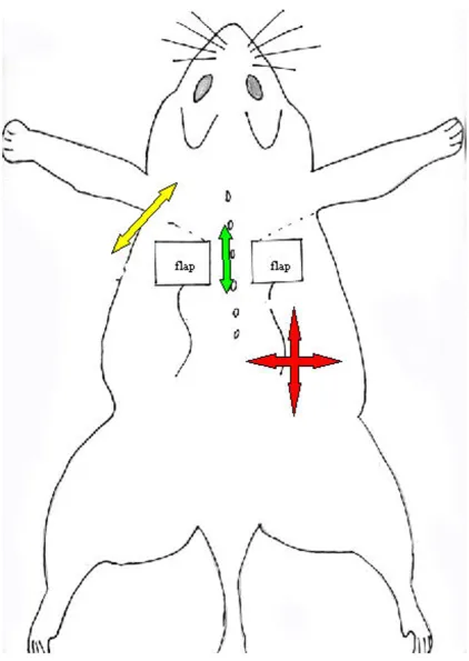
Flap Model
In this study, flaps were based on the caudal dermal pedi-cle without major or minor blood supply (Figure 1). An incision was made on the posterior axillary line, and the main pedicle of the muscle, thoracodorsal artery, was dis-sected. We preferred doing the procedures on latissimus dorsi because it is superficial, large and has a well-known vascular structure.
Elevation of Distal Pedicled Latissimus Musculocutaneous Flap
First, the operational area was shaved. Second, the upper extremity of the rats was abducted at 90°. In this position, the line drawn along the posterior axillary line of the medial side of the dorsum situated along the long axis of the upper extremity formed the lateral side of the flap. The skin island of the flap was a rectangle; about 15 × 10 mm in size and located in the tip of the cephalic part of the area situated below the upper margins. Third, a large, 20 mm, S-shaped incision was made in the caudal side of the skin island to reach latissimus dorsi muscle. Fourth, the skin, panniculus, trapezius and latissimus dorsi muscle in the cephalic side were passed. Fifth, a dissection was made from the cephalic tip towards the caudal side. The dissec-tion was continued in the caudal direcdissec-tion, and a muscle pedicle about 2 cm in width in the lateral and medial sides of the skin island was formed. Sixth, the posterior inter-costal perforator located in the middle of the muscle was excised (Figures 2 and 3). Finally, the flap was sutured to its bed using 4/0 silk.
Vascular Delay
Firstly, the operational area was shaved. Secondly, the upper extremity of the rats was abducted at 90°. Thirdly, a 15 mm incision starting from the point on the axillary line where the upper extremity and the body joined was made. Thus, latissimus dorsi muscle was accessed. Fourthly, the thoracodorsal pedicle of the muscle was found, excised where it enters the muscle and sutured with 6/0 prolene. Finally, the incision was sutured with 4/ 0 silk.
Chemical Delay
The experimental area was shaved and Nitroderm TTS 10 mg was applied. The skin overlying the muscle was cov-ered with a plaster. Nitroderm was renewed every day.
At five days after the operations, macroscopic, micro-scopic, microangiographic and histological examinations were made.
Macroscopic Evaluation
Viable parts of the skin islands in the musculocutaneous flaps were determined and percentages of viable flaps were estimated by a computer-assisted analysis of the images. Since the demarcation between viable and necrotic tissues was easily identified, it was not necessary to use vital dyes to assess flap necrosis. Views of the flaps were transferred to a computer with a digital camera and percentages of necrotic areas were calculated using a com-puter-assisted analysis (with Adobe Photoshop 6.0 soft-ware). The formula below was used: Viable flap area (mm2) / Total flap area (mm2) × 100
Microangiographic Evaluation
First, a catheter was inserted through the femoral veins of the rats and heparin was administered. Second, BaSO4 30% was injected. Finally, the rats were sacrificed and x-rays of the soft tissues were taken using a mammography device.
Histological Evaluation
The flaps were divided into three zones: the area where thoracodorsal artery was entered the muscle was consid-ered as the first zone, one third of the middle of the mus-cle as the second zone and the area where the distal perforators of the muscle entered as the third zone. Biopsy was obtained from each region and stained with hematox-ylene eosine and its histological examination was made.
Statistical Analysis
2-way ANOVA of variance test and Benforini t-test were used to compare the groups. Data were analyzed using SPSS 10.0 software.
Results
Vascular Delay
BMC Surgery 2003, 3 http://www.biomedcentral.com/1471-2482/3/11
Schema of the experimental flap model
Figure 1
strongly enhanced in the middle parts of the flaps in vas-cular delay group. The increase in vasvas-cularization was high in the vascular delay group compared to the control group (Figure 4). Diameter of the vessels in the vascular delay group were longer in the area where choke vessels were located than the control group (Figure 5). Light microscopy demonstrated thickness in the smooth mus-cles of the vessel walls in the middle parts of the flaps and dilatation in the vessel lumens (Figure 6). In addition, there was not necrosis of the muscle in the distal parts.
In Group II, one ear was assigned into five-day vascular delay and the other into the control group. The findings obtained from five-day vascular delay were comparable to those obtained from seven-day delay. There was minimal necrosis on suture lines and hair growth was normal. Necrosis was extensive in the control group. Edema and cyanosis were not observed following five-day vascular delay. Angiography showed an increase in calibres of ves-sels and choke vesves-sels between the neighbouring
perfora-tors. On light microscopy, the findings obtained from five-day vascular delay were comparable to those obtained from seven-day vascular delay.
As to three-day vascular delay, necrosis on suture lines was more extensive, and there was minimal necrosis also in the midline of the flaps. Angiography demonstrated less radioactive medium within the flaps and their edges. Light microscopy showed a slight increase in luminal diameters of chokes vessels. Unlike the other groups, there was not a marked thickness in the muscle of the vessel walls.
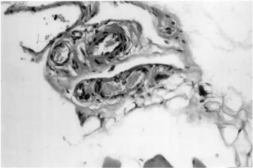
In one-day vascular delay group, necrosis was observed in almost all parts of the flaps. On angiography, vasculariza-tion was hardly visible. Biopsy specimens showed spasm of vessel lumens. In addition, there was myonecrosis in sections obtained from distal parts of the flaps, stria of the muscles disappeared and nuclei were driven towards cell membranes (Figure 7).
Pre-operative view of the flap
Figure 2
BMC Surgery 2003, 3 http://www.biomedcentral.com/1471-2482/3/11
Vascular Delay Combined with Chemical Delay
Since the chemical agent could affect the flap in the oppo-site, only one side of the rats was used. The other side was not assigned into the control group. The control ears in vascular delay groups were considered as the control group of vascular delay combined with chemical delay.
In the rats assigned into one-day vascular delay combined with chemical delay, necrosis was present in almost all parts of the flaps. Angiography showed less radio-opaque medium usually in the periphery. Light microscopy did not demonstrate any spasm of vessels or dilation in vessel lumens.
In the rats assigned into three-day vascular delay com-bined with chemical delay, there was extensive necrosis on suture lines. Hair growth was normal in proximal and middle parts of the flaps. Enhancement of radio-opaque medium was more marked than in one-day vascular delay combined with chemical delay. Histological examination
did not reveal thickness in vessel walls. There was myonecrosis in distal parts of the flaps.
In the rats assigned into five-day vascular delay combined with chemical delay, there were larger viable areas in the middle of the flaps. On angiography, the choke vessels in the middle of the flaps showed a stronger enhancement of radio-opaque medium and an increase in the vascular shadow. Light microscopy demonstrated thickness in ves-sel walls and dilation in vesves-sel lumens.
As to seven-day vascular delay combined with chemical delay, there was minimal necrosis only on suture lines. Microangiography demonstrated a marked vascular shadow due to opening choke vessels (Figure 8). Light microscopy showed thickness in the muscle and dilation in vessel lumens (Figure 9). Macroscopic measurements of viable flap areas in skin islands are shown in the Table 2 and Figure 10.
Elevated flaps
There was a significant difference in the viable flap areas between vascular delay alone and the control group (p < 0.05). The higher rate of flap viability was obtained in seven-day vascular delay alone. However, there was no significant difference in the viability between seven-day vascular delay and five-day vascular delay (p < 0.05), so the earliest time when the flap viability could be obtained is at five days.
The rate of flap viability was significantly higher in the vascular delay combined with chemical delay than the control group (p < 0.05). That is, the combination of vas-cular and chemical delays increased the rate of viability. Nevertheless, there was no significant difference between vascular delay alone and combination of vascular and chemical delays.
As to the question whether vascular delay combined with chemical delay shortened the period of delay, the viability rate was higher with five day-vascular delay alone than
with one and three-day vascular delay with a significant difference. However, there was no significant difference in the viability rate between five-day combined delays and seven-day combined delays. Thus, chemical delay did not significantly decrease the delay period.
Discussion
Latissimus dorsi musculocutaneous flap can be elevated reversely and thus can be used for many purposes. Latissimus dorsi flap based on the secondary segmental vessels, which are termed "reversed"[9,10] or "distally based" [6] latissimus dorsi flaps, have been widely used to repair defects of the spinal, lumbar, and sacral regions [11], but in some critical conditions such as cardiomyo-plasty, delay is preferred to minimize the possibility of distal necrosis and to harvest a larger and a safer flap [12,13]. Delay increases the duration of flap viability, improves the viability and makes the flap circulation safer [14]. The pathogenesis of delay has not been understood although many theories have been proposed. The
Microangiographic view of the flap in Group Ivdc
Figure 4
BMC Surgery 2003, 3 http://www.biomedcentral.com/1471-2482/3/11
procedure of delay can be performed in a few ways such as surgical, vascular, and chemical. In the former, margins of the flap are excised, the flap is elevated from its bed and the pedicle of the flap is ligated. Although the effect of vas-cular delay on skin flaps is clear, there are few studies on the effect of vascular delay on muscle flaps. It is accepted that ischemia plays an important role in the delay. Indeed, increased pCO2 levels have been shown to increase flap survival. In addition, it has been emphasized that ischemia has a strong influence also on revascularization [15]. In a study on dorsal and ventral aspects of pigs by Reinisch, importance of arterio-venous shunts has been underlined [16]. Yang and Morris described flaps on rats: one is ligation of the perforators adjacent to the base of the flap and the other is ligation of all perforators. They claimed that the most effective technique was limited vas-cular delay [17]. Özgentas¸ demonstrated on a rat TRAM flap model that the survival rate of the skin island signifi-cantly improved when superficial inferior epigastric ves-sels and deep superior epigastric vesves-sels (dominant
pedicle) were ligated one week before the elevation of the flap [18]. Finseth and Cutting used a different method to ligate pedicles in double pedicled epigastric skin flaps in the rats and showed the effect of delay clearly [19].
Another alternative of delay is chemical delay. Pang and Hendel noted that decreased levels of vasoconstructive neurohumoral substances play an important role in the early stage of delay and that local vasoconstrictor sub-stances are released in the later stages [20,21]. The drugs which decrease the vasoconstrictor effect of vasoactive substances are daxazosin mesylate and nitroglycerin [7]. Daxazosin mesylate is a selective blocker of adrenergic alpha-1 receptor in the smooth muscle cells of vessel walls. Although the drug has an effect both on arteries and veins, it mainly causes dilatation of the veins. Nitroglycerin directly relaxes smooth muscles. Its primary effect is dilatation of smooth muscles although it may affect both veins and arteries [20].
Microangiographic view of the flap in Group Ivd
Figure 5
Musculocutaneous flaps are compound flaps, which are supplied by one or more vascular pedicles and which involve subcutaneous fat tissue of the muscle fascia and the skin. The pedicles entering between the origo and insertion of the muscle consist of the vessels supplying the muscle. So that the muscle can be used as a flap, the whole muscle or a part of it should be mobilized. The surgical procedure performed for the mobilization of the muscle should not disrupt the sanguination of the muscle. When necrosis develops in a muscle following resection of its pedicle, that pedicle must be the major or the dominant pedicle. Minor or nondominant pedicles are small vascu-lar structures connected to muscles.
Mathes et al in their studies on dogs showed that the via-bility rate of gracili muscle was 99% with vascular delay and 69% without vascular delay [22]. Callegari et al dem-onstrated that choke vessels were dilated in latissimus dorsi, rectus abdominus and sartorius muscles of dogs fol-lowing a three-week delay [23]. Boyd et al noted that
liga-tion of the main vascular pedicle in myocutaneous flaps elevated from rectus abdominus muscle of pigs increased the blood flow and survival of the flap [24]. There were attempts to increase the skin island in models of rectus abdominus in rats and rabbits [19]. In this study, vascular delay in which first thoracodorsal artery, the main vascu-lar pedicle of latissimus dorsi flap in the rats, was ligated and then separated had a strong effect on the flap survival and the flap could survive on the nondominant vascular pedicle.
Although reversed pedicled latissimus dorsi flap is widely used in the clinical practice, its use and limitations are still debated. In the present study, the muscle and the skin tissues of the flap were elevated en block preserving only one or two segmentary vessels opposite the thoracodorsal artery; the main pedicle of the flap, and the largest reversed flap was able to repair defects. That is, like a major pedicled flap, the whole flap can be used only in the distal part of the conventionally used area.
Microscopic view of the flap in Group Ivd (Hematoxylene Eosin staining × 400)
Figure 6
BMC Surgery 2003, 3 http://www.biomedcentral.com/1471-2482/3/11
Microangiographic findings showed that vascular delay caused an increased vascularization in the mid part of the flap. Light microscopy demonstrated a thickness in the choke vessel walls and dilatation in the vessel lumens, a finding which confirms the hypothesis of vascular terri-tory expansion proposed by Boyd et al. [24] Consistent with the results of this study, the previous studies revealed that the delay in which both the artery, the veins and the nerve were dissected contributed more to the healing than the one in which either the artery, or the veins, or the nerve alone was dissected.
The mechanism of the increased vascularization was beyond the scope of this study. Rather, we aimed to reveal the philosophy on the use of increased vascularization in the clinical practice. In the light of the ideas mentioned so far, calibers of choke vessels between posterior intercostals territories are increased when thoracodorsal artery is ligated and the vascular pedicle which has not been dominant before reaches a sufficient size to supply
the flap. Despite beneficial effects of delay, its clinical use has turned out to be disadvantageous with the advent of microsurgery. It is desirable that the period of delay should be short. Various optimal periods of delay have been reported and the period of delay varies with the vas-cular structure and metabolic differences. In this study on distal pedicled latissimus dorsi flap in rats, we found no significant difference between seven-day and five-day delays. However, the above mentioned durations should not be considered as optimal periods of delay as it may take a longer time to confirm the viability of the skin island in this flap. The viability rates with vascular delay were significant compared to controls. It was striking that histological examination demonstrated thickness in ves-sel walls following dilatation in vesves-sel lumens. This may be that ligation of the main vascular pedicle causes the muscle to put pressure on choke vessels, which causes dil-atation in the vessel lumens and the vessel lumens hyper-trophy in response. The period of delay can be shortened only if we understand what takes place after the procedure
Microscopic view of the flap in Group Ivd (Hematoxylene Eosin staining × 400)
Figure 7
of delay is performed and till the choke vessels between the neighboring vessels are opened. It has been shown that vasoconstrictor substances play a major role in the early stage of the delay and vasodilator agents in the later stages. There have been many studies on the effects of chemicals on the duration of vasoconstriction, tolerance against ischemia and changes in the vascular anatomy. The final effect of vascular delay is a change in vascular anatomy of the flap due to opening choke vessels. Further-more, it has been shown to increase flap survival in arteri-alized venous flaps. In this study, the delay period was not shortened by combination of the delay procedures when compared with vascular delay only. However, compared to vascular delay alone, nitroderm was useful in improve-ment of flap survival although it did not shorten the delay period. In addition, it contributed to surgical delay. In the literature, Cho et al concluded that surgical delay increased the survival rate of the arterialized venous flaps in proportion to delay period. The combination of surgi-cal and chemisurgi-cal delays provided a significantly higher
survival rate than surgical delay alone (p < 0.001), and the delay period could be shortened. [7]
However, in this study, nitroderm was not found to shorten the delay period. It can be explained by the fact that muscle flaps are deeper than arterialized venous flaps and skin flaps. The aim of this study was to investigate whether nitroderm shortened the delay period. We did not attempt to investigate whether it was useful in myocu-taneous flaps. Prolonged application of nitroderm could have affected the flap survival.
Conclusions
The hypothesis of displacement of vascular pedicle in myocutaneous flaps was tested. A new rotational arc was created in latissimus dorsi musculocutaneous flap by delay procedures as a new flap experimental model. Although vascular delay achieved this end, nitroderm as a chemical agent did not shorten the delay period. How-ever, nitroderm provided better results on histological
Microangiographic view of the flap in Group IIIvdc7
Figure 8
BMC Surgery 2003, 3 http://www.biomedcentral.com/1471-2482/3/11
examination and microangiography. It is possible that vascular delay provides sufficient blood supply and thus flap survival improves. In addition, the arc of the muscle flap can be changed and the flap can be used for various purposes. We claim that this approach can be applied to other muscle flaps.
Competing interests
None declared.Authors' contributions
E. Copcu designed the study and prepared the manuscript. A. Aktas perofrmed the operations. N. Sivrioglu per-formed the statistical analysis. Y. Oztan participated in the
Microscopic view of the flap in Group IIIvdc7 (Hematoxylene Eosin staining × 400)
Figure 9
Microscopic view of the flap in Group IIIvdc7 (Hematoxylene Eosin staining × 400)
Table 2: Mean percentages of viable flap areas in the skin islands
Groups Length of Delays (Days)
1 3 5 7
Control 23 ± 5 30 ± 9 28 ± 7 27 ± 8
Vascular 35 ± 8 48 ± 8 69 ± 13 80 ± 9
Combined (vascular + chemical)
design of the study. All authors read and approved the final manuscript.
References
1. Mathes SJ, Nahai F: Classification of the vascular anatomy of muscles: experimental and clinical correlation.Plast Reconstr Surg 1981, 67:177-87.
2. Boyd JB, Markland B, Dorion D, Pang CY, Morris S: Surgical aug-mentation of skin blood flow and viability in a pig musculocu-taneous flap model.Plast Reconstr Surg 1990, 86(4):731-8. 3. Caffee HH: Transfer of the scapular flap on a reverse
latis-simus muscle pedicle.Plast Reconstr Surg 1998, 101:85-9. 4. Luce EA, Walsh J: Wound closure of the myelomeningocoele
defect.Plast Reconstr Surg 1985, 75:389-93.
5. Nomori H, Hasegawa T, Kobayashi R: The "reversed" latissimus dorsi muscle flap with conditioning delay for closure of a lower thoracic tuberculous empyema.Thorac Cardiovasc Surg
1994, 42:182-4.
6. Scheflan M, Mehrhof AI Jr, Ward JD: Meningomyelocele closure with distally based latissimus dorsi flap.Plast Reconstr Surg 1984, 73:956-9.
7. Cho BC, Lee MS, Lee JH, Byun JS, Baik BS: The effects of surgical and chemical delay procedures on the survival of arterialized venous flaps in rabbits.Plast Reconstr Surg 1998, 102(4):1134-43. 8. Karacaoglu E, Yuksel F, Turan SO, Zienowicz RJ: Chemical delay:
an alternative to surgical delay experimental study.Ann Plast Surg 2002, 49(1):73-80.
9. Bostwick J, Stevenson TR, Nahai F, Hester TR, Coleman JJ, Jurkiewicz MJ: Radiation to the breast. Complications amenable to sur-gical treatment.Ann Surg 1984, 200:543-53.
10. Stevenson TR, Rohrich RJ, Pollock RA, Dingman RO, Bostwick J 3rd: More experience with the "reverse" latissimus dorsi muscu-locutaneous flap: precise location of blood supply.Plast Recon-str Surg 1984, 74:237-43.
11. Yamamoto N, Igota S, Izaki H, Arai K: "Reverse turnover" trans-fer of a latissimus dorsi muscle flap to a large lumbar defect.
Plast Reconstr Surg 2001, 107:1496-9.
12. Chachques JC, Grandjean P, Nataf P, Mihaileanu S, Perier P, Bourgeois I, Carpentier A: Dynamic cardiomyoplasty: a surgical approach for ventricular assistance.Int J Artif Organs 1988, 11(6):469-74. 13. Wan C, Maldonado C, Papanicolaou G, Anderson GL, Overgoor M,
Kon M, Barker JH: Reducing the vascular delay period in latis-simus dorsi muscle flaps for use in cardiomyoplasty.Plast Reconstr Surg 2002, 15;109(5):1630-7.
14. Dhar SC, Taylor GI: The delay phenomenon: the story unfolds.
Plast Reconstr Surg 1974, 54:585.
15. McFarlane RM, DeYoung G, Henry RA: Prevention of necrosis in experimental pedicle flaps with hyperbaric oxygen.Surg Forum
1965, 16:481-2.
16. Reinisch JF: The pathophysiology of skin flap circulation. The delay phenomenon.Plast Reconstr Surg 1974, 54(5):585-98. 17. Yang D, Morris SF: Comparison of two different delay
proce-dures in a rat skin flap model.Plast Reconstr Surg102(5):1591-7. 18. Ozgentas HE, Shenaq S, Spira M: Study of the delay phenomenon in the rat TRAM flap model. Plast Reconstr Surg 1994, 94(7):1018-24.
19. Finseth F, Cutting C: An experimental neurovascular island skin flap for the study of the delay phenomenon.Plast Reconstr Surg
1978, 61(3):412-20.
20. Finseth F, Adelberg MG: Prevention of skin flap necrosis by a course of treatment with vasodilator drugs.Plast Reconstr Surg
1978, 61(5):738-43.
Comparison of the flap survivals (%)
Figure 10
Publish with BioMed Central and every scientist can read your work free of charge "BioMed Central will be the most significant development for disseminating the results of biomedical researc h in our lifetime."
Sir Paul Nurse, Cancer Research UK
Your research papers will be:
available free of charge to the entire biomedical community
peer reviewed and published immediately upon acceptance
cited in PubMed and archived on PubMed Central
yours — you keep the copyright
Submit your manuscript here:
http://www.biomedcentral.com/info/publishing_adv.asp
BioMedcentral
BMC Surgery 2003, 3 http://www.biomedcentral.com/1471-2482/3/11
21. Hendel PM, Lilien DL, Buncke HJ: A study of the pharmacologic control of blood flow to delayed skin flaps using xenon wash-out. Part II.Plast Reconstr Surg 1983, 71(3):399-407.
22. Mathes SJ, Vasconez LO: Myocutaneous free-flap transfer. Ana-tomical and experimental considerations.Plast Reconstr Surg
1978, 62(2):162-6.
23. Manchot C: In The cutaneous arteries of human body Edited by: Morain WD. NewYork:Springer Verlag; 1983.
24. Boyd JB, Markland B, Dorion D, Pang CY, Morris S: Surgical aug-mentation of skin blood flow and viability in a pig musculocu-taneous flap model.Plast Reconstr Surg 1990, 86(4):731-8.
Pre-publication history
The pre-publication history for this paper can be accessed here: