R E S E A R C H
Open Access
Computational identification of surrogate genes
for prostate cancer phases using machine
learning and molecular network analysis
Rudong Li
1†, Xiao Dong
1†, Chengcheng Ma
1†and Lei Liu
2,3** Correspondence:[email protected] †Equal contributors
2Shanghai Center for Bioinformatics Technology (SCBIT), Shanghai 201203, China
3
Institutes for Biomedical Sciences, Fudan University, Shanghai 200031, China
Full list of author information is available at the end of the article
Abstract
Background:Prostate cancer is one of the most common malignant diseases and is characterized by heterogeneity in the clinical course. To date, there are no efficient morphologic features or genomic biomarkers that can characterize the phenotypes of the cancer, especially with regard to metastasis–the most adverse outcome. Searching for effective surrogate genes out of large quantities of gene expression data is a key to cancer phenotyping and/or understanding molecular mechanisms underlying prostate cancer development.
Results:Using the maximum relevance minimum redundancy (mRMR) method on microarray data from normal tissues, primary tumors and metastatic tumors, we identifed four genes that can optimally classify samples of different prostate cancer phases. Moreover, we constructed a molecular interaction network with existing bioinformatic resources and co-identifed eight genes on the shortest-paths among the mRMR-identified genes, which are potential co-acting factors of prostate cancer. Functional analyses show that molecular functions involved in cell communication, hormone-receptor mediated signaling, and transcription regulation play important roles in the development of prostate cancer.
Conclusion:We conclude that the surrogate genes we have selected compose an effective classifier of prostate cancer phases, which corresponds to a minimum characterization of cancer phenotypes on the molecular level. Along with their molecular interaction partners, it is fairly to assume that these genes may have important roles in prostate cancer development; particularly, the un-reported genes may bring new insights for the understanding of the molecular mechanisms. Thus our results may serve as a candidate gene set for further functional studies.
Background
Prostate cancer is one of the most frequently-occurred malignant diseases affecting human health and life qualities [1]. In this cancer, metastasis (i.e. tumor cells escaping from the pri-mary tissue and eventually colonizing a distant site) reflects the most adverse phase, which commonly results in disruption of a complex set of biological processes, causing severe bone pain and spinal cord complications [2,3]. Due to the heterogeneity of the disease, there are currently no reliable morphologic features or genetic/genomic biomarkers that can effectively discriminate tissue-confined primary and/or metastatic tumors, thus less is known for the mechanisms underlying the development of metastatic disease.
Many efforts have been devoted to revealing the molecular mechanisms underlying the disease progression and/or identifying genetic/genomic surrogates for the tumor pheno-types. In most of the studies, the phenotype of a tumor is defined by its phase [4,5]; and identification of molecular surrogates underlying the different tumor phases is facilitated by classification of samples from the respective phases (i.e. normal prostate, primary tumor, and metastatic tumor). Since the different phases constitute the process of disease progression, the surrogates (i.e. set of genes) that distinguish the phases (or classify sam-ples from different phases) would certainly provide insights for understanding the molecu-lar mechanisms of disease progression. For prostate cancer, gene expression microarray studies have characterized expression profiles of primary cancers, metastatic cancers and normal tissues [6-8]; in some cases, correlations between gene expressions and cancer phases have been revealed [9]. The studies have further led to the finding that differential gene expression profiles hold for metastatic androgen ablation resistant prostate cancer (AARPC) and androgen-dependent metastatic cancers [10]. In general, these results have gained important insights about metastatic prostate cancer, regarding to the changes in expressions of genes involved in various biological processes, e.g. signal transduction, cell cycle, cell adhesion, migration and mitosis, etc. [11,12]. Nonetheless, one important prob-lem remains: previous studies describe the correlations of expression profiles and disease phases in terms of hundreds of genes, whereas they seldom provide a convenient molecu-lar measure (i.e. minimum predictor gene set) for accurate classification of prostate cancer phases, especially with respect to metastasis. Such a predictor gene set would be a better highlight for the mechanisms of prostate cancer.
To address this issue, we herein adopt a two-step pipeline widely-used in previous studies, which includes machine learning to identify disease-related genes and pathway analysis to reveal molecular interactions among the genes [13-16]. First, we utilize the machine learn-ing strategy for accurate classification of prostate cancer phenotypes based on gene expres-sion microarray data. Specifically, we use the minimum redundancy–maximum relevance method (mRMR), a robust method with a broad spectrum of applications [13,17], to serve our goal of identifying a largest-parsimony (i.e. minimum) surrogate (i.e. gene set) for pros-tate cancer phases. Moreover, in order to focus more on the issue of metastasis, we not only consider gene expression data of normal and (tissue-confined) primary prostate tumor sues [7], but also include a previously published dataset of metastatic tumor samples (i.e. tis-sue samples excluding potentially uninformative stromal genes) in our study [11].
Furthermore, genes/proteins usually co-function with their interaction partners; thus molecular interaction partners of disease-related genes are also candidates for further studies. For this purpose, we pinpoint the identified surrogate genes in a molecular inter-action network constructed based on STRING (Search Tool for the Retrieval of Interact-ing Genes), which is a database providInteract-ing resources of molecular interaction information [18]; and we then identify by the shortest-path analysis a set of potential co-acting factors, which may serve as candidate causal genes for further experimental studies.
Materials and methods
Data source
samples, which contained 77 normal tissues (NTs, including both normal prostate tis-sues free of pathological alterations from organ donor and normal tistis-sues adjacent to tumor), 66 primary prostate tumors (PTs) and 24 metastatic tumors (MTs). All tissue samples were acquired from the Health Sciences Tissue Bank of the University of Pittsburgh Medical Center under stringent Institutional Review Board guidelines with appropriate informed consent [11]. The data were downloaded from NCBI Gene Expression Omnibus (GEO) with accession number GSE6919. The normalized expression data were obtained directly from the GEO website, in which the data were normalized by global scaling and analyzed with Microarray Suite version 5.0 (MAS 5.0) using Affymetrix default settings. In our machine learning procedure, we did not combine the expressions from probes to genes; instead we obtained results at the probe level directly. We focused our analysis on probes corresponding to protein coding genes.
Algorithm of mRMR & prediction engine
The minimum redundancy – maximum relevance (mRMR) algorithm was utilized herein to select surrogate genes for prostate cancer progression. The major steps of mRMR implementation were the same as we previously described [13]. The algorithm aimed to balance features’ relevance to the prediction target and the redundancy be-tween features. Both relevance and redundancy were quantified with mutual informa-tion (MI), estimated as,
I x;ð yÞ ¼−1
2ln 1−ρðx;yÞ
2
ð1Þ
where I represented the MI and ρ was the correlation coefficient between the vari-ablesxandy.
First, assume that ywas the input variable, and X= {x1,…,xn } was the set of input features. Givenxias the feature with the highest MI with the input variable, the feature set (S) at the current step was then initialized byxi. Second, we selected the feature xj with the best balance between highest relevance and lowest redundancy and added it to S. It was achieved by maximizing the scoreqas follows,
q¼I xj;y
− 1
S
j j
X
xk∈S
I xj;xk
ð2Þ
We repeated the above steps until a desired solution length was reached. The mRMR algorithm was implemented using the R package“mRMRe”[19].
We predicted the phenotype of an individual in three ways: 1) the phenotype of its near-est neighbor; 2) the most-occurring phenotype of its five nearnear-est neighbors; 3) the pheno-type of its nearest clustering center of each phenopheno-type group (for detailed results, see Additional file 1: Table S1). According to Chou et al.’s studies [17,20], the distance be-tween two individuals was calculated as follows,
d ið1;i2Þ ¼1− e1⋅e2
e1
j j⋅j je2 ð
3Þ
Validation & incremental feature selection
We used jackknife validation to estimate the prediction accuracy of the selected fea-tures. The advantages of jackknife comparing with other validation methods, such as independent-dataset validation and sub-dataset validation, were discussed previously [17,20]. In jackknife validation, givenXsamples of a known outcome variable andN se-lected features, for each sample we compared the known outcome with an estimated outcome, which was computed based on the restX- 1 samples. We defined the accur-acy of a prediction using the following formula,
Accuracy¼ TPþTN
TPþTNþFPþFN ð4Þ
whereTP,TN,FP and FNrepresented the numbers of true positives, true negatives, false positives and false negatives, respectively.
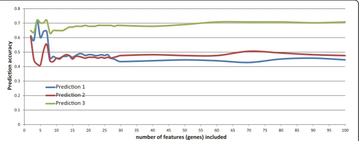
Furthermore, Incremental Feature Selection (IFS) was used to determine the number of features for optimal prediction (Figure 1). As previously described [13], forN= 1 to 400 required number of features, each feature set was computed by mRMR and the prediction accuracy was estimated using Jackknife validation. The set with the best pre-diction accuracy and smallest feature number was regard as the final feature set. In this study, a set with four genes was chosen and its prediction accuracy is 0.7202.
Molecular interaction network & shortest-path analysis
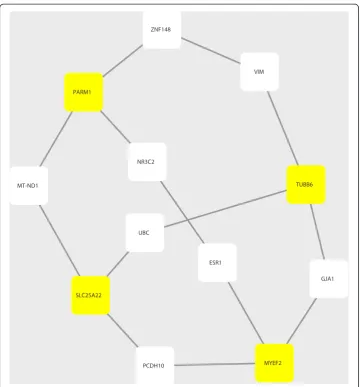
To reveal possible functional implications of the mRMR-selected genes, we explored the shortest-paths among the genes in a background molecular network constructed using the protein-protein interaction (PPI) data from STRING database (version 9.1) (http://string-db.org) [18]. To identify the shortest-path between two genes/proteins, we used Dijkstra’s algorithm and implemented it in the R package “igraph” [21]. The resulting sub-network of PPIs representing the shortest-paths among the four mRMR-selected genes (Figure 2) was visualized using Cytoscape (version 3.0.1) [22].
GO and KEGG pathway annotation
We carried out functional annotation for all the genes identified by mRMR and shortest-path analysis based on GO and KEGG shortest-pathways. The functional annotations were imple-mented using the web service of DAVID tools (version 6.7) [23], by which existence of gene enrichments to certain functional modules/pathways could also be observed.
Results
A set of four genes presents the best accuracy for predictions of NTs vs. MTs and PTs vs. MTs
NT, PT and MT in Additional file 2: Table S2; we specifically discuss the functions of the genes in later sections.
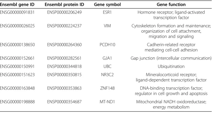
A PPI sub-network provides additional insights for the prostate cancer-related genes Furthermore, we constructed an molecular interaction network with the PPI data from STRING. We traversed all pairs of any two genes from the four surrogate (protein cod-ing) genes identified by mRMR as described above; and we then calculated the shortest paths between any pair of two proteins/genes using the Dijkstra’s algorithm. We even-tually obtained a sub-network of STRING PPIs that contained all these shortest paths (Figure 2). There are a total of 14 protein-protein interactions of 12 proteins, eight of which correspond to genes other than the four mRMR-identified ones and they are all annotated in the Ensemble Biomart database. We list these genes in Table 2 as an ex-tended set of candidate regulatory factors of prostate cancer that are possibly co-acting with the four mRMR-identified surrogate genes.
Functional annotation of the identified genes
Using the functional annotation tool DAVID, we carried out Gene Ontology (GO) and KEGG Pathway annotation for all the 12 identified genes (including the four mRMR-selected genes and another eight ones traced in the shortest-path PPI network). The results show that many genes are functioning to regulate transcription and/or tran-scription factor activity (Table 3), echoing the previous finding that genes expressions in (metastatic) prostate cancer are dictated by distinct transcriptional programs [12]. In addition, the genes are also involved in pathways related to steroid hormone receptor activity (Table 3). This is highly consistent with earlier studies that growth of prostate cancer cells is dependent on the male hormone (i.e. androgen) and overly prolonged changes ofin vivo hormonal level (e.g. androgen deprivation therapy, ADT) causes the emergence of androgen-independent (AI) cancer cells, which result in more malignancy towards advanced or metastatic prostate cancer [24,25]. It is obvious that function of hormone receptor plays a crucial role in prostate cancer progression; and our surrogate gene set captures this reality.
0 0.1 0.2 0.3 0.4 0.5 0.6 0.7 0.8
0 5 10 15 20 25 30 35 40 45 50 55 60 65 70 75 80 85 90 95 100
P
redi
c
o
n accuracy
number of features (genes) included Predicon 1
Predicon 2 Predicon 3
Interestingly, the identified predictor set includes genes (e.g.TUBB6,GJA1) that are an-notated by both GO and KEGG pathways as relating to gap junction and regulation of cell communication (Table 4). In fact, it is long recognized that gap junction-mediated inter-cellular communication is required for inter-cellular normality and breakdown of this commu-nication is a hallmark of cancer [26,27]. Furthermore, earlier studies have shown that intercellular communications and expressions of gap junction-forming proteins are largely reduced or not detected in prostate cancer cells [28,29]. Therefore, our results have faith-fully embodied the impact of cell communication dysregulation in prostate cancer.
Discussion
In the present study, we applied an informatic approach to identify molecular surrogates underlying the different phases of prostate cancer, which would facilitate deciphering the
ESR1
GJA1
MYEF2 UBC
SLC25A22
PCDH10 ZNF148
PARM1
MT-ND1
NR3C2
VIM
TUBB6
mechanism(s) of disease progression. The investigation led four genes into our sight — SLC25A22,TUBB6, MYEF2and PARM1. Data have shown that these genes ensure the sample classification with the accuracy of more than 70% and the genes are annotated to cancer-relevant functions/pathways. Thus the reasonability of our research is suggested. In the results, two of the four genes (TUBB6,PARM1) are supported by literature for their roles in prostate cancer; nonetheless, the other two (SLC25A22, MYEF2) are less known. Therefore, due to the indication of our results, we believe that these two genes may also sustain potentially role(s) during prostate cancer progression and they are worth being fo-cused on for further experimental studies.
SLC25A22 is named as solute carrier family 25 (mitochondrial carrier: glutamate), member 22. It is involved in the transport of glutamate across the inner mitochondrial membrane (accompanied by H+ transportation), which facilitates the malate-aspartate shuttle. The gene has also been validated by another dataset [30]. We hypothesize that the functioning of malate-aspartate shuttle can provide extra energy to cancer cells for gaining the growth advantage against native cells as well as escaping from the original site.
TUBB6 is a gene encoding a subtype ofβ-tubulins, the major constituent of micro-tube, which plays fundamental roles in cell structure maintenance, formation of the mi-totic spindle, transportation of chemicals, etc. Furthermore,TUBB6 is also functionally associated with gap junctional intercellular communication (GJIC). In fact, the respect-ive relationships between both tubulin and GJIC with metastasis of prostate/other cancers have been studied. For instance, the level of tubulin affects the metastasis of colorectal Table 1 The four genes identified by mRMR
Probe ID Ensembl gene ID Ensembl protein ID Gene symbol Gene function
43355_s_at ENSG00000176014 ENSP00000318697 TUBB6 Microtube formation; gap junction (intercellular communication)
55458_at ENSG00000104177 ENSP00000316950 MYEF2 Myelination repression
54033_at ENSG00000169116 ENSP00000370224 PARM1 Telomerase activity upregulation; prostatic cancer cell immortalization
52890_at ENSG00000177542 ENSG00000177542 SLC25A22 Mitochondrial carrier;
energy metabolism
Table 2 Genes on shortest-paths among the four mRMR-identified genes
Ensembl gene ID Ensembl protein ID Gene symbol Gene function
ENSG00000091831 ENSP00000206249 ESR1 Hormone receptor; ligand-activated
transcription factor
ENSG00000026025 ENSP00000224237 VIM Cytoskeleton formation and maintenance;
organization of cell attachment, migration and signaling
ENSG00000138650 ENSP00000264360 PCDH10 Cadherin-related receptor
mediating cell-cell adhesion
ENSG00000152661 ENSP00000282561 GJA1 Gap junction (intercellular communication)
ENSG00000150991 ENSP00000344818 UBC Ubiquitination
ENSG00000151623 ENSP00000350815 NR3C2 Mineralocorticoid receptor;
ligand-dependent transcription factor
ENSG00000163848 ENSP00000353863 ZNF148 DNA-binding transcription factor;
regulator in cell growth and apoptosis
ENSG00000198888 ENSP00000354687 MT-ND1 Mitochondrial NADH oxidoreductase;
carcinoma cells [31]; and breakdowns of GJIC in a variety of cancer cells correlate their metastatic capacity [32-34]. Moreover, studies have also indicated that TUBB6 itself is functionally related with the metastasis of various cancers. For instance, in a study using 60 cancer cell lines with different invasion abilities,TUBB6is identified as an invasion-associated (IA) gene [35]. Moreover, it is also identified as one of the 38 prognostic gene expression signatures of node-positive breast cancer after systemic adjuvant chemotherapy [36]. In addition, Champine et al. have shown that one of the potential mechanisms of BRMS1-mediated metastasis suppression is the suppression ofTUBB6[37].
PARM1is named as prostate androgen-regulated mucin-like protein 1. The gene reg-ulates telomerase protein component 1 (TLP1) expression and telomerase activity, thus enabling certain prostate cells to resist apoptosis. Multiple works have proved that PARM1 is an important causal gene of prostate cancer [38-40]. It contributes to the immortalization of prostatic cancer cells, which enhances the survival advantage against the neighboring native cells and promotes metastasis.MYEF2 is myelin expression fac-tor 2, which functions as a transcription repressor of the myelin basic protein (MBP). Our results underline the importance of the gene for prostate cancer, although no dir-ect relationship betweenMYEF2and the cancer had been established yet.
Our findings have provided a concise picture of the metastasis of prostate cancer. Ac-cording to Valastyan and Weinberg, the metastatic process includes 7 steps [41]: (1) in-vade locally through surrounding extracellular matrix and stromal cell layers, (2) intravasate through blood vessels, (3) survive during the transportation, (4) arrest at distant organ sites, (5) extravasate into the parenchyma of distant tissues, (6) initially survive in these foreign microenvironments in order to form micrometastases, and (7) re-initiate their proliferative programs at metastatic sites. First, as a cancer cell, energy is its priority (i.e. functional relation with SLC25A22). Step (1) and (2) of metastasis need more mobility, for which tubulin will accommodate this task. PARM1 can en-hance the survivability of cancer cells during the transportation and competing with the native cells in the invaded environment.
Table 3 GO annotation for genes co-identified by mRMR and shortest-path analysis
Term Genes Count %*
GO: 0043565 ~ sequence-specific DNA binding ZNF148, ESR1, NR3C2, MYEF2 4 36. 364
GO: 0030528 ~ transcription regulator activity ZNF148, UBC, ESR1, NR3C2, MYEF2 5 45. 455
GO: 0010604 ~ positive regulation of macromolecule metabolic process
ZNF148, UBC, ESR1, GJA1 4 36. 364
GO: 0010647 ~ positive regulation of cell communication UBC, ESR1, GJA1 3 27. 273
GO: 0003700 ~ transcription factor activity ZNF148, ESR1, NR3C2, MYEF2 4 36. 364
GO: 0003707 ~ steroid hormone receptor activity ESR1, NR3C2 2 18. 182
GO: 0004879 ~ ligand-dependent nuclear receptor activity ESR1, NR3C2 2 18. 182
GO: 0005496 ~ steroid binding ESR1, NR3C2 2 18. 182
GO: 0010628 ~ positive regulation of gene expression ZNF148, UBC, ESR1 3 27. 273
GO: 0005198 ~ structural molecule activity VIM, UBC, TUBB6 3 27. 273
*“
%”refers to the percentage of genes in the total gene set.
Table 4 KEGG annotation for genes co-identified by mRMR and shortest-path analysis
Pathway Genes Count %
To investigate the identified surrogate genes further, we have found that they and their co-interacting genes in the PPI network contain existent or potential therapeutic targets for cancers. In fact, Conde-Pueyoet al. suggest thatTUBB6 forms a sythetic le-thal (SL) association with the cancer-related gene BUB1, speculating that treatments targeting the tubulin gene should be more efficient in cancers where BUB1is mutated [42]. Moreover, tubulins are existent targets of anti-cancer drugs, e.g. Paclitaxel and vinca alkaloids (e.g. Vincristine and Vinblastine) in various cancers (including pros-tatic). The drugs disrupt the formations of microtubules/mitotic spindles and hence in-hibit the proliferations and metastases of cancer cells [43,44]. PARM1 is part of the Golgi apparatus that is androgen-responsive, and researches demonstrate that the Golgi apparatus embody new mechanisms of the androgen receptor (AR)-mediated signaling and they are useful biomarkers for prostate cancer diagnosis/prognosis [45]. Moreover, Golgi-targeting drugs have been shown to be effective in both androgen-dependent/-in-dependent prostate cancers [45]. Given this foreground,PARM1may have the potential of a therapeutic target for prostate cancer. It is also noteworthy that although the other two genes we identified (SLC25A22andMYEF2) do not possess direct therapeutic utilities at present, they may somehow implicate theoretical clues for cancer therapies. In fact, members of the SLC25A family (e.g.SLC25A4/5/6) are existent drug targets for the treat-ments of bone metastases in breast cancer and metastatic bone disease [46]; since prostate cancer also exhibit bone metastasis [3],SLC25A22may be worth being examined for po-tential relations to the metastatic properties of prostate cancer. Meanwhile, MYEF2has been characterized as a downstream target modulated by the Wnt/β-catenin pathway; since inhibition of Wnt/β-catenin signaling suppresses a number of cancers (e.g. multiple myeloma, colorectal cancer, etc.) [47], the genes regulated by Wnt/β-catenin may provide insights into the mechanisms of cancer developments and therapies.
1) involving in energy metabolism, which is intuitively crucial to cancer development. In fact, both gap junctions and mitochondria are emerging as therapeutic targets in cancers nowadays [53,54].
The previous work of Chandranet al. has discovered hundreds of genes with differential expression profiles [11]. In order to decipher the disease more concisely, we adopt the mRMR algorithm, which can provide results with the largest parsimony. Our results have showed that prostate cancer samples can be classified with only four genes, indicating that although the cancer is a complex disease with hundreds of differentially-expressed genes, these four genes may be the primary surrogates for the mechanism (s) underlying the dif-ferent cancer phases. Moreover, these four genes (along with their PPI partners) turn out having mechanistic/therapeutic implications in the prostatic or other cancers. Hence, in order to characterize the development of prostate cancer (especially metastasis) and inves-tigate the molecular mechanism (s), these genes could firstly be focused on in further functional experiments.
Conclusion
In all, we have characterized a small-sized predictor gene set for classification of prostate cancer phases. Our results support the roles for specific genes involved in cell communi-cation, hormone-receptor mediated pathways, and transcription regulation in (metastatic) prostate cancer. To our knowledge, the gene set we computed is of the minimal size that can rationally characterize prostate cancer phases; thus we hypothesize that these genes potentially play important roles in the molecular mechanisms of prostate cancer develop-ment. Furthermore, due to the small size, our predictor gene set can be a suitable candi-date list for forthcoming functional experiments; meanwhile, it might possess potential value for other relevant studies (e.g. drug target selection).
Additional files
Additional file 1: Table S1.Prediction accuracies for each individuals in IFS. The file includes detailed prediction data for the normal condition (denoted by“A”), primary tumor (“B”) and metastatic tumor (“C”).“Prediction 1 - 3” have the same meaning as in Figure 1, and the prediction statuses are shown. The file is in the format of electronic data sheet (Microsoft Excel *.xlsx).
Additional file 2: Table S2.Expression profiles of the four mRMR-selected genes. The file shows expression levels of the four mRMR-selected genes in the three class of samples. The file is in the format of Microsoft Excel *.xlsx.
Abbreviations
mRMR:Minimum redundancy - maximum relevance; IFS: Incremental feature selection; PPI: Protein-protein interaction; NT: Normal tissue; PT: (tissue-confined) Primary tumor; MT: Metastatic tumor; SLC25A22: Solute carrier family 25 member 22; TUBB6: Tubulin, beta 6 class V; PARM1: Prostate androgen-regulated mucin-like protein 1; MYEF2: Myelin expression factor 2; PPARγ: Peroxisome proliferator-activated receptor gamma; BRMS1: Breast cancer metastasis suppressor 1.
Competing interests
The authors declare that they have no competing interests.
Authors’contributions
Conceiving the research: RL, XD, CM and LL. Data analysis: RL, XD and CM. Manuscript writing: RL, XD, CM and LL. All authors read and approved the final manuscript.
Acknowledgments
Author details
1
Key Laboratory of Systems Biology, Shanghai Institutes for Biological Sciences (SIBS), Chinese Academy of Sciences (CAS), Shanghai 200031, China.2Shanghai Center for Bioinformatics Technology (SCBIT), Shanghai 201203, China. 3
Institutes for Biomedical Sciences, Fudan University, Shanghai 200031, China.
Received: 18 July 2014 Accepted: 20 August 2014 Published: 23 August 2014
References
1. American Cancer Society:Cancer Facts and Figures, 2006.Atlanta: American Cancer Society; 2006.
2. Stewart DA, Cooper CR, Sikes RA:Changes in Extracellular Matrix (ECM) and ECM-associated proteins in the metastatic progression of prostate cancer.Reprod Biol Endocrinol2004,2:2.
3. Logothetis CJ, Lin SH:Osteoblasts in prostate cancer metastasis to bone.Nat Rev Cancer2005,5(1):21–28. 4. Singh D, Febbo PG, Ross K, Jackson DG, Manola J, Ladd C, Tamayo P, Renshaw AA, D’Amico AV, Richie JP, Lander
ES, Loda M, Kantoff PW, Golub TR, Sellers WR:Gene expression correlates of clinical prostate cancer behavior.
Cancer Cell2002,1(2):203–209.
5. Brawer MK, Deering RE, Brown M, Preston SD, Bigler SA:Predictors of pathologic stage in prostatic carcinoma. The role of neovascularity.Cancer1994,73(3):678–687.
6. Luo JH, Yu YP, Cieply K, Lin F, Deflavia P, Dhir R, Finkelstein S, Michalopoulos G, Becich M:Gene expression analysis of prostate cancers.Mol Carcinog2002,33(1):25–35.
7. Chandran U, Dhir R, Ma C, Michalopoulos G, Becich M, Gilbertson J:Differences in gene expression in prostate cancer, normal appearing prostate tissue adjacent to cancer and prostate tissue from cancer free organ donors.BMC Cancer2005,5(1):45.
8. Chetcuti A, Margan S, Mann S, Russell P, Handelsman D, Rogers J, Dong Q:Identification of differentially expressed genes in organ-confined prostate cancer by gene expression array.Prostate2001,47(2):132–140. 9. Dhanasekaran SM, Barrette TR, Ghosh D, Shah R, Varambally S, Kurachi K, Pienta KJ, Rubin MA, Chinnaiyan AM:
Delineation of prognostic biomarkers in prostate cancer.Nature2001,412(6849):822–826.
10. Holzbeierlein J, Lal P, LaTulippe E, Smith A, Satagopan J, Zhang L, Ryan C, Smith S, Scher H, Scardino P, Reuter V, Gerald WL:Gene expression analysis of human prostate carcinoma during hormonal therapy identifies androgen-responsive genes and mechanisms of therapy resistance.Am J Pathol2004,164(1):217–227. 11. Chandran U, Ma C, Dhir R, Bisceglia M, Lyons-Weiler M, Liang W, Michalopoulos G, Becich M, Monzon F:Gene
expression profiles of prostate cancer reveal involvement of multiple molecular pathways in the metastatic process.BMC Cancer2007,7(1):64.
12. La Tulippe E, Satagopan J, Smith A, Scher H, Scardino P, Reuter V, Gerald WL:Comprehensive gene expression analysis of prostate cancer reveals distinct transcriptional programs associated with metastatic disease.
Cancer Res2002,62(15):4499–4506.
13. Ma C, Dong X, Li R, Liu L:A computational study identifies HIV progression-related genes using mRMR and shortest path tracing.PLoS One2013,8(11):e78057.
14. Li BQ, Huang T, Liu L, Cai YD, Chou KC:Identification of colorectal cancer related genes with mRMR and shortest path in protein-protein interaction network.PLoS One2012,7(4):e33393.
15. Zhang N, Jiang M, Huang T, Cai YD:Identification of influenza a/H7N9 virus infection-related human genes based on shortest paths in a virus-human protein interaction network.Biomed Res Int2014,2014:239462. Epub.
16. Jiang M, Chen Y, Zhang Y, Chen L, Zhang N, Huang T, Cai YD, Kong X:Identification of hepatocellular carcinoma related genes with k-th shortest paths in a protein-protein interaction network.Mol Biosyst2013,
9(11):2720–2728.
17. Chou KC:Some remarks on protein attribute prediction and pseudo amino acid composition.J Theor Biol
2011,273(1):236–247.
18. Franceschini A, Szklarczyk D, Frankild S, Kuhn M, Simonovic M, Roth A, Lin J, Minguez P, Bork P, Von Mering C, Jensen LJ:STRING v9.1: protein-protein interaction networks, with increased coverage and integration.
Nucleic Acids Res2013,41(D1):D808–D815.
19. De Jay N, Papillon-Cavanagh S, Olsen C, El-Hachem N, Bontempi G, Haibe-Kains B:mRMRe: an R package for parallelized mRMR ensemble feature selection.Bioinformatics2013,29(18):2365–2368.
20. Chou KC, Shen HB:Predicting eukaryotic protein subcellular location by fusing optimized evidence-theoretic K-nearest neighbor classifiers.J Proteome Res2006,5(8):1888–1897.
21. Csardi G, Nepusz T:The igraph software package for complex network research.Inter Journal Complex Syst
2006,1695:Article No:1695.
22. Smoot ME, Ono K, Ruscheinski J, Wang PL, Ideker T:Cytoscape 2.8: new features for data integration and network visualization.Bioinformatics2011,27(3):431–432.
23. Huang DW, Sherman BT, Lempicki RA:Systematic and integrative analysis of large gene lists using DAVID bioinformatics resources.Nat Protoc2008,4(1):44–57.
24. Feldman BJ, Feldman D:The development of androgen-independent prostate cancer.Nat Rev Cancer2001,1(1):34–45. 25. McLeod DG:Hormonal therapy: historical perspective to future directions.Urology2003,61(2):3–7.
26. Ruch RJ:The role of gap junctional intercellular communication in neoplasia.Ann Clin Lab Sci1994,24(3):216–231. 27. Hotz-Wagenblatt A, Shalloway D:Gap junctional communication and neoplastic transformation.Crit Rev
Oncogen1993,4(5):541–558.
28. Tsai H, Werber J, Davia MO, Edelman M, Tanaka KE, Melman A, Christ GJ, Geliebter J:Reduced connexin 43 expression in high grade, human prostatic adenocarcinoma cells.Biochem Biophys Res Commun1996,
227(1):64–69.
30. Cermák V, Kosla J, Plachý J, Trejbalová K, Hejnar J, Dvořák M:The transcription factor EGR1 regulates metastatic potential of v-src transformed sarcoma cells.Cell Mol Life Sci2010,67(20):3557–3568.
31. Schaefer KL, Takahashi H, Morales VM, Harris G, Barton S, Osawa E, Nakajima A, Saubermann LJ:PPARγinhibitors reduce tubulin protein levels by a PPARγ, PPARδand proteasome-independent mechanism, resulting in cell cycle arrest, apoptosis and reduced metastasis of colorectal carcinoma cells.Int J Cancer2007,120(3):702–713. 32. Navolotski A, Rumjnzev A, Lu H, Proft D, Bartholmes P, Zanker KS:Migration and gap junctional intercellular
communication determine the metastatic phenotype of human tunor cell lines.Cancer Lett1997,118(2):181–187. 33. Nicolson GL, Dulski KM, Trosko JE:Loss of intercellular junctional communication correlates with metastatic
potential in mammary adenocarcinoma cells.Proc Natl Acad Sci U S A1988,85(2):473–476.
34. Saunders MM, Seraj MJ, Li Z, Zhou Z, Winter CR, Welch DR, Donahue HJ:Breast cancer metastatic potential correlates with a breakdown in homospecific and heterospecific gap junctional intercellular communication.
Cancer Res2001,61(5):1765–1767.
35. Hsu YC, Chen HY, Yuan S, Yu SL, Lin CH, Wu G, Yang PC, Li KC:Genome-wide analysis of three-way interplay among gene expression, cancer cell invasion and anti-cancer compound sensitivity.BMC Med2013,11(1):106. 36. Jézéquel P, Campone M, Roché H, Gouraud W, Charbonnel C, Ricolleau G, Magrangeas F, Minvielle S, Genève J,
Martin AL, Bataille R, Campion L:A 38-gene expression signature to predict metastasis risk in node-positive breast cancer after systemic adjuvant chemotherapy: a genomic substudy of PACS01 clinical trial.
Breast Cancer Res Treat2009,116(3):509–520.
37. Champine PJ, Michaelson J, Weimer BC, Welch DR, DeWald DB:Microarray analysis reveals potential mechanisms of BRMS1-mediated metastasis suppression.Clin Exp Metastasis2007,24(7):551–565.
38. Cornet AM, Hanon E, Reiter ER, Bruyninx M, Nguyen VH, Hennuy BR, Hennen GP, Closset JL:Prostatic androgen repressed message-1 (PARM-1) may play a role in prostatic cell immortalisation.Prostate2003,56(3):220–230. 39. Fladeby C, Gupta SN, Barois N, Lorenzo PI, Simpson JC, Saatcioglu F, Bakke O:Human PARM-1 is a novel
mucin-like, androgen-regulated gene exhibiting proliferative effects in prostate cancer cells.Int J Cancer2008,
122(6):1229–1235.
40. Wang XY, Hao JW, Zhou RJ, Zhang XS, Yan TZ, Ding DG, Shan L:Meta-analysis of gene expression data identifies causal genes for prostate cancer.Asian Pac J Cancer Prev2013,14(1):457–461.
41. Valastyan S, Weinberg RA:Tumor metastasis: molecular insights and evolving paradigms.Cell2011,147(2):275–292. 42. Conde-Pueyo N, Munteanu A, Sole R, Rodriguez-Caso C:Human synthetic lethal inference as potential
anti-cancer target gene detection.BMC Syst Biol2009,3(1):116.
43. Katsetos CD, Dráber P:Tubulins as therapeutic targets in cancer: from bench to bedside.Curr Pharm Des2012,
18(19):2778–2792.
44. Jordan A, Hadfield JA, Lawrence NJ, McGown AT:Tubulin as a target for anticancer drugs: agents which interact with the mitotic spindle.Med Res Rev1998,18(4):259–296.
45. Migita T, Inoue S:Implications of the Golgi apparatus in prostate cancer.Int J Biochem Cell Biol2012,44(11):1872–1876. 46. Rask-Andersen M, Masuram S, Fredriksson R, Schiöth HB:Solute carriers as drug targets: current use, clinical
trials and prospective.Mol Aspects Med2013,34(2–3):702–710.
47. Yao H, Ashihara E, Strovel JW, Nakagawa Y, Kuroda J, Nagao R, Tanaka R, Yokota A, Takeuchi M, Hayashi Y, Shimazaki C, Taniwaki M, Strand K, Padia J, Hirai H, Kimura S, Maekawa T:AV-65, a novel Wnt/β-catenin signal inhibitor, successfully suppresses progression of multiple myeloma in a mouse model.Blood Cancer J2011,1(11):e43. 48. Holst F, Stahl PR, Ruiz C, Hellwinkel O, Jehan Z, Wendland M, Lebeau A, Terracciano L, Al-Kuraya K, Janicke F,
Sauter G, Simon R:Estrogen receptor alpha (ESR1) gene amplification is frequent in breast cancer.Nat Genet
2007,39(5):655–660.
49. Narayan G, Freddy AJ, Xie D, Liyanage H, Clark L, Kisselev S, Un Kang J, Nandula SV, McGuinn C, Subramaniyam S, Alobeid B, Satwani P, Savage D, Bhagat G, Murty VV:Promoter methylation-mediated inactivation of PCDH10 in acute lymphoblastic leukemia contributes to chemotherapy resistance.Genes Chromosomes Cancer2011,
50(12):1043–1053.
50. Ying J, Li H, Seng TJ, Langford C, Srivastava G, Tsao SW, Putti T, Murray P, Chan ATC, Tao Q:Functional epigenetics identifies a protocadherin PCDH10 as a candidate tumor suppressor for nasopharyngeal, esophageal and multiple other carcinomas with frequent methylation.Oncogene2006,25(7):1070–1080. 51. Zhang CZ, Chen GG, Lai PB:Transcription factor ZBP-89 in cancer growth and apoptosis.Biochim Biophys Acta
2010,1806(1):36–41.
52. Semenza GL:Targeting HIF-1 for cancer therapy.Nat Rev Cancer2003,3(10):721–732.
53. Kandouz M, Batist G:Gap junctions and connexins as therapeutic targets in cancer.Expert Opin Ther Targets
2010,14(7):681–692.
54. Don AS, Hogg PJ:Mitochondria as cancer drug targets.Trends Mol Med2004,10(8):372–378. doi:10.1186/1742-4682-11-37