Hypoxia May Increase Rat Insulin mRNA Levels by Promoting
Binding of the Polypyrimidine Tract-binding Protein (PTB) to the
Pyrimidine-rich Insulin mRNA 3
-Untranslated Region
Linda Tillmar and Nils Welsh
Department of Medical Cell Biology, Uppsala University, Uppsala, Sweden
Accepted May 6, 2002
Abstract
Background:Recent reports identify the 3-UTR of insulin mRNA as crucial for control of insulin messenger stability. This region contains a pyrimidine-rich sequence, which is similar to the hypoxia-responsive mRNA-stabilizing ele-ment of tyrosine hydroxylase. This study aimed to deter-mine whether hypoxia affects insulin mRNA levels. Materials and Methods:Rat islets were incubated at nor-moxic or hypoxic conditions and with or without hydro-gen peroxide and a nitric oxide donor. Insulin mRNA was determined by Northern hybridization. Islet homogenates were used for electrophoretic mobility shift assay with an RNA-oligonucleotide, corresponding to the pyrimidine-rich sequence of the 3-UTR of rat insulin I mRNA. The ex-pression of reporter gene mRNA, in islets transfected with reporter gene constructs containing the wild-type or mu-tated insulin mRNA pyrimidine-rich sequences, was mea-sured by semiquantitive RT-PCR.
Correspondence and reprint requests should be addressed to: Linda Tillmar, Department of Medical Cell Biology, Uppsala University, Biomedicum, P.O. Box 571, SE-751 23 Uppsala, Sweden. Phone: 46-(0)18 4714395; fax: 46-(0)18 556401; e-mail: Linda.Tillmar@medcellbiol.uu.se.
Results:Insulin mRNA was increased in response to hy-poxia. This was paralleled by increased binding of the polypyrimidine tract-binding protein (PTB) to the pyrim-idine-rich sequence of the 3-UTR of insulin mRNA, which was counteracted by hydrogen peroxide. The re-porter gene mRNA level containing the wild-type bind-ing site was not increased in response to hypoxia, but mutation of the site resulted in a destabilization of the mRNA.
Conclusions:The complete understanding of different di-abetic conditions requires the elucidation of mechanisms that control insulin gene expression. Our data show that hypoxia may increase insulin mRNA levels by promoting the binding of PTB to the insulin mRNA 3-UTR. Hydro-gen peroxide abolishes the hypoxic effect indicating in-volvement of reactive oxygen species and/or the redox po-tential in the oxygen-signaling pathway.
Introduction
We recently demonstrated that insulin mRNA is highly abundant and constitutes up to 30% of the pancreatic beta cell mRNA (1). The insulin mRNA is also especially long-lived, 29–77 hr, depending on glucose concentration (2), and its stability is therefore likely to be regulated by specific mecha-nisms. A recent study has identified the 3 -untrans-lated region (3-UTR) as critical for beta cell–specific glucose-mediated control of rat insulin II mRNA (3). In addition, we have observed that the 3-UTR of rat insulin mRNA contains an active RNA-stabilizing element, the insulin mRNA 3-UTR pyrimidine-rich sequence (ins-PRS), to which the polypyrimidine tract-binding protein (PTB) binds (1). This binding is increased in response to glucose as well as to reducing agents (1). PTB exists in at least three isoforms, with molecular masses ranging from 55–62 kDa. It belongs to the heterogeneous nuclear ribonucleoprotein (hnRNP) family, and is also known as hnRNP-I (4). In addition to the three
sufficient, for both constitutive and hypoxia-mediated stability of this mRNA (17).
In view of the close homology between TH HIPBS and the insulin mRNA 3-UTR (1), the aim of the pre-sent study was to determine whether insulin mRNA levels are affected by hypoxia and, if so, if this is par-alleled by an altered PTB–ins-PRS interaction.
Material and Methods
Isolation and Culture of Pancreatic Islets
Adult Sprague-Dawley rats from a local colony were killed by cervical dislocation after an intraperitoneal injection of a sodium pentobarbital. The pancreases were taken out and the islets isolated by collagenase digestion (21). The islets were cultured in 5 ml RPMI-1640, supplemented with 10% Fetal Clone II serum (Hyclone Europe Ltd, Cramlington, UK), 2 mM L-glutamine, 100 U/ml benzyl penicillin, and 0.1 mg/ml streptomycin (RPMI-WS). The islets were cultured free floating for 5–10 days with the medium changed every second day (21). Only small or medium sized islets were chosen for experimenta-tion; cells located near the center of large islets often become necrotic due to insufficient diffusion of nu-trients and/or oxygen. To produce hypoxia, 50–100 islets/5 cm2 where cultured under a medium depth of 4–6 mm, in flasks containing 5 ml of culture medium, which were flushed with 1%, 5% or 10% O2/5% CO2/94%, 90%, or 85% N2 for at least 20 min
prior to the addition of islets. The flasks were incu-bated under airtight conditions. The oxygen tension in the culture media, following different gassing time periods and incubation periods, were verified using modified Clark microelectrodes (Unisense, Aarhus, Denmark) (22). These electrodes have a tip with a 2- to 6-m outer diameter and a 1- to 2-m inner diameter. The electrodes were polarized at 0.800 V, which gives a linear response between the oxygen tension and the electrode current. The elec-trodes were calibrated in water saturated with NaS2O5or air at 37C. The baseline deviation of the
recordings was 0.5%/hr. The oxygen tensions were measured in the medium, at a depth of 3 mm, and not in the immediate proximity of the islets. The oxygen tension was recorded in three different flasks per condition. To ensure hypoxia throughout a long-term incubation, flasks with islets incubated for 16–24 hours were gassed, sealed, and incubated in an airtight box, which was also gassed with the same gas mixture as the flasks.
Assessment of Cell Viability in Response to Hypoxia
Islets were incubated for 24 hr at 21%, 10%, 5% or 1% O2 and cell death was determined by staining
with 20 g/ml Hoechst 33342 (bisbenzimide) and 10 g/ml propidium iodide (Sigma-Aldrich, St. Louise, MO, USA) for 10 min at 37C (23). The islets were then examined with a fluorescence microscope, using an UV-2A filter. For the quantification of islet
cell death, islets were trypsinized and analyzed by flow cytometry (FACS-Calibur, Becton-Dickinson, Oxford, UK) (24).
Northern Hybridization Analysis
Rat islets in groups of 50 were incubated for 6, 16 or 24 hr as given in Figure 1. RNA was isolated using the Ultraspec Total RNA Isolation System (Biotech Laboratories, Houston, TX, USA) and ana-lyzed by Northern hybridization. The samples were loaded on a 1.25% agarose gel and run for 1 hr at 100 V. Gels were washed with water to remove formaldehyde, stained with ethidium bromide, and photographed. The RNA was transferred to a nylon membrane and cross-linked by UV irradiation. The filter was then incubated in prehybridization buffer for 3 hr at 42C. The buffer was exchanged for a buffer containing a radiolabeled insulin cDNA (25) and the membrane was incubated overnight at 42C. The insulin probe, pRI7, was labeled with [␣-32P]
dCTP using the Mega Prime DNA labeling kit (Amersham Pharmacia Biotech, Uppsala, Sweden). The filter was washed and exposed to Hyperfilm MP (Amersham Pharmacia Biotech). Insulin mRNA bands were quantified by densitometry and recalcu-lated per amount of 28S rRNA, detected in each lane, using The Kodak Digital Science camera and Kodak Digital Science 1D software (Eastman Kodak Company, Rochester, NY, USA).
Electrophoretic Mobility Shift Assay and Cross-Linking Analysis
The ins-PRS RNA oligonucleotide (Scandinavian Gene Synthesis AB, Köping, Sweden) (1 pmol), was incubated for 45 min at 37C with 50 Ci [␥-32P] ATP (5000 Ci/mmole, Amersham Pharmacia) and 8 units bacteriophage T4 polynucleotide kinase (Sigma-Aldrich). Heating for 2 min at 60C then inactivated the kinase. The radiolabeled RNA-oligonucleotide was purified on Chroma spin-10 columns (Clontech Laboratories Inc., Paolo Alto, CA, USA). The integrity of the RNA oligonucleotide was controlled by elec-trophoresis in a 15% polyacrylamide gel. The RNA oligonucleotide was stored at 20C until use.
Rat islets in groups of 50 were incubated in normoxic or hypoxic (1% or 5% O2) glucose-free
RPMI-WS, containing 2.8 mM glucose, with or without 50 M hydrogen peroxide or 2.5 mM of the nitric oxide donor DETA NONOate (Alexis Biochemicals, San Diego, CA, USA) for 20 min or 1 hr at 37C. After incubation, the islets were col-lected and washed in ice-cold PBS. Following cen-trifugation the cells were homogenized in 50 l homogenization buffer (10 mM HEPES, 5 mM MgCl2, 50 mM KCl, 10% glycerol, and 1 mM
dissolved in SDS-sample buffer. The proteins were separated on a 12% SDS-PAGE and blotted on a nitrocellulose filter. The filters were hybridized with the monoclonal anti-PTB 3 antibody (27). Horseradish peroxidase conjugated anti-mouse antibody (1:1000) was used as secondary antibody, which was detected by the Amersham ECL system (Amersham Pharmacia).
Lipofection of Islet Cells with ins-PRS Reporter Gene Vectors
Double-stranded DNA oligonucleotides with the se-quences of wild-type (wt) PRS or mutant ins-PRS (Table 1) were cloned in to the pCRTM3-CAT vector (Invitrogen, San Diego, CA, USA) down-stream of the coding sequence of the chloram-phenicol acetyltransferase (CAT) reporter gene and upstream of the bovine growth hormone polyadeny-lation signal, as previously described (1). The cells were transfected with 1 g of either empty pCRTM
3-CAT vector, pCRTM3-CAT wild-type ins-PRS,
pCRTM3-CATins-PRS mutant 1 or pCRTM3-CAT
ins-PRS mutant 2. The cells were then resuspended in 5 ml RPMI-WS and maintained in culture for 48 hr at 37C. The cells from each of the four dishes were then further cultured at 37C for 24 hr at normoxia or hypoxia (5% O2) in 5 ml RPMI-WS
containing 2.8 mM glucose. In some experiments, the culture medium was also supplemented with 5g/ml actinomycin D.
Quantification of CAT Reporter Gene Expression by Semiquantitative RT-PCR
The cells were collected from the previous step and the RNA was isolated using the Ultraspec Total RNA Isolation reagent. The RNA was dissolved in 30–32 l RNase-free water. Thirty microliters of the RNA and 1 M oligo (dT)-primer were used for cDNA synthe-sis, in agreement with the You-Prime First-strand Beads protocol (Amersham Pharmacia). The samples were incubated at 37C for 1 hr followed by 65C for 10 min. Some RNA samples were not used for cDNA synthesis but were analyzed in parallel by PCR to electrophoretic mobility shift assay. Nitrite
accu-mulation was measured in the medium at the end of the incubation time, as an indicator of nitric oxide release from the nitric oxide donor. Ten micro-liters of freshly made Griess reagent was added to 100 l of medium (26). The samples were incubated at room temperature for 20 min, after preincubating at 60C for 2 min, and the absorbance was measured at 546 nm on a Beckman DU-62 spectrophotometer (Palo Alto, CA, USA).
The RNA–protein binding reaction was perfor-med as described (1). Duplicates containing 20 l homogenate, 200 ng/ml Escherichia coli tRNA, 10% glycerol, and 1 l radiolabeled RNA-oligonu-cleotide probe (40,000–100,000 cpm/reaction) were prepared. To one of the duplicates 11 mM DTT was added. The reaction mixtures were incu-bated at 30C for 30 min. RNase T1 (20 units/sample) was added and the incubation proceeded for 10 min at 30C. Finally the samples were incubated an-other 10 min with 5 mg/ml heparin. In some cases, the reaction mixtures were divided into two aliquots, one that was cross-linked by exposure to UV radiation (260 nm) for 10 min and then ana-lyzed by boiling the samples in SDS-sample buffer without beta-mercaptoethanol and separated on a 9% SDS-PAGE gel for 1 hr at 160 V. The other aliquot was used directly for nondenaturing gel electrophoresis. In the latter case, the samples were applied on a 7% polyacrylamide gel and elec-trophoresed in 0.5 TBE at 100 V for 1 hr. The free probe was run out of the gel to avoid radioactive contamination when analyzing the ins-PRS–protein complexes. The gel was fixed (30% ethanol, 10% acetic acid), dried and exposed to a film overnight at 70C. The results were analyzed by the Kodak system.
Western Hybridization
Islets in groups of 35 were incubated at normoxia or hypoxia (5% O2) for 1 hr. The islets were
thor-oughly rinsed in ice-cold PBS before they were
Table 1. The sequences of the different ins-PRS probes used and comparison to the TH 3-UTR. Ins-PRS, insulin pyrimidine-rich sequence; TH HIPBS, tyrosine hydroxylase hypoxia inducible protein binding sequence. The core binding sequences for respective mRNA are marked in bold and mutated nucleotides are underlined.
mRNA probe Sequence
TH HIPBS 5CUUUCCCAAAGUCUCCAUCCCCUUCUCCAACCUUUCCUa
Ins-PRS wild-type 5UCCACCACUCCCCGCCCACCCCUCUb
Ins-PRS mutant 1 5UCCACCACUCCCCGCCCACCACUCU
Ins-PRS mutant 2 5UCCACCACUCCCCUCCCACCCCUCU
aFrom Gene Bank accession number M10244, 5is at position 1540, HIPBS is located between 1552 and 1579 (46).
exclude the possibility of DNA contamination. The Touch Down Temperature cycling system (Hybaid, Teddlington, UK) was used for PCR. The sequences of the PCR primers (Cybergene AB, Huddinge, Sweden) were as follows:
CAT forward: 5´GAATGCTCATCCGGAACT CAT reverse: 5´CCAGGGTCAAGGAAGGCACGG GAPDH forward: 5´GACCCCTTCATTGACCTCA GAPDH reverse: 5´CCTTCTCCATGGTGGTGAA
-actin forward:
5´CCTGCTTGCTGATCCACATCT-GCTGG
-actin reverse: 5´CTGACCGAGCGTGGCTAC
For amplification of samples incubated without actinomycin D, the following time course was used: 94C, 9 min and 25 cycles 94C, 30 sec, 55C, 45 sec and 72C, 90 sec (CAT and -actin). When analyzing samples incubated in the presence of actinomycin D: 94C, 9 min and 30 cycles (GAPDH) of 94C, 45 sec, 55C, 45 sec and 72C, 1 min or 30 cycles (-actin) 94C, 30 sec, 55C, 30 sec and 72C, 90 sec were used. The PCR products were run in a 1.5% agarose gel and stained with ethidium bromide for quantification using the Kodak system. CAT mRNA was normalized against -actin or GAPDH.
Statistics
Means SEM were calculated and the Student
paired t-test was performed. When analyzing the in-sulin mRNA levels one-way ANOVA for repeated measurements and Dunnets test was used.
Results
Verification of Culture Medium Oxygen Tension
Normal oxygen tension, 150 mm Hg, was set as 100%. After 10 min of gassing with 5% O2/5%
CO2/85% N2 the oxygen tension in the media of
three separate flasks were 30.3 0.57, which gives an oxygen tension of approximately 45.5 mm Hg. After 20 or 30 min of gassing the tension was 25.8
1.80% (38.8 mm Hg) or 25.0 3.6% (37.5 mm Hg),
respectively. Because 37.5 mm Hg corresponds to 5% O2, 20–30 min of gassing was used in all
exper-iments.
Islet Cell Viability in Response to Hypoxia
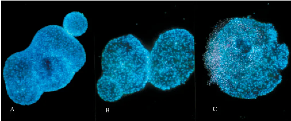
Islets cell death in response to incubation at differ-ent hypoxic conditions for 24 hr was determined by vital staining with Hoechst 33342 and propidium iodide. This method allows the discrimination be-tween necrosis and apoptosis in intact islets (23,24). Inspection of the islets in a fluorescence microscope revealed a moderate increase in propidium iodide positive islet cells, mainly located centrally, in islets incubated at 5% oxygen (Fig. 1). Only small- to medium-sized islets were analyzed because large islets are known to develop central necrosis in tissue culture. At 1% oxygen, the increase in propidium iodide positive cells was more pronounced (Fig. 1). We did not observe any increase in apoptotic cells (i.e., propidium iodide negative cells with increased Hoechst staining and a nuclear morphology typical for apoptosis). The number of cells that had taken up propidium iodide where quantified by flow cyto-metric analysis. The percentage of propidium iodide
Fig. 1. Islet cell viability after 24-hr culturing at different media oxygen concentrations. Islets were incubated for 24 hr at declining oxygen concentrations: (A)21% O2; (B)5% O2; and (C)1% O2. Cell viability was determined by staining with Hoechst
changed by the presence of 2.5 mM of the nitric ox-ide donor DETA NONOate (Fig. 4A). These results indicate that reactive oxygen species and/or the re-dox potential regulate ins-PRS binding.
Inhibition of PTB Binding to ins-PRS Results in mRNA Destabilization
To assess whether hypoxia regulates insulin mRNA stability by controlling PTB binding to ins-PRS in vivo, we lipofected dispersed rat islet cells with pCRTM-CAT vector with or without wild-type or mutated ins-PRS, as described previously (1). Two days following the transfection, the cells were ex-posed for 24 hr to normoxic or hypoxic (5% O2)
conditions at substimulatory glucose concentrations with or without actinomycin D. Similar results positive cells from islets incubated at different
oxy-gen levels were in three separate experiments: 1.4
0.7%, 2.9 0.8%, 4.4 2.3%, and 10.1 3.7% at
21%, 10%, 5% and 1% oxygen, respectively. Be-cause prolonged exposure to pronounced hypoxia (1% O2) increased beta cell death, primary by
accel-erating necrosis (Fig. 1), we chose to expose islets to only mild hypoxia (10% and 5% O2) and to limit the
exposure period to 6 and 16 hr in most experimen-tal setups. However, we cannot exclude that there exists some degree of heterogeneity in the islet pop-ulation, and that a fraction of the islet cells may have become severely anoxic even though the oxygen concentration of the surrounding culturing media was as high as 5%.
Hypoxia Increases Rat Islet Insulin mRNA Levels
It was observed that exposure to 5% of O2 in the
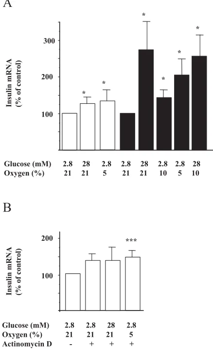
presence of 2.8 mM glucose increased insulin mRNA levels by 33% at 6 hr and by 100% at 16 hr (Fig. 2A). A high glucose concentration (28 mM) in-creased insulin mRNA to a similar extent as that ob-served in response to hypoxia (Fig. 2A). There were no additive effects of glucose (28 mM) and hypoxia (10%) (Fig. 2A). In the presence of actinomycin D, there was no stimulatory effect of hypoxia or 28 mM glucose on insulin mRNA levels (Fig. 2B).
Hypoxia Increases Binding of the PTB to the ins-PRS, but not PTB Expression
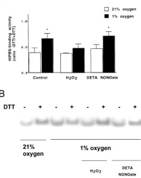
Because glucose and reducing agents stimulate ins-PRS binding activity in vivo and in vitro, respec-tively (1), and because it is known that both glucose and acute hypoxia promote an increased redox po-tential in islet cells (28,29), we next analyzed ins-PRS binding activity in hypoxia-treated islets. Cross-linking experiments showed that DTT and hypoxia (Fig. 3A) increased formation of a 65-kDa complex, which is at the same position as the glucose-induced complex (1). The ins-PRS binding protein was previously identified as PTB (1). Binding of PTB to ins-PRS was equally increased in response to hy-poxia as to DTT (Fig. 3B). Its hyhy-poxia-induced bind-ing activity is probably mediated through posttrans-lational modifications; hypoxia (5% O2) had no effect
on PTB protein expression after 1-hr incubation (data not shown).
Hypoxia-Increased ins-PRS Binding Activity is Counteracted by Hydrogen Peroxide, but not by Nitric Oxide
We next studied whether the oxidizing agent hydro-gen peroxide or a nitric oxide donor, DETA NONOate, could affect alterations in binding activ-ity. The islets were incubated for 20 min at 1% O2
and at a substimulatory glucose concentration. This resulted in increased ins-PRS–binding activity at nonreducing conditions (Fig. 4A and 4B, lane 3 ver-sus lane 1). The hypoxia-induced increase in ins-PRS–binding activity was counteracted by the addi-tion of 50 M hydrogen peroxide, but was not
*
Glucose (mM) 2.8 28 2.8 2.8 28 2.8 2.8 28 Oxygen (%) 21 21 5 21 21 10 5 10
Insulin mRNA (% of control)
*
100 200 300
*
* *
*
A
***
Glucose (mM) 2.8 2.8 28 2.8 Oxygen (%) 21 21 21 5 Actinomycin D - + + +
Insulin mRNA (% of control)
100 200
B
Fig. 2. Hypoxia increases insulin mRNA levels in isolated rat islets. Isolated rat islets were incubated for 6 hr (white bars) or 16 hr (black bars) in the presence of normoxia (21% O2)
or hypoxia (10% or 5% O2) at 2.8 or 28 mM glucose, without
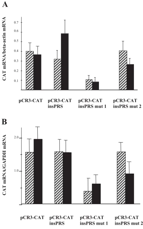
where obtained both with (Fig. 5B) and without actinomycin D (Fig. 5A), indicating that transcrip-tion was not affected by the ins-PRS mutatranscrip-tion. We observed that the reporter gene mRNA containing the wild-type rat insulin I ins-PRS was equally abundant as the mRNA lacking ins-PRS (Fig. 5). However, a mutation of one of the critical pyrim-idines to a purine in the ins-PRS core-binding site resulted in a marked destabilization of the mRNA, whereas a mutation of a purine to a pyrimidine outside the core-binding site had little effect (Fig. 5). As expected, hypoxia did not affect reporter mRNA levels in cells transfected with pCR3TM-CAT lacking wild-type or mutated ins-PRS. In cells transfected
with wild-type ins-PRS-pCR3TM-CAT, however,
there was a nonsignificant trend to higher reporter mRNA levels in response to hypoxia, in the absent
of actinomycin D. Nevertheless, this suggests that ins-PRS alone is not sufficient to confer hypoxia mediated mRNA stabilization, which is in line with our previous results showing the same for glucose-regulated mRNA stability (1).
Discussion
We currently observe that hypoxia increases insulin mRNA levels in beta cells. In the presence of actomycin D there were no effects of hypoxia on in-sulin mRNA levels. This could possibly be ex-plained by the previous finding that an actinomycin Fig. 3. Hypoxia increases ins-PRS–protein binding activity.
The 32P-labelled insulin mRNA-oligonucleotide, ins-PRS, was
incubated with rat islet extract, from islets incubated for 1 hr in normoxic or hypoxic (5% O2) 2.8 mM glucose-containing
med-ium. The reactions were performed with (11 mM) or without DTT. In (A),the samples were cross-linked by exposure to UV radiation (260 nm) for 10 min and then analyzed by boiling in SDS-sample buffer without beta-mercaptoethanol and separa-tion on a SDS-PAGE gel for 1 hr at 160 V. In (B),the samples were directly separated with nondenaturating polyacrylamide gel electrophoresis. Results shown are representative for three separate observations.
Fig. 4. Hypoxia-induced increase in ins-PRS–protein binding activity is counteracted by hydrogen peroxide but not affected by nitric oxide. The binding activity was measured by EMSA. The 32P-labelled insulin mRNA-probe,
ins-PRS, was incubated with rat islets extract, from islets incubated for 20 min in normoxic or hypoxic (1% O2) 2.8 mM
glucose medium, with or without hydrogen peroxide (50 M) or the nitric oxide donor DETA NONOate (2.5 mM). The reactions were performed with (11 mM) or without DTT and separated with nondenaturating polyacrylamide gel
course must be extended over several days to detect a decrease in insulin mRNA. Unfortunately, culture of islets for several days in the presence of transcrip-tion inhibitors affects the islets cell viability nega-tively. Thus, we cannot exclude that the increase in insulin mRNA by a 16-hr exposure to hypoxia in-volves transcriptional effects.
Our previous findings that glucose-induced changes in insulin mRNA stability have a major im-pact on insulin mRNA levels and that glucose
pro-moted binding of PTB to the insulin mRNA 3-UTR
(1) prompted us to next investigate whether this mRNA–protein interaction is stimulated also by hy-poxia. Indeed, the hypoxia-induced increase in in-sulin mRNA levels was accompanied by increased binding of PTB to the pyrimidine-rich sequence of the insulin 3-UTR, which we refer to as the ins-PRS. Further, mutation of the core PTB-binding site resulted in dramatic drop in reporter gene mRNA expression, at both normoxia and hypoxia. Intro-duction of the wild-type ins-PRS did not signifi-cantly affect the reporter gene mRNA expression. These results are in line with our previous findings on glucose-induced ins-PRS-PTB binding (1). As we concluded then, the ins-PRS-PTB binding is probably not sufficient, but indispensable, for con-stitutive as well as regulated insulin mRNA stabil-ity. Therefore, it is likely that additional proteins and mRNA regions need to interact to obtain full regulation of mRNA stability.
Despite recent advances, the details of the mam-malian oxygen sensor system remain largely un-known. Hypoxia probably activates several intracel-lular signaling pathways (30–33), of which one may involve changes in the intracellular redox potential. The activity of hypoxia-inducible factor 1 (HIF-1), a transcription factor that is critical for hypoxic induc-tion of a number of physiologically important genes, is enhanced in response to an increased redox po-tential of the cell (34). Thus, it is conceivable that the insulin-producing cell utilizes changes in the re-dox potential to mediate the hypoxia-induced effects upon mRNA stability and transcription. Indeed, it is well established that glucose increases the redox po-tential of insulin-producing cells (28). Moreover, moderate hypoxia is known to increase the redox potential mainly due to a lowered production of re-active oxygen radicals and a decreased oxidation of NADH in islets mitochondria (29). In our system, the hypoxia-activated PTB binding to ins-PRS was inhibited by exogenous hydrogen peroxide. Induc-tion of hypoxia-induced genes is in other systems inhibited by hydrogen peroxide, and a pharmaco-logically achieved decrease of endogenous hydrogen peroxide levels is known to induce TH and EPO mRNA (reviewed in 30,32). Thus, hydrogen perox-ide has been proposed to serve as an intracellular signaling molecule sensitive to changes in oxygen pressure (30,32,33). The details are not clear, but cytochrome b might have an activating effect on a D–induced inhibition of transcription of RNA
de-grading factors results in insulin mRNA stabiliza-tion (2), which could have masked the hypoxia-induced effects. Hypoxia-hypoxia-induced stabilization of VEGF-mRNA is known to occur in the presence of actinomycin D, indicating that activation of tran-scription is not necessary for this particular event (29). Similar experiments cannot easily be per-formed when studying insulin mRNA in islets tis-sue for two reasons. First, it is difficult to obtain large amounts of islets, which are required for such time course studies. Second, the long half-life of in-sulin mRNA, 29–77 hr (2), implies that the time Fig. 5. Mutation of the ins-PRS leads to reporter CAT mRNA destabilization. Rat islets cells were transfected with four different constructs. The empty pCRTM3-CAT vector, the pCRTM3-CAT vector with wild-type, mutant 1, or mutant 2
ins-PRS inserted in the 3-UTR of the vector. Two days after the transfection, the islets were incubated for 24 h in 2.8 mM glucose during normoxic (hatched bars 21% O2) or hypoxic
(black bars 5% O2) conditions, without (A)or with
(B)5 g/ml actinomycin D. RT-PCR was performed for CAT, -actin, and GAPDH mRNA. Results are mean SEM for three to four experiments.
pCR3-CAT pCR3-CAT insPRS
pCR3-CAT insPRS mut 1
pCR3-CAT insPRS mut 2
0.1 0.2 0.3 0.4 0.5 0.6 0.7
CAT
mRNA/beta
-actin mRNA
A
B
CAT mRNA/GAPDH mRNA
pCR3-CAT pCR3-CAT insPRS
pCR3-CAT insPRS mut 1
pCR3-CAT insPRS mut 2
2.0
particular NAD(P)H oxidase, which as a conse-quence generates hydrogen peroxide in an oxygen-dependent manner. By this mechanism, hypoxia would result in declining levels of hydrogen perox-ide, leading to a shift toward a more reduced state (30,33). The lack of hydrogen peroxide may affect the phosphorylation of proteins and/or shift protein thiols to reduced forms, thereby stimulating the HIF-1 nucleic acid binding capacity (30) and other cellular functions that respond to hypoxia.
Given the similarities between nitric oxide and oxygen (they both bind to ferrous atoms in heme proteins), it is possible that nitric oxide could either mimic or inhibit hypoxia-induced signals. Nitric ox-ide has in fact been demonstrated to inhibit hy-poxia-induced TH (35), erythropoietin (EPO) (36,37) and VEGF (38) gene transcription and to de-crease TH mRNA stability (35). This effect is proba-bly achieved by a nitric oxide–mediated inhibition of the HIF-1 DNA-binding activity (35,39). HIF-1 also regulates nitric oxide synthase (NOS) gene transcription and high levels of nitric oxide may in some tissues exert a negative feedback on inducible NOS gene expression via HIF-1 regulation (33). Nitric oxide can probably interfere with hypoxia signaling pathways in other ways than binding heme proteins. Indeed, a short-time exposure to ni-tric oxide has been shown to inhibit the formation of reactive oxygen species (40). Nitric oxide may in-hibit cytochrome oxidase activity by competing with oxygen, leading to a decreased production of reac-tive oxygen species (31). Because hydrogen perox-ide often prevents a hypoxic response, it could in this context be anticipated that nitric oxide mimics hypoxia. Indeed, nitric oxide has also been reported to enhance c-fos, EPO (35), and VEGF (41) mRNA expression. These contradictory roles of nitric oxide could possibly be explained by tissue-, dose-, and time-related factors. In the present study, however, we could not detect any significant effects on PTB-ins–PRS binding by the nitric oxide donor DETA NONOate. Because only acute effects of nitric oxide were presently assessed, the possibility remains that nitric oxide might alter ins-PRS–PTB interactions over a longer time period. This notion fits well with recent findings showing increased PTB expression in rat islets exposed for 24 hr to nitric oxide–inducing cytokines or chemically derived nitric oxide (14).
The physiologic role of insulin during hypoxia is unclear. On the one hand, it is known that exer-cise-induced hypoxia is associated with decreased insulin release, hypoglycemia, and increased in-sulin-independent glucose uptake (42). In this situ-ation, increased insulin production would clearly not be beneficial. On the other hand, high altitude– associated hypoxia in humans results in hyperinsu-linemia and hyperglycemia, which is probably due to an impaired insulin action (43). Similar results were shown in newborn calves breathing hypoxic gas for 2 hr. In this study, it was concluded that
blood glucose and plasma insulin were increased due to insulin resistance (44). Furthermore, in fast-ing rats exposed to hypoxia for 48 hr, plasma glu-cose and insulin concentrations were significantly increased (45). Thus, increased insulin release may be an appropriate response in certain hypoxic situ-ations that are associated with decreased insulin sensitivity.
It is also unclear whether the presently observed hypoxia-induced increase in insulin mRNA levels results in an increased release in insulin. The gen-eral opinion seems to be that severe hypoxia and the subsequent reduction of intracellular ATP/ADP ratio is restraining the ATP demanding secretion of in-sulin (46–50). Several studies have shown that the first phase of insulin secretion often remains intact while the second phase of secretion declines. At what oxygen tension this occurs depends on whether whole islets or single cells are studied. Islets are affected at oxygen tension below 60–30 mm Hg (48,49,51), whereas single cells from primary islets or  cell lines are containing normal second phase secretion at oxygen levels above 7–12 mm Hg (52,53). Further investigations will hopefully estab-lish at which hypoxic conditions, if any, increased
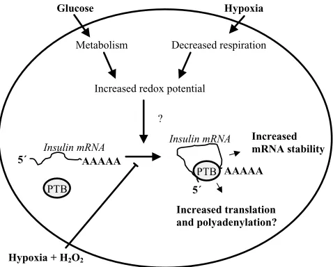
Fig. 6. Drawing showing the putative role of PTB in the regulation of insulin mRNA stability. We hypothesize that glucose and hypoxia increase binding of PTB to the pyrimidine-rich sequence in ins-PRS by their ability to increase the redox potential of the cell. Interaction between PTB and the ins-PRS leads to an increased stability of the mRNA. Because PTB has been shown, in other systems, to participate in both trans-lation and polyadenytrans-lation processes, it is possible that PTB might act by forming a loop of the insulin mRNA. Circulariza-tion of RNA is known to promote both higher messenger sta-bility, as well as a more efficient translation. So far the hypoxia signaling pathways leading to increase ins-PRS–PTB binding are unknown. Because exogenous addition of hydrogen perox-ide (H2O2) decreased the hypoxia-induced ins-PRS–PTB
inter-action, it is possible that the changes of the redox potential and/or reactive oxygen species mediate the hypoxia- and glucose-induced signals.
Increased mRNA stability Glucose Hypoxia
Increased translation and polyadenylation?
Hypoxia + H2O2
Insulin mRNA
Metabolism Decreased respiration
Increased redox potential
?
AAAAA 5´
Insulin mRNA
PTB 5´
AAAAA
insulin mRNA levels results in an increased release of insulin. In this context, it could be speculated that hypoxia-induced insulin mRNA stabilization is of minor physiologic importance and that the  cell instead utilizes a phylogenetically older signaling system to achieve glucose-regulated insulin gene ex-pression.
We have previously identified an interaction be-tween the rat insulin I 3-UTR and the protein PTB. This binding seems to be involved in regulation of insulin mRNA stability, and can be enhanced by glucose and reducing compounds (1). We show here that hypoxia increases insulin mRNA levels in -cells in vitro. In addition, PTB binding to the ins-PRS was stimulated in parallel, and we conclude that this latter event is necessary, but not sufficient, for maintained mRNA stability. Finally, the hypoxia-induced PTB and ins-PRS interaction was prevented by hydrogen peroxide, implicating the involvement of reactive oxygen species and the importance of the redox status for regulated PTB binding (Fig. 6). Future studies using nonrodent islets will hopefully clarify whether hypoxia-induced stabilization of in-sulin mRNA is a rodent-specific phenomenon. In addition, it should also be determined whether the (rodent) insulin-producing cell promotes glucose-induced stabilization of insulin mRNA by at least in part the same mechanisms as those induced by hy-poxia and through which signaling pathways this is mediated.
Acknowledgments
The excellent technical assistance of Ing-Marie Mörsare and Ing-Britt Hallgren is gratefully acknowledged. We also thank Per-Ola Carlsson for performing the oxygen tension measurements and David Helfman and colleagues for the generous PTB antibody gift.
This work was supported by grants from the Swedish Medical Research Council (109, 12X-11564, 72P-12995), the Swedish Diabetes Association, the Nordic Insulin Fund, the Juvenile Diabetes Foun-dation International, and the Family Ernfors Fund.
References
1. Tillmar L, Carlsson C, Welsh N. (2002) Control of insulin mRNA stability in rat pancreatic islets: regulatory role of a 3 -UTR pyrimidine-rich sequence. J. Biol. Chem.277:1099–1106. 2. Welsh M, Nielsen DA, MacKrell AJ, Steiner DF. (1985) Con-trol of insulin gene expression in pancreatic beta cells and in an insulin-producing cell line, RIN-5F cells. II. Regulation of insulin mRNA stability. J. Biol. Chem.260:13590–13594. 3. Wicksteed B, Herbert TP, Alarcon C, Lingohr MK, Moss LG,
Rhodes CJ. (2001) Cooperativity between the preproinsulin mRNA untranslated regions is necessary for glucose-stimulated translation. J. Biol. Chem.276:22553–22558. 4. Li HP, Huang P, Park S, Lai MM. (1999) Polypyrimidine
tract-binding protein binds to the leader RNA of mouse hepatitis virus and serves as a regulator of viral transcription. J. Virol.
73:772–777.
5. Ashiya M, Grabowski PJ. (1997) A neuron-specific splicing switch mediated by an array of pre-mRNA repressor sites:
evidence of a regulatory role for the polypyrimidine tract binding protein and a brain-specific PTB counterpart. RNA3: 996–1015.
6. Markovtsov V, Nikolic JM, Goldman JA, Turck CW, Chou MY, Black DL. (2000) Cooperative assembly of an hnRNP complex induced by a tissue-specific homolog of polypyrim-idine tract binding protein. Mol. Cell. Biol.20:7463–7479. 7. Sickinger S, Schweizer M. (1999) A high affinity binding site
for the polypyrimidine tract binding protein (PTB) is located in the 5-untranslated region of the rat proteinase alpha1-inhibitor 3 variant I gene. Biol. Chem.380:1217–1223. 8. Kim JH, Hahm B, Kim YK, Choi M, Jang SK. (2000)
Protein-protein interaction among hnRNPs shuttling between nu-cleus and cytoplasm. J. Mol. Biol.298:395–405.
9. Moreira A, Takagaki Y, Brackenridge S, Wollerton M, Manley JL, Proudfoot NJ. (1998) The upstream sequence element of the C2 complement poly(A) signal activates mRNA 3end formation by two distinct mechanisms. Genes Dev. 12: 2522–2534.
10. Lou H, Helfman DM, Gagel RF, Berget SM. (1999) Polypyrimidine tract-binding protein positively regulates inclusion of an alternative 3-terminal exon. Mol. Cell. Biol.19: 78–85.
11. Cote CA, Gautreau D, Denegre JM, Kress TL, Terry NA, Mowry KL. (1999) A Xenopus protein related to hnRNP I has a role in cytoplasmic RNA localization. Mol. Cell.4:431–437. 12. Wagner EJ, Garcia-Blanco MA. (2001) Polypyrimidine tract
binding protein antagonizes exon definition. Mol. Cell. Biol.21: 3281–3288.
13. Webb GC, Akbar MS, Zhao C, Steiner DF. (2000) Expression profiling of pancreatic beta cells: glucose regulation of secre-tory and metabolic pathway genes. Proc. Natl. Acad. Sci. U.S.A.
97:5773–5778.
14. John NE, Andersen HU, Fey SJ, et al. (2000) Cytokine- or chemically derived nitric oxide alters the expression of pro-teins detected by two-dimensional gel electrophoresis in neonatal rat islets of Langerhans. Diabetes49:1819–1829. 15. Leffers H, Dejgaard K, Celis JE. (1995) Characterisation of
two major cellular poly(rC)-binding human proteins, each containing three K-homologous (KH) domains. Eur. J. Biochem.230:447–453.
16. Makeyev AV, Liebhaber SA. (2000) Identification of two novel mammalian genes establishes a subfamily of KH-domain RNA-binding proteins. Genomics67:301–316. 17. Paulding WR, Czyzyk-Krzeska MF. (1999) Regulation of
ty-rosine hydroxylase mRNA stability by protein-binding, pyrimidine-rich sequence in the 3-untranslated region. J. Biol. Chem.274:2532–2538.
18. Czyzyk-Krzeska MF, Bendixen AC. (1999) Identification of the poly(C) binding protein in the complex associated with the 3untranslated region of erythropoietin messenger RNA.
Blood93:2111–2120.
19. Yu J, Russell JE. (2001) Structural and functional analysis of an mRNP complex that mediates the high stability of human beta-globin mRNA. Mol. Cell. Biol.21:5879–5888.
20. Holcik M, Liebhaber SA. (1997) Four highly stable eukary-otic mRNAs assemble 3untranslated region RNA- protein complexes sharing cis and trans components. Proc. Natl. Acad. Sci. U.S.A. 94:2410–2414.
21. Andersson A. (1978) Isolated mouse pancreatic islets in cul-ture: effects of serum and different culture media on the in-sulin production of the islets. Diabetologia14:397–404. 22. Carlsson PO, Palm F, Andersson A, Liss P. (12001) Markedly
decreased oxygen tension in transplanted rat pancreatic islets irrespective of the implantation site. Diabetes 50:489–495. 23. Saldeen J. (2000) Cytokines induce both necrosis and
apop-tosis via a common bcl-2- inhibitable pathway in rat insulin-producing cells. Endocrinology141:2003–2010.
24. Welsh, N. (2000) Assessment of apoptosis and necrosis in isolated islets of Langerhans: methodological considerations.
Biochem. Res.3:189–200.
full-length complementary DNAs corresponding to rat in-sulins I and II. Proc. Natl. Acad. Sci. U.S.A.76:5036–5040. 26. Green LC, Wagner DA, Glogowski J, Skipper PL, Wishnok
JS, Tannenbaum SR. (1982) Analysis of nitrate, nitrite, and [15N] nitrate in biological fluids. Anal. Biochem. 126: 131–138.
27. Grossman JS, Meyer MI, Wang YC, Mulligan GJ, Kobayashi R, Helfman DM. (1998) The use of antibodies to the polypyrimidine tract binding protein (PTB) to analyze the protein components that assemble on alternatively spliced pre-mRNAs that use distant branch points. RNA4:613–625. 28. Malaisse WJ, Malaisse-Lagae F, Sener A. (1984) Coupling
factors in nutrient-induced insulin release. Experientia 40: 1035–1043.
28. Berne C, Brolin SE, Agren A. (1973) Influence of ischemia on the levels of reduced pyridine nucleotides in the pancreatic islets. Horm. Metab. Res.5:141–142.
29. Levy AP, Levy NS, Goldberg MA. (1996) Hypoxia-inducible protein binding to vascular endothelial growth factor mRNA and its modulation by the von Hippel-Lindau protein. J. Biol. Chem.271:25492–25497.
30. Czyzyk-Krzeska MF. (1997) Molecular aspects of oxygen sensing in physiological adaptation to hypoxia. Respir. Physiol.
110:99–111.
31. Semenza GL. (1999) Perspectives on oxygen sensing. Cell98: 281–284.
32. Wenger RH. (2000) Mammalian oxygen sensing, signaling and gene regulation. J. Exp. Biol. 203:1253–1263.
33. Lopez-Barneo J, Pardal R, Ortega-Saenz P. (2001) Cellular mechanism of oxygen sensing. Annu. Rev. Physiol. 63:259–287. 34. Huang LE, Arany Z, Livingston DM, Bunn HF. (1996) Activa-tion of hypoxia-inducible transcripActiva-tion factor depends pri-marily upon redox-sensitive stabilization of its alpha sub-unit. J. Biol. Chem.271:32253–32259.
35. Adhikary G, Premkumar DR, Prabhakar NR. (2000) Dual in-fluence of nitric oxide on gene regulation during hypoxia.
Adv. Exp. Med. Biol.475:285–292.
36. Todorov V, Gess B, Godecke A, Wagner C, Schrader J, Kurtz A. (2000) Endogenous nitric oxide attenuates erythropoietin gene expression in vivo. Pflugers Arch.439:445–448.
37. Schobersberger W, Hoffmann G, Fandrey J. (1996) Nitric ox-ide donors suppress erythropoietin production in vitro.
Pflugers Arch.432:980–985.
38. Liu Y, Christou H, Morita T, Laughner E, Semenza GL, Kourembanas S. (1998) Carbon monoxide and nitric oxide suppress the hypoxic induction of vascular endothelial growth factor gene via the 5 enhancer. J. Biol. Chem. 273: 15257–15262.
39. Huang LE, Willmore WG, Gu J, Goldberg MA, Bunn HF. (1999) Inhibition of hypoxia-inducible factor 1 activation by carbon monoxide and nitric oxide. Implications for oxygen sensing and signaling. J. Biol. Chem. 274:9038–9044. 40. Genius J, Fandrey J. (2000) Nitric oxide affects the
produc-tion of reactive oxygen species in hepatoma cells: implica-tions for the process of oxygen sensing. Free Radic. Biol. Med.
29:515–521.
41. Ankoma-Sey V, Wang Y, Dai Z. (2000) Hypoxic stimulation of vascular endothelial growth factor expression in activated rat hepatic stellate cells. Hepatology31:141–148.
42. Zierler K. (1999) Whole body glucose metabolism. Am. J. Physiol.276:E409–E426.
43. Larsen JJ, Hansen JM, Olsen NV, Galbo H, Dela F. (1997) The effect of high altitude hypoxia on glucose homeostasis in men. J. Physiol. 504: 241–249.
44. Cheng N, Cai W, Jiang M, Wu S. (1997) Effect of hypoxia on blood glucose, hormones, and insulin receptor functions in newborn calves. Pediatr. Res.41:852–856.
45. Pison CM, Chauvin C, Perrault H, et al. (1998) In vivo hy-poxic exposure impairs metabolic adaptations to a 48 hour fast in rats. Eur. Respir. J.12:658–665.
46. Narimiya M, Yamada H, Matsuba I, Ikeda YU, Tanese T, Abe M. (1982) The effect of hypoxia on insulin and glucagon se-cretion in the perfused pancreas of the rat. Endocrinology 111: 1010–1014.
47. Malaisse WJ, Rasschaert J, Zahner D, Sener A. (1988) Hexose metabolism in pancreatic islets: the Pasteur effect. Diabetes Res.
7:53–58.
48. Ohta M, Nelson D, Nelson J, Meglasson MD, Erecinska M. (1990) Oxygen and temperature dependence of stimulated insulin secretion in isolates rat islets of Langerhans. J. Biol. Chem. 265:17525–17532.
49. Dionne KE, Colton CK, Yarmush ML. (1993) Effects of hy-poxia on insulin secretion by isolated rat and canine islets of Langerhans. Diabetes42:12–21.
50. Papas KK, Long Jr RC, Sambanis A, Constantinidis I. (1999) Development of a Bioartificial pancreas: II. Effects of oxygen on long-term entrapment TC3 cell cultures. Biotechnol. Bioeng.
66:231237.
51. Dionne KE, Colton CK, Yarmush ML. (1991) A microperfu-sion system with environmental control for studying insulin secretion by pancreatic tissue. Biotechnol. Prog.7:359–368. 52. Dionne KE, Colton CK, Yarmush ML. (1989) Effect of oxygen
on isolates pancreatic tissue. ASAIO Transactions35:739–741. 53. Papas KK, Long Jr RC, Sambanis A, Constantinidis I. (1996)