R E S E A R C H
Open Access
Clinical implementation of whole-genome array
CGH as a first-tier test in 5080 pre and postnatal
cases
Sang-Jin Park
1†, Eun Hye Jung
1, Ran-Suk Ryu
1, Hyun Woong Kang
2, Jung-Min Ko
3, Hyon J Kim
3,
Chong Kun Cheon
4, Sang-Hyun Hwang
5and Ho-Young Kang
1*†Abstract
Background:Array comparative genomic hybridization (CGH) is currently the most powerful method for detecting chromosomal alterations in pre and postnatal clinical cases. In this study, we developed a BAC based array CGH analysis platform for detecting whole genome DNA copy number changes including specific micro deletion and duplication chromosomal disorders. Additionally, we report our experience with the clinical implementation of our array CGH analysis platform. Array CGH was performed on 5080 pre and postnatal clinical samples from patients referred with a variety of clinical phenotypes.
Results:A total of 4073 prenatal cases (4033 amniotic fluid and 40 chorionic villi specimens) and 1007 postnatal cases (407 peripheral blood and 600 cord blood) were studied with complete concordance between array CGH, karyotype and fluorescencein situhybridization results. Among 75 positive prenatal cases with DNA copy number variations, 60 had an aneuploidy, seven had a deletion, and eight had a duplication. Among 39 positive postnatal cases samples, five had an aneuploidy, 23 had a deletion, and 11 had a duplication.
Conclusions:This study demonstrates the utility of using our newly developed whole-genome array CGH as first-tier test in 5080 pre and postnatal cases. Array CGH has increased the ability to detect segmental deletion and duplication in patients with variable clinical features and is becoming a more powerful tool in pre and postnatal diagnostics.
Background
Array comparative genomic hybridization (CGH) was developed as a genome wide screening strategy for detect-ing DNA copy number changes mainly in chromosomal disorders and cancer research [1-4]. Chromosomal abnormalities are a major cause of congenital and develop-mental abnormalities in human genetic diseases including dysmorphic features, mental retardation and developmen-tal delay, and multiple congenidevelopmen-tal anomalies. Karyotype analysis has been the gold standard for pre and postnatal diagnosis. Using traditional cytogenetic techniques, chro-mosomal imbalances such as aneuploidies and segmental abnormalities must be larger than about 3~5 Mb to be detected by GTG banding. In the past two decades,
various traditional banding techniques have been com-bined with molecular cytogenetic technologies to improve the resolution at which genomic changes can be detected. Array CGH technology has higher resolution and excellent throughput when compared to conventional and molecu-lar cytogenetics. In array CGH, genomic DNA from the patient and reference are labeled with different fluorescent dyes and co-hybridized to an array matrix containing cloned DNA. The content of an array may include specific targeted regions of the genome or the entire genome arrayed on a single glass slide. Similar to conventional CGH, genome imbalances are quantified by analyzing the ratio of the two fluorescent hybridizing signals. The reso-lution of array CGH is determined by the size and number of the clones placed on the array to interrogate genome copy number changes. Whole genome or targeted array CGH is a powerful tool to accurately detect subtelomeric rearrangements. Array CGH has higher resolution and * Correspondence: [email protected]
†Contributed equally
1MG MED, Inc., Seoul, Korea
Full list of author information is available at the end of the article
powerful clinical utility when compared with conventional and other molecular cytogenetic technologies. Array CGH has successfully detected DNA copy number changes, and several groups have studied the clinical application of this technology in both prenatal and postnatal samples [5-9]. Recently, The International Standard Cytogenomic Array (ISCA) Consortium reported consensus statement on the use of chromosomal microarray as a first-tier diagnostic test in the evaluation of individuals with developmental delays and/or congenital anomalies [10,11]. Our group successfully developed and validated a bacterial artificial chromosome (BAC) based array CGH analysis platform including analysis software [12]. This BAC array CGH analysis platform is based on 1440 fluorescencein situ hybridization (FISH) verified BAC clones that were selected among 100,000 BAC clones constructed by the Asian Genome Project [13] and validated by end-sequen-cing and FISH. This array CGH contains whole genomic regions including 356 major cell-growth related genes and over 40 known DNA copy number change disorders.
In this study, we applied our newly developed array CGH platform to 5080 clinical pre and postnatal cases and identified 114 abnormal cases with 75 prenatal and 39 postnatal cases. Our results support previous reports of the utility of array CGH for detecting chromosomal DNA copy number variations in prenatal and postnatal clinical cases.
Materials and methods 1) Clinical samples
We analyzed the results obtained from 5080 clinical cases referred to MG MED laboratories for array CGH analysis between April 2007 and December 2009. We studied 4073 prenatal cases (4033 Amniotic Fluid [AF] and 40 Chorionic Villi [CV]) and 1007 postnatal cases (407 Peripheral Blood [PB] and 600 Cord Blood [CB]). The indications for the 4073 prenatal cases were family history, advanced maternal age, fetal ultrasound anoma-lies, elevated serum alpha fetoprotein, and parental anxi-ety. The indications for the 407 postnatal cases were mainly developmental delay, mental retardation, and multiple congenital abnormalities. The 600 postnatal cord blood samples were collected from the cord blood bank for detecting genomic imbalances, which were sub-mitted by obstetricians, pediatricians, and geneticists. All samples were well prepared for experiments using pre-viously described methods [12]. Appropriate ethical approval was obtained, and informed consent for the genetic testing was obtained from all patients.
2) Development of the array CGH analysis platform
We developed an BAC based array CGH analysis plat-form for detecting genomic imbalances in human genetic diseases [12]. Our array CGH chip consists of
1440 non-overlapping BAC clones (MACArray Karyo 1440 BAC-chip, Macrogen, Seoul, South Korea). A total of 1440 clone locations are shown on this website (http://www.macrogen.co.kr/eng/biochip/genelist_over-view.html), which were selected among 100,000 BAC clones constructed by the Asian Genome Project [13,14] and carefully mapped, end-sequenced, and fluorescently labeled by FISH. All clones were two-end sequenced using an ABI PRISM 3700 DNA Analyzer (Applied Bio-systems, Foster City, CA), and their sequences were Blast analyzed and mapped according to their positions as described in the University of California, Santa Cruz (UCSC) human genome database (http://www.genome. ucsc.edu). Confirmation of the locus specificity of the chosen clones was performed by removing multiple loci-binding clones by individual examination using standard FISH procedures as described previously.
3) Array CGH and data analysis
DNA was extracted from AF, CB, PB and CV using the PureGene kit (Gentra Systems, Minneapolis, MN). After extracting the DNA, we labeled 50~500 ng of both test and reference DNA with Cy 3- and Cy 5-dCTP (Perkin Elmer) by a random priming method using Exo-Klenow Fragment (Invitrogen, Carlsbad, CA) for 16 hr. labeled according to the manufacturer’s recommendations. Reported here are pre and postnatal cases tested using the BAC chip. The array CGH chip data were analyzed using the chromofluor image analysis system (Array Analysis; Macrogen, South Korea). The slides contained 1440 human BAC clones including specific loci of more than 40 chromosomal disorders and 356 cell growth related genes from BAC libraries at a resolution average of 2.3 Mb for the entire genome. The human DNA source for making the BAC library was human sperm derived from a Korean man. Each BAC clone was repre-sented on an array as triplicate spots, and each array was scanned using a GenePix4000B scanner (Axon Instruments, Foster City, CA, USA) and analyzed with array software (MAC VIEWER, Macrogen, South Korea). Green (test) to red (reference) (G/R) ratios were automatically determined for each sample, and the nor-malized G/R ratio represented the relative average num-ber of copies of the sequence for those spots that were selected as controls. Spots with G/R ratios more than the mean plus 2.5 standard deviations (1.25) were con-sidered amplifications or gains of the indicated copy number; less than the mean minus 2.5 deviations (-0.75) were considered losses of the copy number.
4) Karyotype and FISH with region-specific probes analyses
fluid and peripheral blood samples obtained from the patient and available parents. Metaphase preparations were analyzed by G-banding techniques. FISH studies on interphase or metaphase spreads with specific probes were performed following the manufacturer’s protocols (Macrogen). FISH was performed with specific BAC clones to confirm the array CGH analysis. FISH was performed mainly with two color probes; the specific BAC probes labeled with Cy3 (red) and the control probe (green) with FITC. The specific BAC probes were constructed using BAC clone DNA (NCBI build35). FISH was performed on metaphase chromosomes cul-tured from amniotic fluid and peripheral blood lympho-cytes from each patients and available parents. Image acquisition of metaphase cells and subsequent karyotyp-ing were performed uskaryotyp-ing the CytoVision system (Applied Imaging, Santa Clara, CA, USA). A karyotype was characterized according to the conventions of the International System for Human Cytogenetic Nomencla-ture (ISCN, 2009).
Results
Array CGH is currently the most powerful method for simultaneously detecting genomic alterations. We per-formed whole genome array CGH using CGH array slides containing 1440 clones including about 40 chro-mosomal disorders specific loci and 356 cell growth related genes from BAC libraries in 5080 pre and post-natal cases (Table 1). We simultaneously performed FISH and a G-banding analysis to confirm abnormal array CGH results. In all prenatal cases, we performed array CGH analysis and concurrent karyotype analysis. We detailed genotype-phenotype correlations as far as possible in postnatal cases.
Of 4073 prenatal cases (4033 AF and 40 CV speci-mens), we identified 75 positive cases (75/4073 = 1.8%) with DNA copy number variations; 60 had an aneu-ploidy, seven had a deletion, and eight had a duplication (Table 2). Thirty-six cases had an autosomal aneuploidy, and 24 cases had a sex chromosome aneuploidy. The detection rate of overall chromosomal rearrangements
including balanced translocations and inversions by kar-yotype analysis was 3.8% (155/4073). In prenatal cases, we identified deletion/micro deletion breakpoints (2q13, 7q11.23, 17p11.2, Xp22.31, and Xq24qter) and duplica-tion/micro duplication chromosomes (1q42q44, 15q11.2q12, 21q11.2, Xp22.31, Xp21.2, and Xq27.2qter). A heterozygous micro deletion at 2q13, which includes the Joubert syndrome (2q13 homozygous deletion) criti-cal region, was observed in two cases. The array CGH results were normal in two marker chromosome cases. The marker chromosomes seemed to consist of mainly heterochromatin or this may have been due to the lim-ited array CGH coverage.
A total of 1007 postnatal cases (407 PB and 600 CB) were studied with complete concordance between array CGH, karyotype and FISH results. Among the postnatal cases with 39 positive cases, five had an aneuploidy, 23 had a deletion, and 11 had a duplication (Table 3). We identi-fied breakpoints on 17 types of deletion/micro deletion and seven types of duplication/micro duplication chromosomes. The most common indications for referral in the 407 PB cases were developmental delay, mental retardation, conge-nital abnormalities, and dysmorphic features. We identified 34 deletion/duplication cases (34/407 = 8.3%) in 407 PB cases with clinical indications. In 600 CB cases for screen-ing genomic imbalances from the cord blood bank, we found four deletion (15q11.2, 22q11.2, and Xp11.2petr) cases and one duplication (2q13) case (5/600 = 0.83%). Thirty-four cases had segmental gains or losses associated with chromosomal deletion (micro deletion) or duplication (micro duplication), including Wolf-Hirschhorn syndrome (WHS)(4p16.3), Cri-du-chat syndrome (5p15.3), Soto’s syn-drome (5q35.3), William’s syndrome (7q11.23), Digeorge syndrome 2 (10p14), Prader Willi/Angelman syndrome (15q11.2), Digeorge syndrome (22q11.2), steroid sulfatase deficiency (Xp22.31), Cat eye syndrome (22q11.2), and Emanuel syndrome (22q11, 11q23). We performed array CGH, and the molecular cytogenetic results are shown in Figure 1 (examples of prenatal deletion/duplication cases) and 2 (examples of postnatal deletion/duplication cases). We also found clinical features in the postnatal cases. For
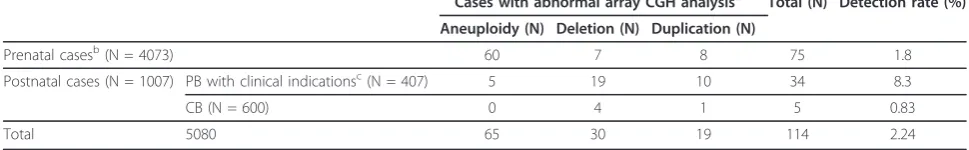
Table 1 Summary of array CGH analysis in 5080 cases
Cases with abnormal array CGH analysisa Total (N) Detection rate (%)
Aneuploidy (N) Deletion (N) Duplication (N)
Prenatal casesb(N = 4073) 60 7 8 75 1.8
Postnatal cases (N = 1007) PB with clinical indicationsc(N = 407) 5 19 10 34 8.3
CB (N = 600) 0 4 1 5 0.83
Total 5080 65 30 19 114 2.24
PB, Peripheral Blood; CB, Cord Blood for banking in general population
a
Karyotype and FISH analyses performed with array CGH anaysis. FISH analyses were performed using specific bacterial artificial chromosome (BAC) clones.
b
AF, Amniotic Fluid (N = 4033); CV, Chorionic Villi (N = 40)
c
example, the postnatal case 10 patient with Sotos syndrome was a 1.1-year-old male with a birth weight of 4.1 kg. He had typical facial features including macrocephaly, sparse-ness of frontotemporal hair, high bossed forehead, a long narrow face and prominent narrow jaw, developmental delay, overgrowth, abnormally large hands, and a café au lait spot. We identified not only simple deletion and dupli-cation but also multiple copy number abnormalities includ-ing various chromosomal rearrangements (postnatal cases 4, 24, 27, and 30). Additionally, postnatal case 4 was identi-fied with multiple rearrangements. Although we found a 2q14q21.1 deletion using array CGH, we were able to char-acterize accurate cytogenetic features as 46,XY, der(2)t(2;6) (q21.1;q23)inv(2)(q14.1q37) and der(6)t(2;6)(q21.1;q23) by concurrent karyotype analysis (data not shown). In case 30,
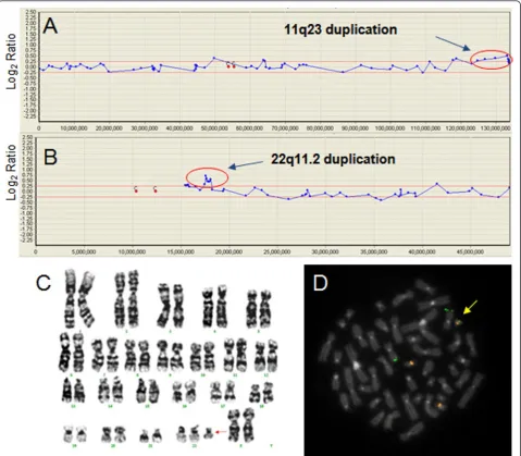
we identified two chromosomes involved as a small super-numerary marker chromosome (sSMC) as 47,XX, +der(22) t(11;22)(q23;q11) with mental retardation (Figure 2).
Discussion
Array CGH offers a high-resolution genome analysis in a clinical setting [15]. Currently, many commercial and academic laboratories have used BAC-based array, oligo-nucleotide-based array or SNP-based arrays. Array reso-lution is dependent on the number and types of probes used and how they are designed on the genome. Current commercial BAC arrays (targeted or whole genome) generally have 600~5200 BAC clones (e.g. PerkinElmer). Our whole-genome BAC array CGH platform is based on 1440 fluorescence in situ hybridization (FISH)
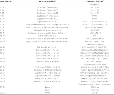
Table 2 Summary of array CGH and cytogenetic analyses in 4073 prenatal casesa
Case (number) Array CGH analysisb Cytogenetic analysesc
Aneuploidy
1 (1) Duplication of whole chr.13 Trisomy 13
2 (7) Duplication of whole chr.18 Trisomy 18
3 (28) Duplication of whole chr.21 Trisomy 21
4 (6) Duplication of whole chr.X 47,XXX
5 (7) Duplication of whole chr.X 47,XXY
6 (1) Duplication of whole chr.X Mos 47,XXY [18]/48,XXY,+17 [2]
7 (1) Copy number ratio of less than one copy loss at chr. 9 Mos 47,XXX [29]/48,XXX,+9 [11] 8 (1) Copy number ratio of less than one copy loss at chr.X Mos 47,XXY [32]/46,XY [8]
9 (1) Duplication of whole chr.X and 18 48,XXY,+18
10 (1) Duplication of long arm at Xq10qter/whole chr. 9 47,X,i(X)(q10),+9
11 (4) Deletion of whole chr.X 45,X
12 (1) Copy number ratio of less than one copy loss at chr.X Mos 45,X [22]/46,XX [8] 13 (1) Copy number ratio of less than one copy loss at chr.X Mos 45,X [10]/46,XX [20] Deletion/Microdeletion
14 (2) Deletion of 0.5Mb at 2q13 46,XX,ish del(2)(q13q13)(NPHP1-)
15 (1) Deletion of 0.4Mb at 7q11.23 46,XY.ish del(7)(q11.23q11.23)(ELN-) 16 (2) Deletion of 0.4Mb at 17p11.2 46,XX.ish del(17)(p11.2p11.2)(D17S29-)
Deletion of 0.4Mb at 17p11.2 46,XY.ish del(17)(p11.2p11.2)(D17S29-) 17 (1) Deletion of 0.5Mb at Xp22.31 46,XY.ish del(X)(p22.31p22.31)(STS-)
18 (1) Deletion of 25Mb at Xq24qter 46,X,del(X)(q24qter)
Duplication/Microduplication 19 (1) Duplication of 10Mb at 1q42q44 46,XY.ish dup(1)(q42.1q44)(D1S491+) 20 (2) Duplication of 1.3Mb at 15q11.2q12 46,XX.ish dup(15)(q11.2q12)(SNRPN+) 21 (2) Duplication of 1.5Mb at 22q11.2 46,XY.ish dup(22)(q11.2q11.2)(COMT+) 22 (1) Duplication of 0.5Mb at Xp22.31 46,XX.ish dup(X)(p22.31p22.31)(KAL1+) 23 (1) Duplication of 0.5Mb at Xp21.2 46,XY.ish dup(X)(p21.2p21.2)(DMD+) 24 (1) Duplication of 5Mb at Xq27.2qter. 46,XY,ish dup(X)(q27.2qter)(DX904+)
Small supernumerary marker chromosome
25 (2) Normal 47,XX,+mar
Normal 47,XY,+mar
26 (80) Normal Othersd
a
Data compiled from array CGH and cytogenetic analyses in 4033 AF and 40 CV by the major indications for prenatal testing such as advanced maternal age, fetal ultrasound anomalies, and elevated serum alpha fetoprotein .blog2mean green/red (G/R) ratios more than the mean + 2.5 SD (~ 0.25) were considered high
amplifications or gains of the indicated copy number, and less than the mean -2.5 SD (~ -2.5) were considered high losses of the copy number.c
performed by karyotype and FISH analyses. FISH analyses were performed using specific BAC clones.d
verified BAC clones, which were selected among 100,000 BAC clones constructed by the Asian Genome Project [13,14] and validated by end-sequencing and FISH. Therefore, our abnormal array CGH results were able to
be confirmed by FISH, that BAC clones were available to clinically relevant turnaround times.
In this study, we showed the utility of our whole-gen-ome BAC-based array CGH platform in a large
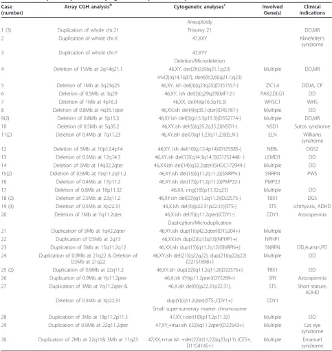
Table 3 Summary of array CGH and cytogenetic analyses in 1007 postnatal casesa
Case (number)
Array CGH analysisb Cytogenetic analysesc Involved
Gene(s)
Clinical indications
Aneuploidy
1 (3) Duplication of whole chr.21 Trisomy 21 DD,MR
2 Duplication of whole chr.X 47,XXY Klinefelter’s
syndrome
3 Duplication of whole chr.Y 47,XYY
Deletion/Microdeletion
4 Deletion of 15Mb at 2q14q21.1 46,XY, der(2)t(2;6)(q21.1;q23) Multiple DD,MR inv(2)(q14.1q37), der(6)t(2;6)(q21.1;q23)
5 Deletion of 1Mb at 3q23q25 46,XY. ish del(3)(q23q25)(D3S1557-) ZIC1,4 DD,IA, CP 6 Deletion of 0.5Mb at 3q29 46,XY, ish del(3)(q29q29)(MF12-) PAK2,DLG1 DD
7 Deletion of 1Mb at 4p16.3 46,XX, del(4)(p16.3p16.3) WHSC1 WHS
8 Deletion of 0.8Mb at 4q35.1qter 46,XX.ish del(4)(q35.1qter)(D4S187-) Multiple DD 9(2) Deletion of 0.8Mb at 5p15.3 46,XY.ish del(5)(p15.3p15.3)(D5S2774-) Multiple DD,MR 10 Deletion of 0.5Mb at 5q35.2 46,XY.ish del(5)(q35.2q35.2)(NSD1-) NSD1 Sotos syndrome 11(2) Deletion of 0.4Mb at 7q11.23 46,XY.ish del(7)(q11.23q11.23)(ELN-) ELN Williams
syndrome 12 Deletion of 5Mb at 10p12.4p14 46,XY. ish del(10)(p12.4p14)(D10S585-) NEBL DGS2 13 Deletion of 0.5Mb at 12q14.3 46,XY.ish del(12)(q14.3q14.3)(D12S1448- ) LEMD3 DD 14 Deletion of 5Mb at 14q32.2qter 46,XX.ish del(14)(q32.2qter)(SHGC172944-) Multiple DD 15(2) Deletion of 0.5Mb at 15q11.2q11.2 46,XY.ish del(15)(q11.2q11.2)(SNRPN-) SNRPN PWS 16 Deletion of 0.4Mb at 17p11.2 46,XY.ish del(17)(p11.2p11.2)(PMP22-) PMP22
17 Deletion of 0.8Mb at 18p11.32 46,XX, ring(18)(p11.32q23) Multiple DD
18 (2) Deletion of 2.5Mb at 22q11.2 46,XY.ish del(22)(q11.2q11.2)(D22S75-) TBX1 DGS 19 (3) Deletion of 0.5Mb at Xp22.31 46,X.ish del(X)(p22.31p22.31)(STS-) STS ichthyosis, ADHD 20 Deletion of 1Mb at Yq11.2qter 46,X.ish del(Y)(q11.2qter)(CDY1-) CDY1 Azoospermia
Duplication/Microduplication
21 Duplication of 5Mb at 1q42.2qter 46,XY.ish dup(1)(q42.2qter)(D1S204+) Multiple 22 Duplication of 0.5Mb at 2q13 46,XX.ish dup(2)(q13q13)(NPHP1+) NPHP1
23 Duplication of 3Mb at 15q11.2q12 46,XX.ish dup(15)(q11.2q12)(SNRPN+) SNRPN DD,Autism,PD 24 Duplication of 0.9Mb at 21q22 & Deletion of
0.5Mb at 21q22
46,XY.ish del(21)(q22q22), dup(21)(q22q22) (D21S1898+)
Multiple DD
25 (2) Duplication of 0.9Mb at 22q11.2 46,XY.ish dup(22)(q11.2q11.2)(D22S75+) TBX1 DD 26 Duplication of 0.9Mb at Yp11.2pter 46,X.ish i(Y)(p11.2pter)(DYS289+) SRY Azoospermia 27 Duplication of 3Mb at Yq11.2qter & 46,X.ish del(X)(p22.31p22.31), STS Short stature,
ADHD Deletion of 0.5Mb at Xp22.31 dup(Y)(q11.2qter)(STS-,CDY1+) CDY1
Small supernumerary marker chromosome
28 Duplication of 3Mb at 18p11.2p11.3 47,XY,+der(18)(p11.2p11.32) Multiple DD 29 Duplication of 0.9Mb at 22q11.2qter 47,XY,+mar.ish i(22)(q11.2qter)(D22S43+) Multiple Cat eye
syndrome 30 Duplication of 2Mb at 22q11& 3Mb at 11q23 47,XX,+mar.ish +der(22)t(11;22)(q23;q11) (CES+,
D11S4145+)
Multiple Emanuel syndrome
DD, developmental delay; MR, mental retardation; IA, imperforated anus; CP, cleft palate; WHS, Wolf-Hirschhorn syndrome; DGS, DiGeorge syndrome; PWS, Prader-Willi syndrome; ADHD, Attention Deficit Hyperactivity Disorder; PD, Pigmentation disorder
a
Data compiled from 407 PB and 600 CB.
b
log2mean green/red ratios more than the mean +2.5 SD (~ 0.25) were considered high amplifications or gains of the indicated copy number, and less than the
mean -2.5 SD (~ -0.25) were considered high losses of the copy number.
c
collection of clinical cases. Previous prenatal studies reported chromosomal abnormalities in ~1.3% of cases [16]. Furthermore, previous postnatal studies have shown that array CGH with cytogenetic analysis detects clinically significant chromosome abnormalities in ~7% of children referred for cytogenetic testing [16]. In this study, the prenatal detection rate was 1.8% (75/4073) and the postnatal detection rate with clinical indications was 8.3% (34/407) using array CGH (Table 1). These results are consistent with previous reports [17]. In
We also identified 15q11.2 and 22q11.2 duplications (cases 20 and 21). These duplication copy number varia-tion phenotypes appear to be generally mild and highly variable; findings ranged from apparently normal to mental retardation, growth retardation, and autism (15q11.2 duplication). Therefore, prenatal array CGH results must be evaluated whether the results show benign variants or pathogenic variants. Currently, many useful copy number variation data base websites are available. For example, the Database of Genomic Var-iants (http://projects.tcag.ca/variation/), DECIPHER (http://decipher.sanger.ac.uk/), and Genereviews (http:// www.genetests.org/), which is a more clinically useful
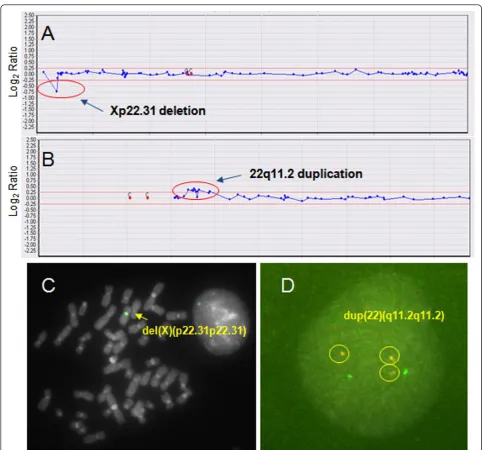
website that lists the associated genetic diseases includ-ing phenotypes, genotypes, and clinical management of pathogenic copy number variant diseases. Many known micro deletion syndromes are expressed by incomplete penetrance [19], whereas duplications generally produce milder and, therefore, less identifiable phenotypes than counterpart deletions [20]. A previous study suggested an effective general algorithm for clinical testing using array CGH [21]. Moreover, practical guidelines for array CGH have been issued by the American College of Medical Genetics [22,23]. It is important that, with the exception of purported benign copy number variables (CNVs), all regions showing abnormal copy number Figure 2Array CGH with cytogenetics validation data for case 30 (Table 3). (A), (B) The array CGH results for chromosomes 11 and 22. Arrows indicates duplication of 11q23 and 22q11.2. (C) G-banding karyotype result of 47,XX, +der(22)t(11;22)(q23;q11). Arrow indicates the marker
findings be characterized by FISH and parental studies. An increase in the prenatal detection rate of chromo-some abnormalities would benefit patients seeking genetic testing prior to delivery [24,25].
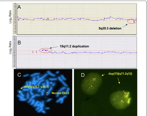
In postnatal cases, we identified breakpoints on 17 types of deletion/micro deletion, seven types of duplica-tion/micro duplication chromosomes and three sSMC types. Among the 39 positive postnatal cases, five had an aneuploidy, 23 had a deletion, and 11 had a duplica-tion (Table 3). Most postnatal cases had clinical indica-tions with developmental delay or mental retardation. Therefore, detection rates of chromosomal abnormalities using array CGH analysis are generally higher than pre-natal cases. In this study, we analyzed various patients with clinical indications and/or unknown specific indica-tions. We observed a 3q24 deletion in a patient (case 5) with developmental delay, imperforated anus, and cleft palate. Deletion of the 3q24-q26 region including the Dandy-Walker malformations (DWM) critical region (3q24) appears to be associated with a somewhat similar constellation of findings including craniofacial dys-morphism (broad and depressed nasal bridge and low set posteriorly rotated ears), mental retardation, conge-nital heart defects, and central nervous system malfor-mations [26]. A previous study reported the critical region associated with DWM, which encompasses the ZIC1 and ZIC4 genes, by mapping the 3q24 interstitial deletions in several individuals with DWM [27]. In case 10, we identified a micro deletion of the 5q35 region including the NSD1 gene in one Korean patient with Sotos syndrome (Figure 3). The most common mutation (50%) was a micro deletion of the 5q35 region including
NSD1 in Japanese patients with Sotos syndrome, while, in the UK, approximately 70% of patients with Soto syn-drome haveNSD1point mutations, and only 10% of the patients have a micro deletion of the 5q35 region [28]. Therefore, further studies will be necessary for a better understanding of Sotos syndrome in Korean patients. The most commonly observed deletions were in the Xp22.31 region. Our patients with Xp22.31 (cases 19 and 27) appeared to be associated with ichthyosis, short stature, and attention deficit hyperactivity disorder (ADHD). The Xp22.31 deletion, causing loss of function of the STS gene, is associated with steroid sulfatase defi-ciency in males. The STS gene encodes steroid sulfatase, a membrane-bound microsomal enzyme that is ubiqui-tously expressed and hydrolyzes several 3-beta-hydro-xysteroid sulfates, which serve as metabolic precursors for estrogens, androgens, and cholesterol [29]. A pre-vious study reported on a patient with a Xp22.3 intersti-tial deletion that had ichthyosis, dysmorphic features, and mental retardation with ADHD [30]. We identified duplication cases at 15q11.2q12 with developmental delay, autism, and pigmentation disorder (Figure 3). A
previous study reported that this patient had infantile autism with cytogenetic abnormalities on chromosomal region 15q11-q13, as reported in patients with autistic disorder [31]. We identified three marker chromosome origins (11, 18, and 22 chromosome segments). sSMC origin analysis is very important for evaluating clinical phenotypes. The marker chromosome origin and size appear to result in variable phenotypes [32]. In our study, we identified a 22q11.2 duplication including the CES gene and two chromosomes involved in derivative translocation marker chromosome (22q11&11q23 dupli-cation) (Figure 2). This sSMC, der(22)t(11;22)(q23;q11), is related to Emanuel syndrome, which is characterized by severe mental retardation, microcephaly, failure to thrive, preauricular tag or sinus, ear anomalies, and cleft or high-arched palate [33]. These array CGH and other cytogenetic findings are consistent with the clinical phe-notypes of Cat eye syndrome and Emmanuel syndrome with mental retardation. We also identified CNVs (2q13 duplication and 4q32 duplication) in phenotypically nor-mal individuals from cord bank blood. The clinical sig-nificance of the 2q13 duplication is still emerging, as these copy number variations are also found in phenoty-pically normal and control individuals [34]. Duplication of 4q32 is a benign copy number variation represented in a copy number variation database (http://projects. tcag.ca/variation/).
And also, it is important to address some of the lim-itations of array CGH before this test is considered for clinical diagnosis as a first line test. Array CGH does not detect polyploidy, balanced translocations, inversions and low level mosaicims. Marker chromosomes may also be missed, depending on the size, marker composi-tion and array coverage of the specific chromosomal region present on the marker chromosome. And also we must consider various limitations of array CGH testing (e.g. point mutations, uniparental disomy and appropri-ate turnaround times). Therefore, genetics laboratories must be capable of performing array CGH and concur-rent conventional cytogenetic analyses (G-banding and FISH) including molecular genetic analyses (DNA sequencing etc.) and also clinical geneticists offer appro-priate genetic counseling including interpretation of results and limitations of test for patients and family members.
Conclusions
characterize various chromosomal disorders. Further-more, the newly developed array CGH analysis platform will lead to a new understanding of genomic disorders with DNA copy number changes and their relationship to genotype and phenotype. Additionally, functional stu-dies based on the identity of the involved genes are necessary for a further understanding of the mechanism related to contiguous genes on deletion and duplication loci in the development and/or progression of various DNA copy number change disorders.
Acknowledgements
We thank Dr. He doo Chung for his contribution to clinical validation and our clinical colleagues for supporting this work. We are grateful to other laboratory members for their enthusiastic participation.
Author details
1MG MED, Inc., Seoul, Korea.2MACROGEN, Inc, Seoul, Korea.3Department of
Medical Genetics, Ajou University School of Medicine, Suwon, Korea.
4Department of Pediatrics, School of Medicine, Pusan National University
Children’s Hospital, Yangsan, Korea.5Department of Laboratory Medicine, Center for Diagnostic Oncology, National Cancer Center, Gyeonggi-do, Korea.
Authors’contributions
SJP drafted the manuscript and analysed the data for the paper. EHJ performed array CGH. RSR performed karyotyping and FISH. HWK performed FISH, relevant confirmations and participated in validation work on the BAC array CGH platform. JMK, HJK, CKC and SHH provided clinical information, evaluation and advice. HYK conceived of the study, performed data analysis and also approved the manuscript. All authors read and approved the final manuscript.
Competing interest statement
The authors declare that they have no competing interests.
Received: 30 March 2011 Accepted: 9 May 2011 Published: 9 May 2011
References
1. Park SJ, Jeong SY, Kim HJ:Y chromosome loss and other genomic alterations in hepatocellular carcinoma cell lines analyzed by CGH and CGH array.Cancer Genet Cytogenet2006,166:56-64.
2. Bejjani BA, Shaffer LG:Clinical utility of contemporary molecular cytogenetics.Annu Rev Genomics Hum Genet2008,9:71-86.
3. Shinawi M, Cheung SW:The array CGH and its clinical applications.Drug
Discov Today2008,13:760-770.
4. Albertson DG, Pinkel D:Genomic microarrays in human genetic disease and cancer.Hum Mol Genet2003,12:R145-152, Spec No 2.
5. Lu X, Shaw CA, Patel A, Li J, Cooper ML, Wells WR, Sullivan CM, Sahoo T, Yatsenko SA, Bacino CA,et al:Clinical implementation of chromosomal microarray analysis: summary of 2513 postnatal cases.PLoS One2007,2: e327.
6. Shao L, Shaw CA, Lu XY, Sahoo T, Bacino CA, Lalani SR, Stankiewicz P, Yatsenko SA, Li Y, Neill S,et al:Identification of chromosome
abnormalities in subtelomeric regions by microarray analysis: a study of 5,380 cases.Am J Med Genet A2008,146A:2242-2251.
7. Rickman L, Fiegler H, Shaw-Smith C, Nash R, Cirigliano V, Voglino G, Ng BL, Scott C, Whittaker J, Adinolfi M,et al:Prenatal detection of unbalanced chromosomal rearrangements by array CGH.J Med Genet2006, 43:353-361.
8. Evangelidou P, Sismani C, Ioannides M, Christodoulou C, Koumbaris G, Kallikas I, Georgiou I, Velissariou V, Patsalis PC:Clinical application of whole-genome array CGH during prenatal diagnosis: Study of 25 selected pregnancies with abnormal ultrasound findings or apparently balanced structural aberrations.Mol Cytogenet2010,3:24.
9. Ahn JW, Mann K, Walsh S, Shehab M, Hoang S, Docherty Z, Mohammed S, Mackie Ogilvie C:Validation and implementation of array comparative genomic hybridisation as a first line test in place of postnatal karyotyping for genome imbalance.Mol Cytogenet2010,3:9. 10. Miller DT, Adam MP, Aradhya S, Biesecker LG, Brothman AR, Carter NP,
Church DM, Crolla JA, Eichler EE, Epstein CJ,et al:Consensus statement: chromosomal microarray is a first-tier clinical diagnostic test for individuals with developmental disabilities or congenital anomalies.Am
J Hum Genet2010,86:749-764.
11. Manning M, Hudgins L:Array-based technology and recommendations for utilization in medical genetics practice for detection of chromosomal abnormalities.Genet Med2010,12:742-745.
12. Choe J, Kang JK, Bae CJ, Lee DS, Hwang D, Kim KC, Park WY, Lee JH, Seo JS: Identification of origin of unknown derivative chromosomes by array-based comparative genomic hybridization using pre- and postnatal clinical samples.J Hum Genet2007,52:934-942.
13. Kim JI, Ju YS, Park H, Kim S, Lee S, Yi JH, Mudge J, Miller NA, Hong D, Bell CJ,et al:A highly annotated whole-genome sequence of a Korean individual.Nature2009,460:1011-1015.
14. Park H, Kim JI, Ju YS, Gokcumen O, Mills RE, Kim S, Lee S, Suh D, Hong D, Kang HP,et al:Discovery of common Asian copy number variants using integrated high-resolution array CGH and massively parallel DNA sequencing.Nat Genet2010,42:400-405.
15. Cheung SW, Shaw CA, Yu W, Li J, Ou Z, Patel A, Yatsenko SA, Cooper ML, Furman P, Stankiewicz P,et al:Development and validation of a CGH microarray for clinical cytogenetic diagnosis.Genet Med2005,7:422-432. 16. Shaffer LG, Bejjani BA, Torchia B, Kirkpatrick S, Coppinger J, Ballif BC:The
identification of microdeletion syndromes and other chromosome abnormalities: cytogenetic methods of the past, new technologies for the future.Am J Med Genet C Semin Med Genet2007,145C:335-345. 17. Shaffer LG, Coppinger J, Alliman S, Torchia BA, Theisen A, Ballif BC,
Bejjani BA:Comparison of microarray-based detection rates for cytogenetic abnormalities in prenatal and neonatal specimens.Prenat
Diagn2008,28:789-795.
18. Otto EA, Helou J, Allen SJ, O’Toole JF, Wise EL, Ashraf S, Attanasio M, Zhou W, Wolf MT, Hildebrandt F:Mutation analysis in nephronophthisis using a combined approach of homozygosity mapping, CEL I endonuclease cleavage, and direct sequencing.Hum Mutat2008, 29:418-426.
19. Slavotinek AM:Novel microdeletion syndromes detected by chromosome microarrays.Hum Genet2008,124:1-17.
20. Mencarelli MA, Katzaki E, Papa FT, Sampieri K, Caselli R, Uliana V, Pollazzon M, Canitano R, Mostardini R, Grosso S,et al:Private inherited microdeletion/microduplications: implications in clinical practice.Eur J
Med Genet2008,51:409-416.
21. Edelmann L, Hirschhorn K:Clinical utility of array CGH for the detection of chromosomal imbalances associated with mental retardation and multiple congenital anomalies.Ann N Y Acad Sci2009,1151:157-166. 22. Shaffer LG, Beaudet AL, Brothman AR, Hirsch B, Levy B, Martin CL,
Mascarello JT, Rao KW,et al:Microarray analysis for constitutional cytogenetic abnormalities.Genet Med2007,9:654-662.
23. Shaffer LG:American College of Medical Genetics guideline on the cytogenetic evaluation of the individual with developmental delay or mental retardation.Genet Med2005,7:650-654.
24. Darilek S, Ward P, Pursley A, Plunkett K, Furman P, Magoulas P, Patel A, Cheung SW, Eng CM:Pre- and postnatal genetic testing by array-comparative genomic hybridization: genetic counseling perspectives.
Genet Med2008,10:13-18.
25. Sahoo T, Cheung SW, Ward P, Darilek S, Patel A, del Gaudio D, Kang SH, Lalani SR, Li J, McAdoo S,et al:Prenatal diagnosis of chromosomal abnormalities using array-based comparative genomic hybridization.
Genet Med2006,8:719-727.
26. Sudha T, Dawson AJ, Prasad AN, Konkin D, de Groot GW, Prasad C:De novo interstitial long arm deletion of chromosome 3 with facial dysmorphism, Dandy-Walker variant malformation and hydrocephalus.
Clin Dysmorphol2001,10:193-196.
27. Grinberg I, Northrup H, Ardinger H, Prasad C, Dobyns WB, Millen KJ: Heterozygous deletion of the linked genes ZIC1 and ZIC4 is involved in Dandy-Walker malformation.Nat Genet2004,36:1053-1055.
28. Kurotaki N, Stankiewicz P, Wakui K, Niikawa N, Lupski JR:Sotos syndrome common deletion is mediated by directly oriented subunits within inverted Sos-REP low-copy repeats.Hum Mol Genet2005,14:535-542. 29. Van Esch H, Hollanders K, Badisco L, Melotte C, Van Hummelen P,
Vermeesch JR, Devriendt K, Fryns JP, Marynen P, Froyen G:Deletion of VCX-A due to NAHR plays a major role in the occurrence of mental retardation in patients with X-linked ichthyosis.Hum Mol Genet2005, 14:1795-1803.
30. Lonardo F, Parenti G, Luquetti DV, Annunziata I, Della Monica M, Perone L, De Gregori M, Zuffardi O, Brunetti-Pierri N, Andria G, Scarano G:Contiguous gene syndrome due to an interstitial deletion in Xp22.3 in a boy with ichthyosis, chondrodysplasia punctata, mental retardation and ADHD.
Eur J Med Genet2007,50:301-308.
31. Ouldim K, Natiq A, Jonveaux P, Sefiani A:Tetrasomy 15q11-q13 Diagnosed by FISH in a Patient with Autistic Disorder.J Biomed Biotechnol2007, 2007:61538.
32. Crolla JA, Youings SA, Ennis S, Jacobs PA:Supernumerary marker chromosomes in man: parental origin, mosaicism and maternal age revisited.Eur J Hum Genet2005,13:154-160.
33. Kurahashi H, Shaikh TH, Hu P, Roe BA, Emanuel BS, Budarf ML:Regions of genomic instability on 22q11 and 11q23 as the etiology for the recurrent constitutional t(11;22).Hum Mol Genet2000,9:1665-1670. 34. Rudd MK, Keene J, Bunke B, Kaminsky EB, Adam MP, Mulle JG,
Ledbetter DH, Martin CL:Segmental duplications mediate novel, clinically relevant chromosome rearrangements.Hum Mol Genet2009,
18:2957-2962.
doi:10.1186/1755-8166-4-12